
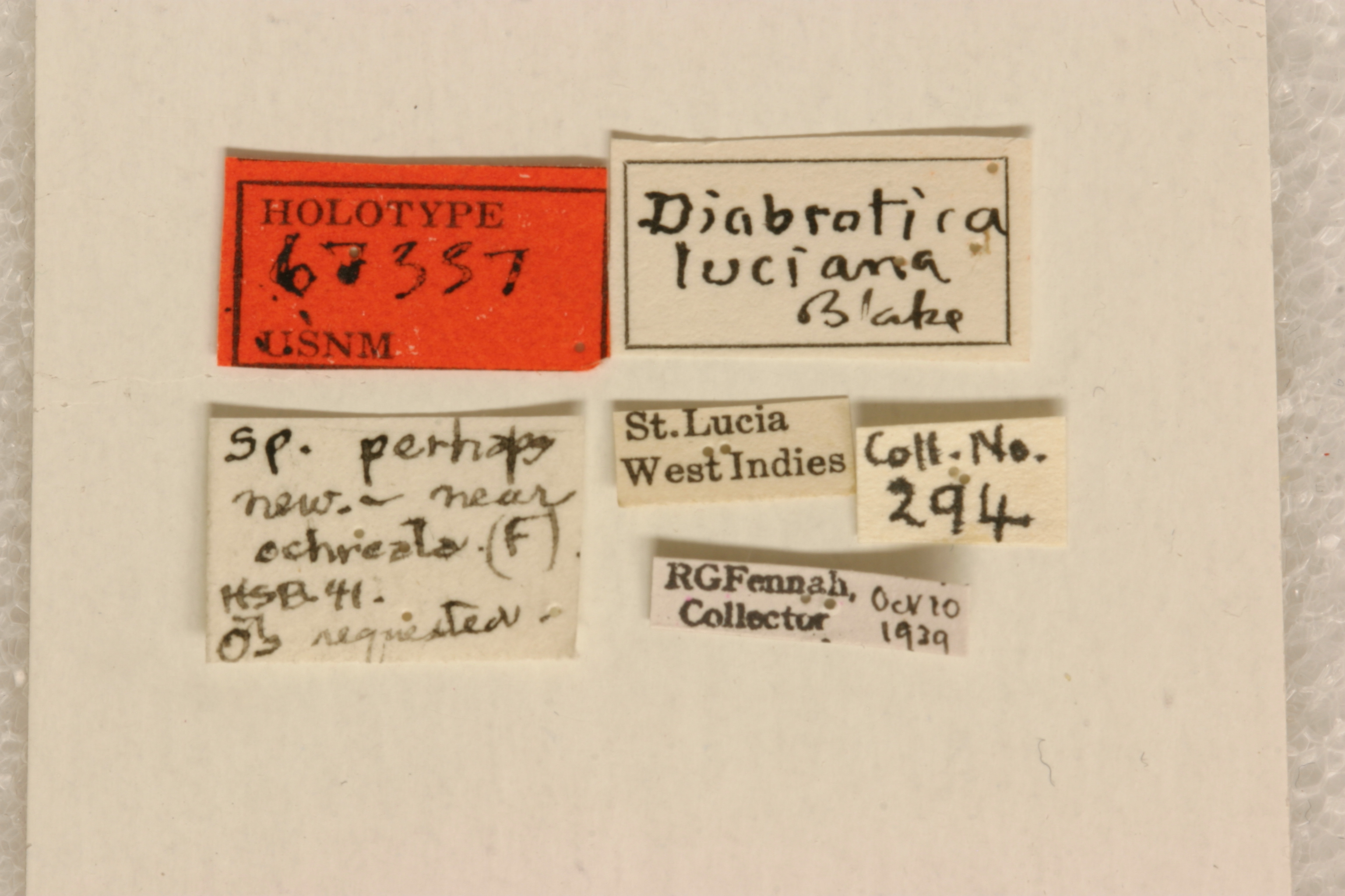
Diabrotica luciana Blake 1965: 104
St. Lucia, British West Indies
USNM, holotype, female, verified
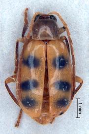
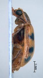
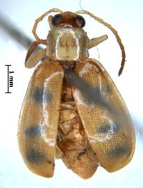
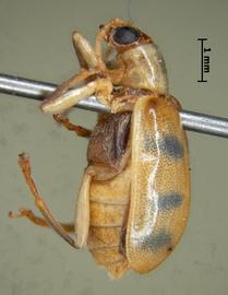
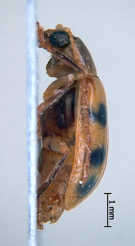
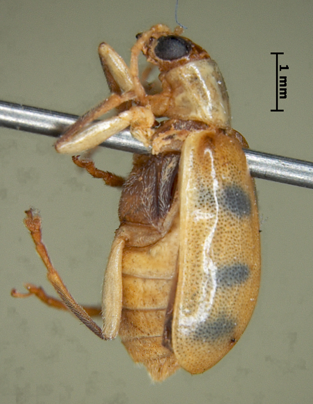
Body length 5.3-5.6 mm. Body width 2.9-3.0 mm. Head basic color yellow. Antennae filiformfiliform:
slender antennae with antennomeres of similar shape
, bi- or tricolored, antennomeres 1-3 yellow, antennomeres 4-8 gradually infuscated, antennomeres 9-11 yellow ocher. Maxillary palpi yellow or yellow ocher, labrumlabrum:
the "upper lip" of beetles, a movable sclerite joined under clypeus
yellow ocher. Pronotumpronotum:
the notum of the prothorax with highly sclerotized pronotal disc
green or pale olivine, subquadrate, nonfoveate, not shagreened. Scutellumscutellum:
small, usually triangular shield between the bases of elytra
black or amber brown. Elytra yellow or rufous, maculatemaculate:
(here) marked by maculae or patches of a different shape and size, usually clearly separated from each other
, with with fuzzy-edged round olive maculae on each elytronelytron:
<em>(pl. elytra)</em> the fore highly sclerotized wing of beetle
. Elytral epipleura completely green. Sutural anglesutural angle:
the posterior angle or apex of the elytron near the suture
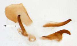
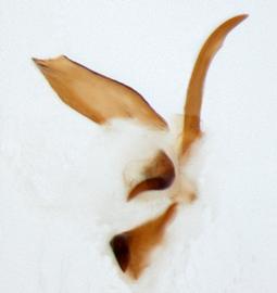
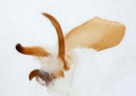
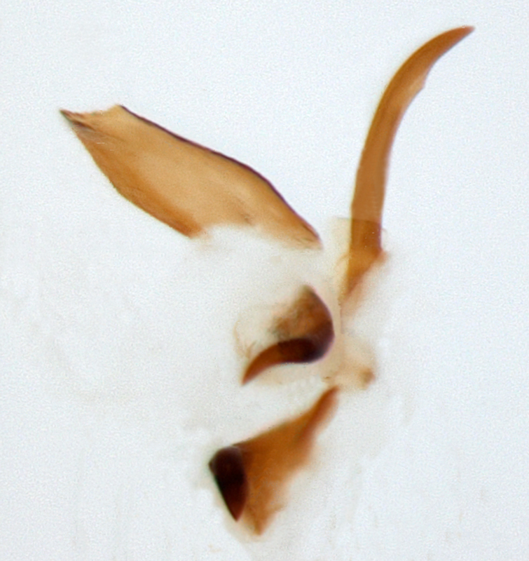
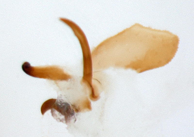
of elytra round, punctation dense, fine. Abdomen pale olivine or green. Tarsi yellow or yellow ocher, tibiae bicolored, yellow, outer edge with piceous or testaceous line, or extensively darkened. Femora uniform pale olivine. Aedeagusaedeagus:
the main sclerotized part of the male genitalia; "aedeagus" is used here instead of "median lobe of aedeagus"
symmetric, with four internal sac scleritessclerites:
(here) the sclerotized hooks, spines or plates in the internal sac
.
British West Indies (St. Lucia)
Unknown
Diabrotica luciana Blake is very similar to D. fucata (Fabricius). They can be separated by the following features: there are three maculae on the elytra of D. luciana, but two macula in D. fucata; internal sac sclerite 4c in D. fucata is longer than in D. luciana.
Authors: A. Derunkov, A. Konstantinov, A. Tishechkin, L. Hartje, and A.J. Redford
Last updated Feb. 12, 2015
idtools.org | tool images at ITP Node

