
Phytophthora pini
|
Phytophthora spp. in subclade 2c: portion of the seven-loci ML phylogeny featuring the type cultures of 212 described species (by T. Bourret). Notice the position of P. pini Ex-type CBS 181.25 = S&T BL 48. Gloria Abad, USDA S&T.
|
|
Phytophthora spp. in subclade 2c: Morphological Tabular key (PDF) and Tabular key legends (PDF) in IDphy2 KEY SECTION. Notice the data of P. pini Ex-type CBS 181.25 = S&T BL 48. Gloria Abad, USDA S&T.
|
|
Phytophthora pini (CPHST BL 48) colonies of the ex-type grown for 7 days on (a) V8® Agar, (b) potato dextrose agar, and (c) malt extract agar; photo by Krysta Jennings and Leandra Knight, USDA-APHIS-PPQ |
|
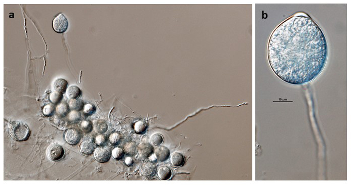
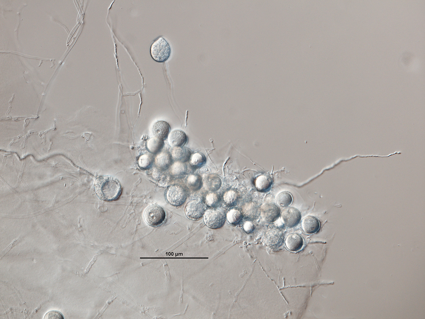
Phytophthora pini (ex-type CPHST BL 48) asexual phase formed on V8 agar flooded with soil extract: (a) sporangium originated in single long sporangiophore, oospores at bottom, (b) semipapillate persistent sporangium in single long sporangiophore; photos by G.Abad, USDA-APHIS-PPQ. |
|
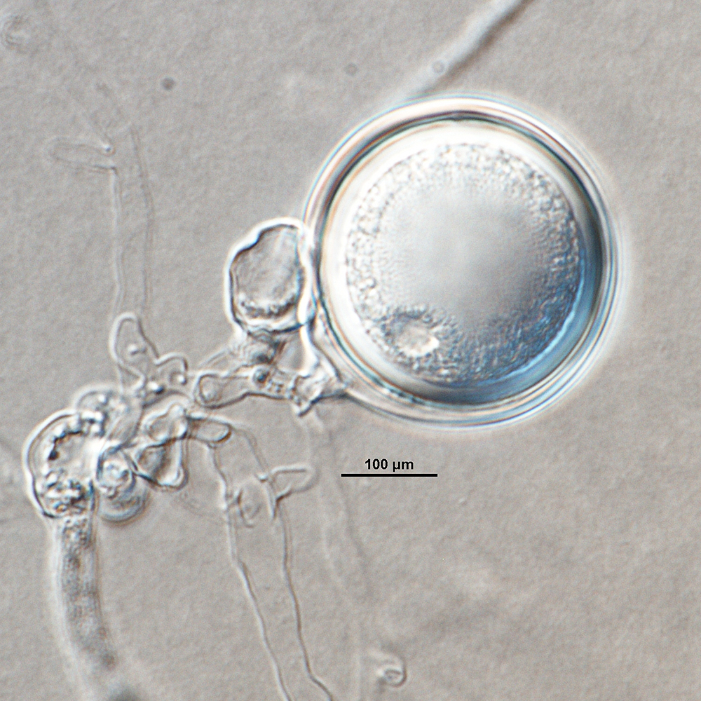
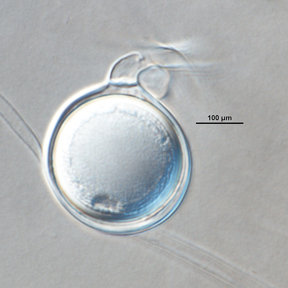
Phytophthora pini (ex-type CPHST BL 48) sexual phase formed on V8 agar flooded with soil extract: (a) smooth-walled oogonium with comma-like oogonial stalk, paragynous antheridium, and semi-aplerotic oospore, (b) plerotic oospore in smooth-wall oogonium with paragynous antheridium; photos by Gloria Abad, USDA-APHIS-PPQ. |
|
Phytophthora pini (ex-type CPHST BL 48) sexual phase formed on V8 agar flooded with soil extract: plerotic oospore in smooth-wall oogonium with paragynous antheridium; photo by Gloria Abad, USDA-APHIS-PPQ. |
|
Phytophthora pini (ex-type CPHST BL 48) sexual phase formed on V8 agar flooded with soil extract: smooth-walled oogonium with comma-like oogonial stalk, paragynous antheridium, and semi-aplerotic oospore; photo by Gloria Abad, USDA-APHIS-PPQ. |
|
Phytophthora pini (ex-type CPHST BL 48) asexual phase formed on V8 agar flooded with soil extract: semipapillate persistent sporangium in single long sporangiophore; photo by G.Abad, USDA-APHIS-PPQ. |
|
Phytophthora pini (ex-type CPHST BL 48) asexual phase formed on V8 agar flooded with soil extract: sporangium originated in single long sporangiophore, oospores at bottom; photo by G.Abad, USDA-APHIS-PPQ. |
.JPG)
.JPG)


Name and publication
Phytophthora pini Leonian (1925)
Leonian LH. 1925. Physiological studies on the genus Phytophthora. American Journal of Botany 12: 444–498.
Nomenclature
Mycobank
Synonym: Phytophthora citricola
Typification
from Leonian (1925)
Holotype: UNITED STATES OF AMERICA, isolated from the roots of pinus (Pinus resinosa) in Minnesota by Roy G. Pierce in 1925 and sent to Leonian by Annie R. Gravatt; No. 372 (Leonian); VTMH 11737 (holotype) a dried culture deposited at the Massey Herbarium of Virginia Polytechnic Institute and State University in Blacksburg, Virginia
Isotype: CBS H-7639 deposited by L.H. Leonian, April 1925
Ex-isotype: CBS 181.25
Ex-isotype in other collections
(ET) CBS 181.25, NRRL 64190, ATCC 64532, CABI IMI77970 (PA), WPC P3274, S&T BL 48 (Abad), 45F1 (Hong), p343 (Gallegly)
Molecular identification
Voucher sequences for barcoding genes (ITS rDNA and COI) of the ex-type (see Molecular protocols page)
Phytophthora pini isolate CPHST BL 48 (= P3274 WPC) = ITS rDNA MG865565, COI MH136957
Voucher sequences for Molecular Toolbox with seven genes (ITS, β-tub, COI, EF1α, HSP90, L10, and YPT1
(see Molecular protocols page) (In Progress)
Voucher sequences for Metabarcoding High-throughput Sequencing (HTS) Technologies [Molecular Operational Taxonomic Unit (MOTU)]
(see Molecular protocols page) (In Progress)
Sequences with multiple genes for ex-type in other sources
- NCBI: Phytophthora pini CPHST BL 48
- NCBI: Phytophthora pini CBS 181.25
- NCBI: Phytophthora pini CBS18125
- EPPO-Q-bank: Phytophthora pini CBS 181.25
- BOLDSYSTEMS: Phytophthora pini OOMYA035-07 = CBS 181.25 (barcoding COI & ITS)
Position in multigenic phylogeny with 7 genes (ITS, β-tub, COI, EF1α, HSP90, L10, and YPT1)
Clade clade:
a taxonomic group of organisms classified together on the basis of homologous features traced to a common ancestor
2c
Morphological identification
Colonies and cardinal temperatures
Colony colony:
assemblage of hyphae which usually develops form a single source and grows in a coordinated way
morphology after 7 days of growth on V8 agar and potato dextrose agar with no distinct pattern, and on malt extract agar with chrysanthemum pattern. Minimum temperature for growth is 5°C, optimum 25°C, and maximum 30°C.
Conditions for growth and sporulation
Gametangia formed in different agar including V-8 clarified as well as in hemp seed agar, and in soil solution or in distilled water.
Asexual phase
SporangiaSporangia:
sac within which zoospores form, especially when water is cooled to about 10°C below ambient temperature; in solid substrates, sporangia usually germinate by germ tubes
semipapillatesemipapillate:
pertaining to the production of shallow having papilla that are not well developed, shallow and less nipple-like than fully papillate structures
; persistentpersistent:
pertaining to sporangia that remain attached to the sporangiophore and do not separate or detach easily (cf. caducous)
; ovoidovoid:
egg-shaped, with the widest part at the base of the sporangium and the narrow part at the apex
, globoseglobose:
having a rounded form resembling that of a sphere
, ellipsoidellipsoid:
refers to a solid body that forms an ellipse in the longitudinal plane and a circle in cross section; many fungal spores are ellipsoidal or elliptic
, bluntly ellipsoidellipsoid:
refers to a solid body that forms an ellipse in the longitudinal plane and a circle in cross section; many fungal spores are ellipsoidal or elliptic
, and irregular shapes (30–90 µm L x 22–50 µm W); produced in unbranched or in simple sympodial loose sporangiophoresporangiophore:
the hyphal strand on which the sporangium is formed; may be branched or unbranched to form compound sympodia or simple sympodia
. Hyphal swellings small and irregular. ChlamydosporesChlamydospores:
an asexual spore with a thickened inner wall that is delimited from the mycelium by a septum; may be terminal or intercalary, and survives for long periods in soil
absent.
Sexual phase
Homothallic. Oogonia smooth-walled (22–42 µm diam.) sometimes with slight tapered basetapered base:
pertaining to the base of a sporangium or oogonium; funnel-shaped
; antheridiaantheridia:
the male gametangium; a multinucleate, swollen hyphal tip affixed firmly to the wall of the female gametangium (the oogonium)
predominantly paragynousparagynous:
pertaining to the sexual stage in which the antheridium is attached to the side of the oogonium (cf. amphigynous)
, occasionally two or three antheridiaantheridia:
the male gametangium; a multinucleate, swollen hyphal tip affixed firmly to the wall of the female gametangium (the oogonium)
per oogoniumoogonium:
the female gametangium in which the oospore forms after fertilization by the antheridium
may be observed; oosporesoospores:
zygote or thick-walled spore that forms within the oogonium after fertilization by the antheridium; may be long-lived
predominantly pleroticplerotic:
pertaining to an oospore that fills the oogonium (cf. aplerotic)
(20–34 µm diam.).
Specimen(s) evaluated
Phytophthora pini ex-type CPHST BL 48, duplicate of P3274 (World Phytophthora Collection), which is a duplicate of ex-type CBS 181.25
Hosts and distribution
Distribution: widespread
Substrate: roots
Disease note: root rot, canker
Host: Pinaceae
Retrieved February 01, 2018 from U.S. National Fungus Collections Nomenclature Database.
Additional references and links
- SMML USDA-ARS: Phytophthora pini
- EPPO Global Database: Phytophthora pini
- Forest Phytophthoras of the world: Phytophthora pini
- CABI Digital Library: Phytophthora pini
- Encyclopedia of Life (EOL): Phytophthora pini
- Index Fungorum (IF): Phytophthora pini
- Google All Phytophthora pini
- Google Images Phytophthora pini
- Google Scholar Phytophthora pini
Fact sheet author
Z. Gloria Abad, Ph.D., USDA-APHIS-PPQ-S&T Plant Pathogen Confirmatory Diagnostics Laboratory (PPCDL), United States of America.
