
Phytophthora lateralis
|
Phytophthora spp. in subclade 8c: portion of the seven-loci ML phylogeny featuring the type cultures of 212 described species (by T. Bourret). Notice the position of P. lateralis Ex-type CBS 168.42 = S&T BL 42. Gloria Abad, USDA S&T.
|
|
Phytophthora spp. in subclade 8c: Morphological Tabular key (PDF) and Tabular key legends (PDF) in IDphy2 KEY SECTION. Notice the data of P. lateralis Ex-type CBS 168.42 = S&T BL 42. Gloria Abad, USDA S&T.
|
|
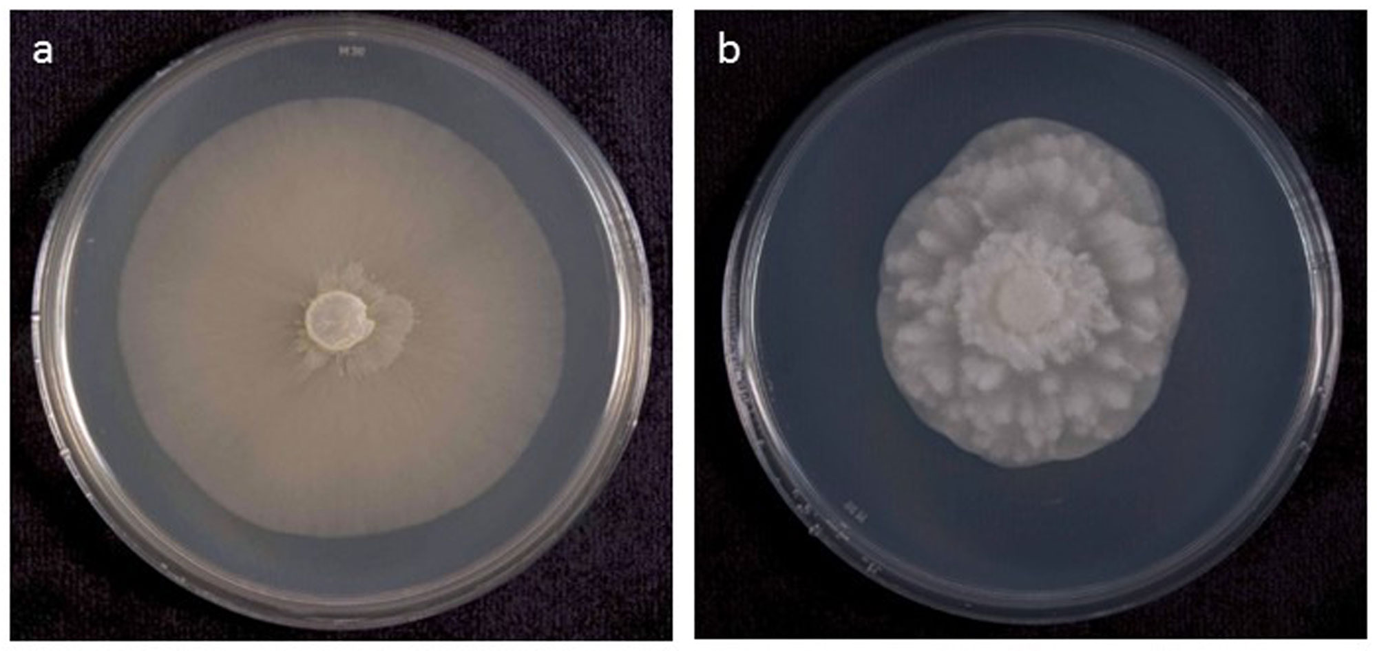
Phytophthora lateralis (P3888 WPC) colony of selected specimen grown for 7 days on (a) V8 agar and (b) potato dextrose agar; photos by Yilmaz Balci, USDA-APHIS-PPQ |
|
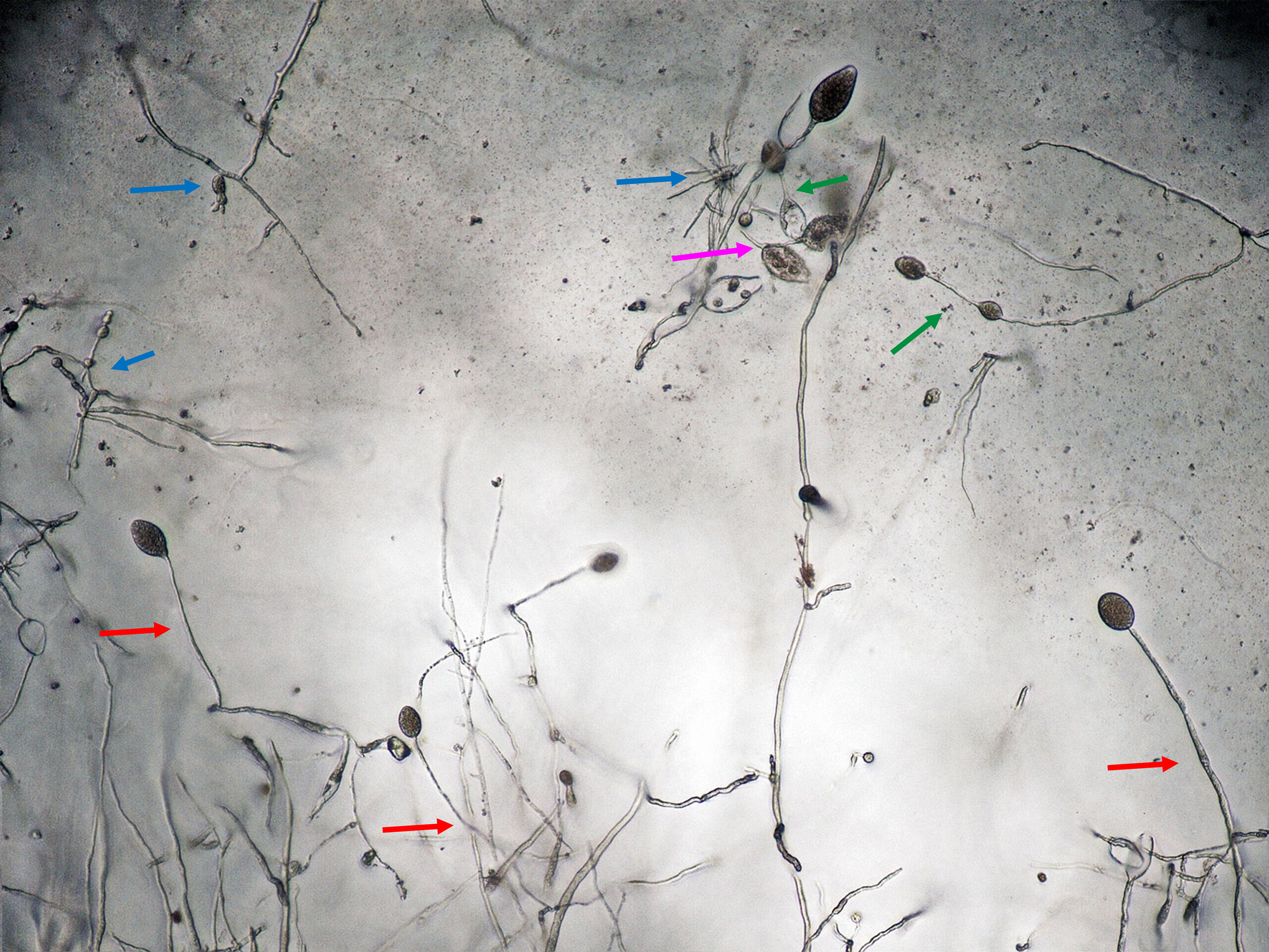
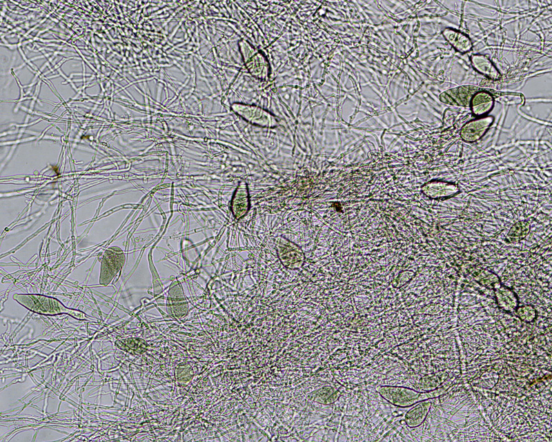
P. lateralis asexual phase of selected specimen P3888 (WPC) showing sporangia produced in sporangiophores: individual long (red arrows), simple sympodial (purple arrow), external proliferation (green arrows); hyphal swellings (blue arrows); photo by Gloria Abad, USDA-APHIS-PPQ. |
|
Phytophthora lateralis lobate and irregular hyphal swellings produced by selected specimen P3888 (WPC) in hemp seed agar media; photos by Gloria Abad, USDA-APHIS-PPQ. |
|
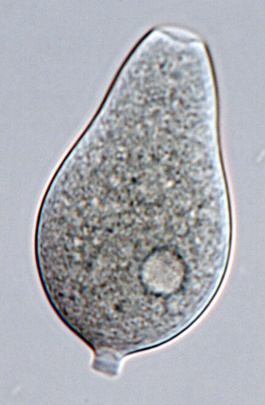
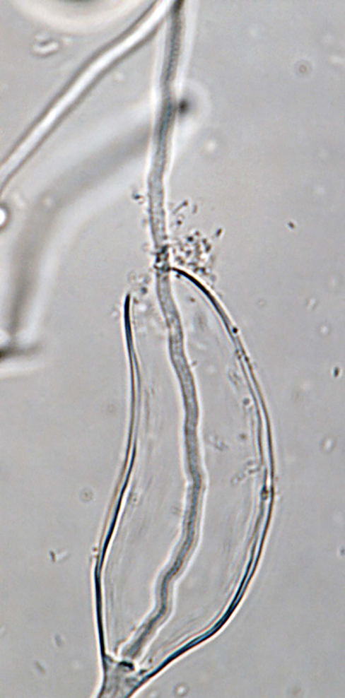
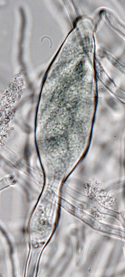
Phytophthora lateralis selected specimen (P11090 WPC) asexual phase: (a, b) sporangia in sporangiophores; (c, d) persistent sporangia; (e) caducous sporangium with short pedicel; (f) sporangium with internal proliferation; photos by G. Abad, USDA-APHIS-PPQ. |
|
Phytophthora lateralis selected specimen (P11090 WPC) asexual phase: caducous sporangium with short pedicel; photo by G. Abad, USDA-APHIS-PPQ. |
|
Phytophthora lateralis selected specimen (P11090 WPC) asexual phase: sporangia in sporangiophores; photo by G. Abad, USDA-APHIS-PPQ. |
|
Phytophthora lateralis selected specimen (P11090 WPC) asexual phase: sporangium with internal proliferation; photo by G. Abad, USDA-APHIS-PPQ. |
|
Phytophthora lateralis selected specimen (P11090 WPC) asexual phase: persistent sporangium; photo by G. Abad, USDA-APHIS-PPQ. |
|
Phytophthora lateralis selected specimen (P11090 WPC) asexual phase: persistent sporangium; photo by G. Abad, USDA-APHIS-PPQ. |
|
Phytophthora lateralis selected specimen (P11090 WPC) asexual phase: sporangia in sporangiophores; photo by G. Abad, USDA-APHIS-PPQ. |
.JPG)
.JPG)


Name and publication
Phytophthora lateralis Tucker & Milbrath (1942)
Tucker CM and Milbrath JA. 1942. Root rot of Chamaecyparis caused by a species of Phytophthora. Mycologia 34: 94–103.
Nomenclature
Mycobank
Typification
from Tucker and Milbrath (1942)
Type: UNITED STATES OF AMERICA, Port Orford, Cedar (Chamaecyparis lawsoniana), Oregon, CBS-H-7643
Ex-type: CBS 168.42
Ex-type in other collections
(ET) CBS 168.42, NRRL 64131, ATCC 58816 (MCI), CABI IMI40503 (PA), WPC P3361 P3917, S&T BL 42 (Abad), DSM 62687, VKM F-1835, IMB 10780, PD 06 03209088
Molecular identification
Voucher sequences for barcoding genes (ITS rDNA and COI) of the ex-type (see Molecular protocols page)
Phytophthora lateralis isolate CPHST BL 42 (= P3361 WPC) = ITS rDNA MG865522, COI MH136917
Voucher sequences for Molecular Toolbox with seven genes (ITS, β-tub, COI, EF1α, HSP90, L10, and YPT1
(see Molecular protocols page) (In Progress)
Voucher sequences for Metabarcoding High-throughput Sequencing (HTS) Technologies [Molecular Operational Taxonomic Unit (MOTU)]
(see Molecular protocols page) (In Progress)
Sequences with multiple genes for ex-type in other sources
- NCBI: Phytophthora lateralis CPHST BL 42
- NCBI: Phytophthora lateralis CBS 168.42
- EPPO-Q-bank: Phytophthora lateralis CBS 168.42
- BOLDSYSTEMS: Phytophthora lateralis OOMYA028-07 = CBS 168.42 (barcoding COI & ITS); OOMYA522-08 = CBS 168.42 (barcoding ITS)
Position in multigenic phylogeny with 7 genes (ITS, β-tub, COI, EF1α, HSP90, L10, and YPT1)
Clade 8c
Genome sequence
Phytophthora lateralis strain ex-type CBS 168.42. Accession genome ASM50020v2, BioProject PRJNA190827, Tree Aggressors Identification using Genomic Approaches (2016), Feau et al 2016
Morphological identification
Colonies and cardinal temperatures
Colony colony:
assemblage of hyphae which usually develops form a single source and grows in a coordinated way
morphology after 7 days of growth on V8-A and MEA with no distinctive pattern, on PDA coarsely radiate. Minimum temperature for growth is 3°C, optimum 20°C, and maximum 25°C.
Conditions for growth and sporulation
Sporangia are not produced on agar media, but produced relatively abundantly on washed mycelial mats transferred from 7-day pea broth cultures (20°C) to sterile distilled water and incubated 7 days at 20°C. ChlamydosporesChlamydospores:
an asexual spore with a thickened inner wall that is delimited from the mycelium by a septum; may be terminal or intercalary, and survives for long periods in soil
abundant in agar cultures and liquid media.
Asexual phase
SporangiaSporangia:
sac within which zoospores form, especially when water is cooled to about 10°C below ambient temperature; in solid substrates, sporangia usually germinate by germ tubes
nonpapillatenonpapillate:
pertaining to the production of a non-distinct, or inconspicuous, papilla at the distal end of the sporangium (cf. papillate and semipapillate)
; persistentpersistent:
pertaining to sporangia that remain attached to the sporangiophore and do not separate or detach easily (cf. caducous)
; ovoidovoid:
egg-shaped, with the widest part at the base of the sporangium and the narrow part at the apex
, obovoidobovoid:
inversely egg-shaped; ovoid, but with the widest part at the apex
, obpyriformobpyriform:
inversely pear-shaped, i.e. with the widest part at the point of attachment (cf. pyriform)
, elongate (24–76 L x 19–41 W µm); originated unbranched in long sporangiophores, by external proliferationexternal proliferation:
formation of a sporangium after a sporangiophore has emerged from beneath and external to an empty sporangium that has previously emitted its zoospores (cf. internal proliferation)
and on simple sympodial sporangiophores. Hyphal swellings globose, catenulatecatenulate:
having a chain-like form
, or with irregular shape. ChlamydosporesChlamydospores:
an asexual spore with a thickened inner wall that is delimited from the mycelium by a septum; may be terminal or intercalary, and survives for long periods in soil
subspherical to spherical, occasionally ovoidovoid:
egg-shaped, with the widest part at the base of the sporangium and the narrow part at the apex
to irregular; individual or distant catenulated; lateral, terminal, or intercalaryintercalary:
positioned within a hypha (cf. terminal)
(20–77 µm diam).
Sexual phase
Sterile.
Note: Original publication of Tucker, C.M.; Milbrath, J.A. 1942 referred to oogoniaoogonia:
the female gametangium in which the oospore forms after fertilization by the antheridium
, antheridiaantheridia:
the male gametangium; a multinucleate, swollen hyphal tip affixed firmly to the wall of the female gametangium (the oogonium)
, and oosporesoospores:
zygote or thick-walled spore that forms within the oogonium after fertilization by the antheridium; may be long-lived
as unknown. Isolate of the ex-type CBS 168.42 evaluated by Q-Bank has not produced oosporesoospores:
zygote or thick-walled spore that forms within the oogonium after fertilization by the antheridium; may be long-lived
, and the same results were found for the duplicate of the ex-type CPHST BL 42.
Most typical characters
Phytophthora lateralis can be distinguished by the shape of the sporangiasporangia:
sac within which zoospores form, especially when water is cooled to about 10°C below ambient temperature; in solid substrates, sporangia usually germinate by germ tubes
, hyphal swellings, and typical chlamydosporeschlamydospores:
an asexual spore with a thickened inner wall that is delimited from the mycelium by a septum; may be terminal or intercalary, and survives for long periods in soil
.
Specimen(s) evaluated
Phytophthora lateralis ex-type CPHST BL 42 = P3361 (World Phytophthora Collection), duplicate of ex-type CBS 168.42
Additional well-authenticated specimens evaluated:
P. lateralis P3888 (World Phytophthora Collection)
P. lateralis P11090 (World Phytophthora Collection)
Hosts and distribution
Distribution: widespread. Reports from New Zealand and North Carolina are doubtful.
Substrate: roots, crown, bark, occasionally on foliage
Disease note: root and crown rot, girdling, death
Host: Chamaecyparis lawsoniana (Lawson cypress aka Port Orford cedar), C. lateralis (Cupressaceae), rarely, Taxus brevifolia (Pacific yew, Taxaceae). Hosts in other families can be infected when inoculated.
Retrieved January 31, 2018 from U.S. National Fungus Collections Nomenclature Database.
Additional references and links
- SMML USDA-ARS: Phytophthora lateralis
- EPPO Global Database: Phytophthora lateralis
- Forest Phytophthoras of the world: Phytophthora lateralis
- CABI Digital Library: Phytophthora lateralis
- Forest Reserach UK: Phytophthora lateralis
- Encyclopedia of Life (EOL): Phytophthora lateralis
- Index Fungorum (IF): Phytophthora lateralis
- Google All Phytophthora lateralis
- Google Images Phytophthora lateralis
- Google Scholar Phytophthora lateralis
Fact sheet author
Z. Gloria Abad, Ph.D., USDA-APHIS-PPQ-S&T Plant Pathogen Confirmatory Diagnostics Laboratory (PPCDL), United States of America.
