
Phytophthora drechsleri (in progress - Abad et al. 2023b)
|
Phytophthora spp. in subclade 8a: portion of the seven-loci ML phylogeny featuring the type cultures of 212 described species (by T. Bourret). Notice the position of P. drechsleri selected specimen CBS 292.35 = S&T BL 17. Gloria Abad, USDA S&T.
|
|
Phytophthora spp. in subclade 8a: Morphological Tabular key (PDF) and Tabular key legends (PDF) in IDphy2 KEY SECTION. Notice the data of P. drechsleri selected specimen CBS 292.35 = S&T BL 17. Gloria Abad, USDA S&T.
|
|
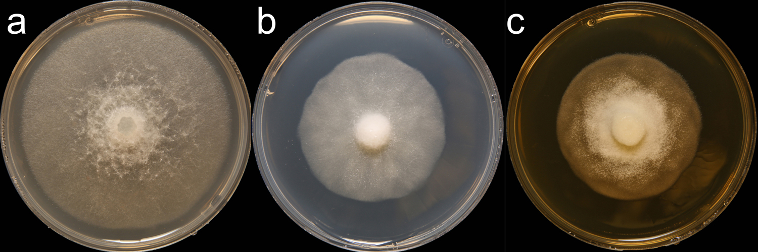
Phytophthora dreschsleri (CPHST BL 140) colonies of selected specimen #2 grown for 7 days on (a) V8® Agar, (b) potato dextrose agar, and (c) malt extract agar; photo by Krysta Jennings and Leandra Knight, USDA-APHIS-PPQ |
|
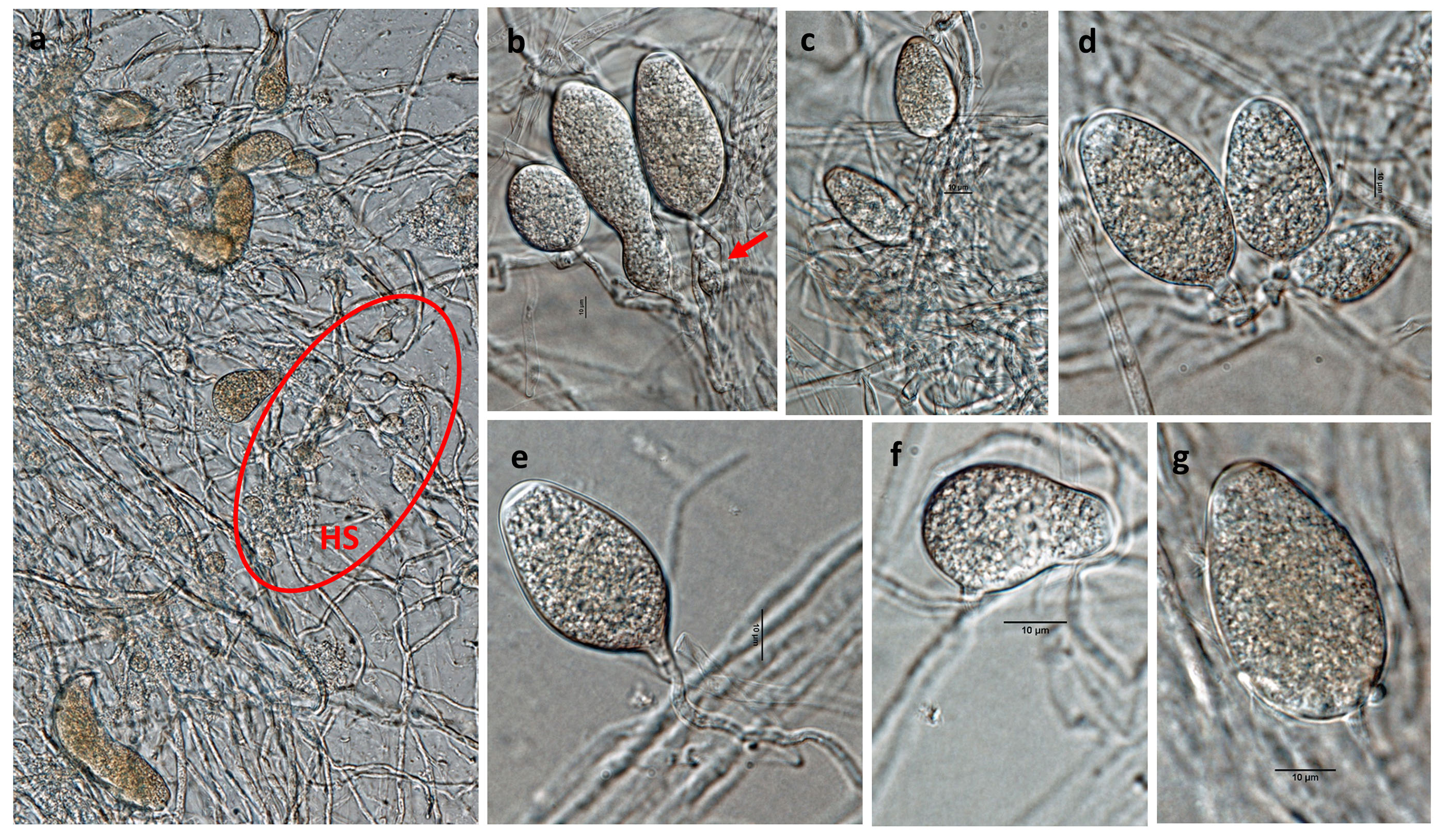
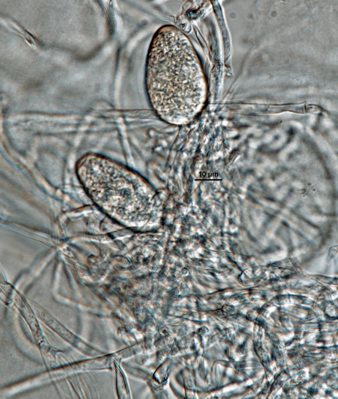
Phytophthora drechsleri (CPHST BL 140, selected specimen) asexual phase: (a) sporangia and hyphal swellings (HS marked in red area); (b-g) different shapes of nonpapillate persistent sporangia; (b) hyphal swellings in sporangiophore (red arrow); (e) sporangium with tapered base; photos by Gloria Abad, USDA-APHIS-PPQ. |
|
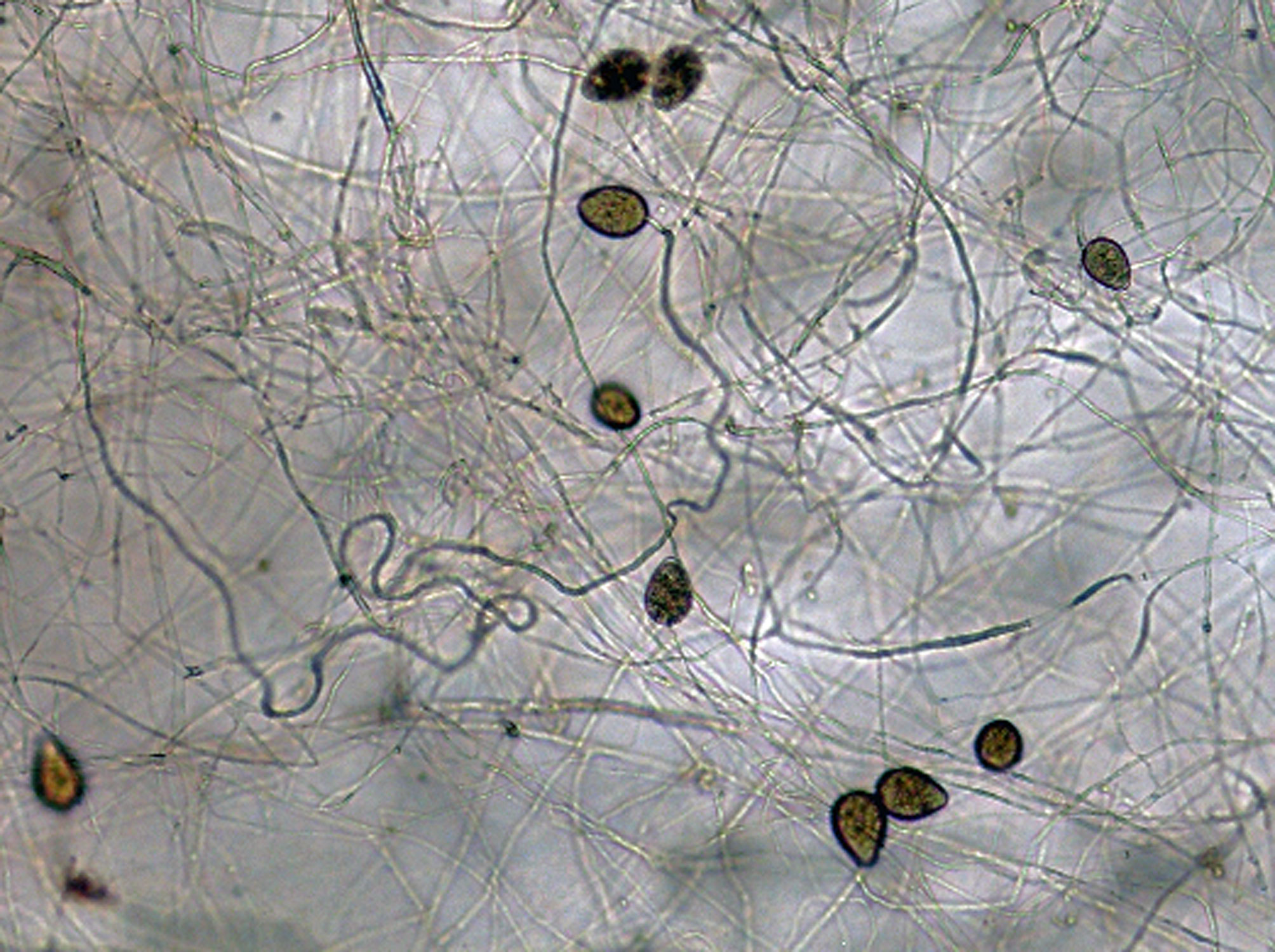
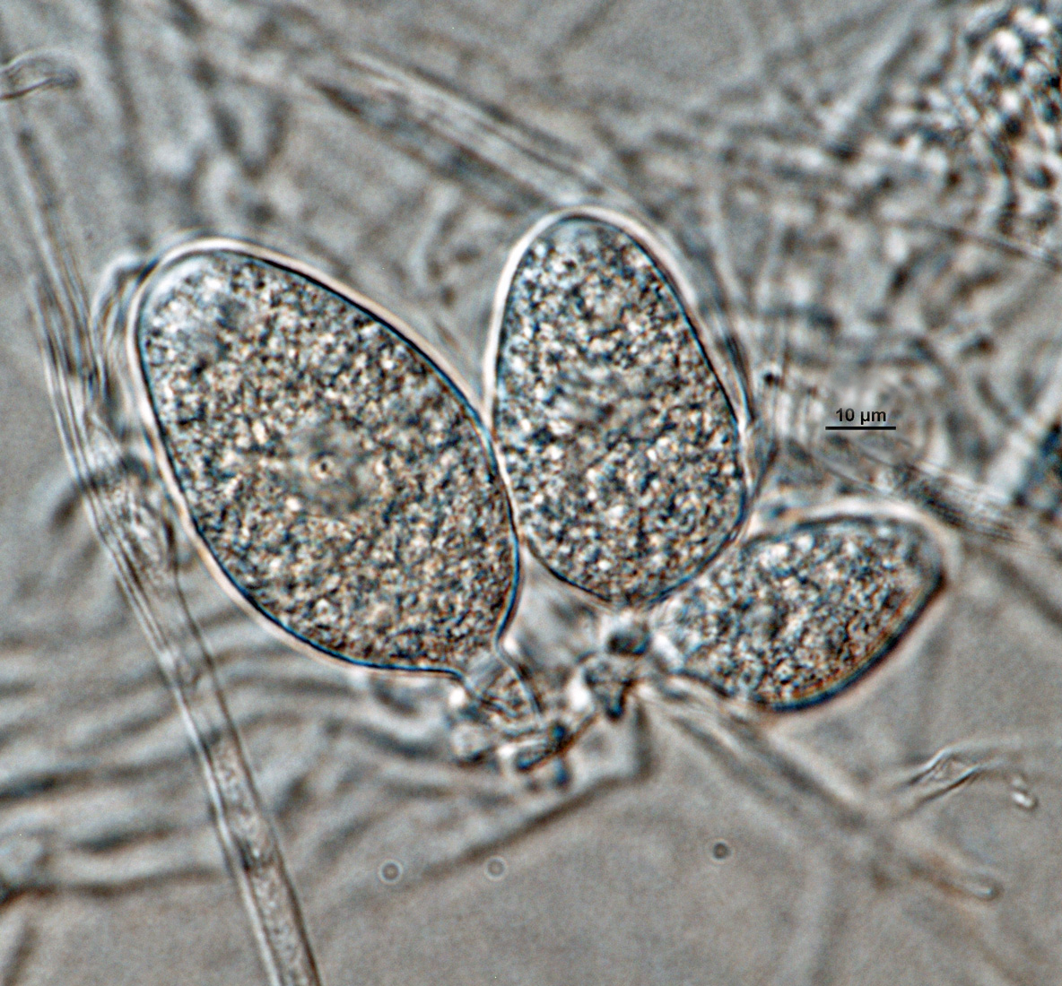
Phytophthora drechsleri (CPHST BL 140) selected specimen asexual phase: nonpapillate persistent sporangia originated in unbranched sporangiophores; photos by Gloria Abad, USDA-APHIS-PPQ |
|
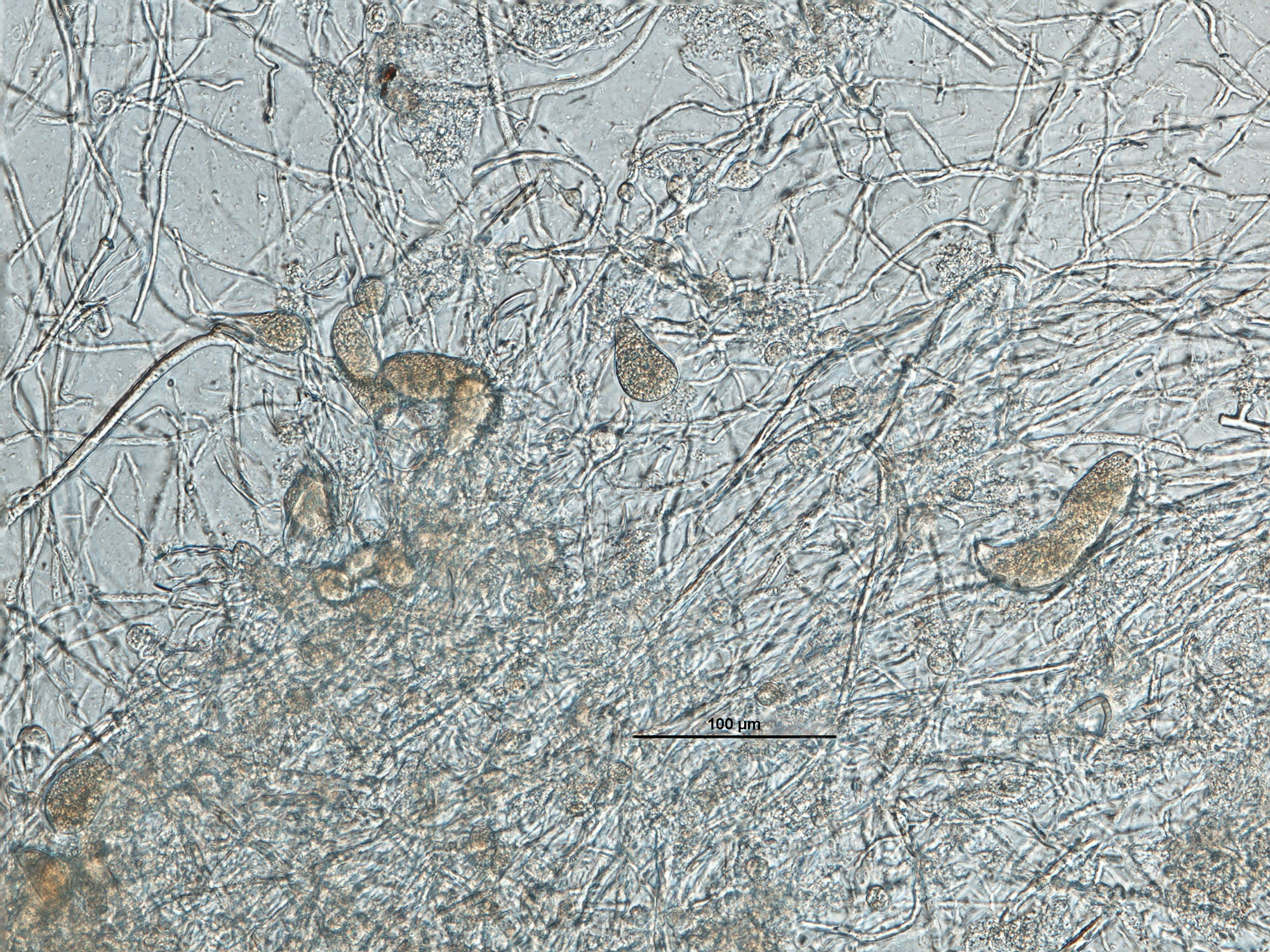
Phytophthora drechsleri (CPHST BL 17) selected specimen asexual phase (a, b): globose, catenulate (rosary shape), and frequently clustered hyphal swellings; photos by G. Abad, USDA-APHIS-PPQ |
|
Phytophthora drechsleri (CPHST BL 140, selected specimen) asexual phase: nonpapillate persistent sporangia originated in unbranched sporangiophores; photo by Gloria Abad, USDA-APHIS-PPQ. |
|
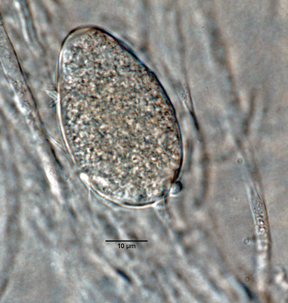
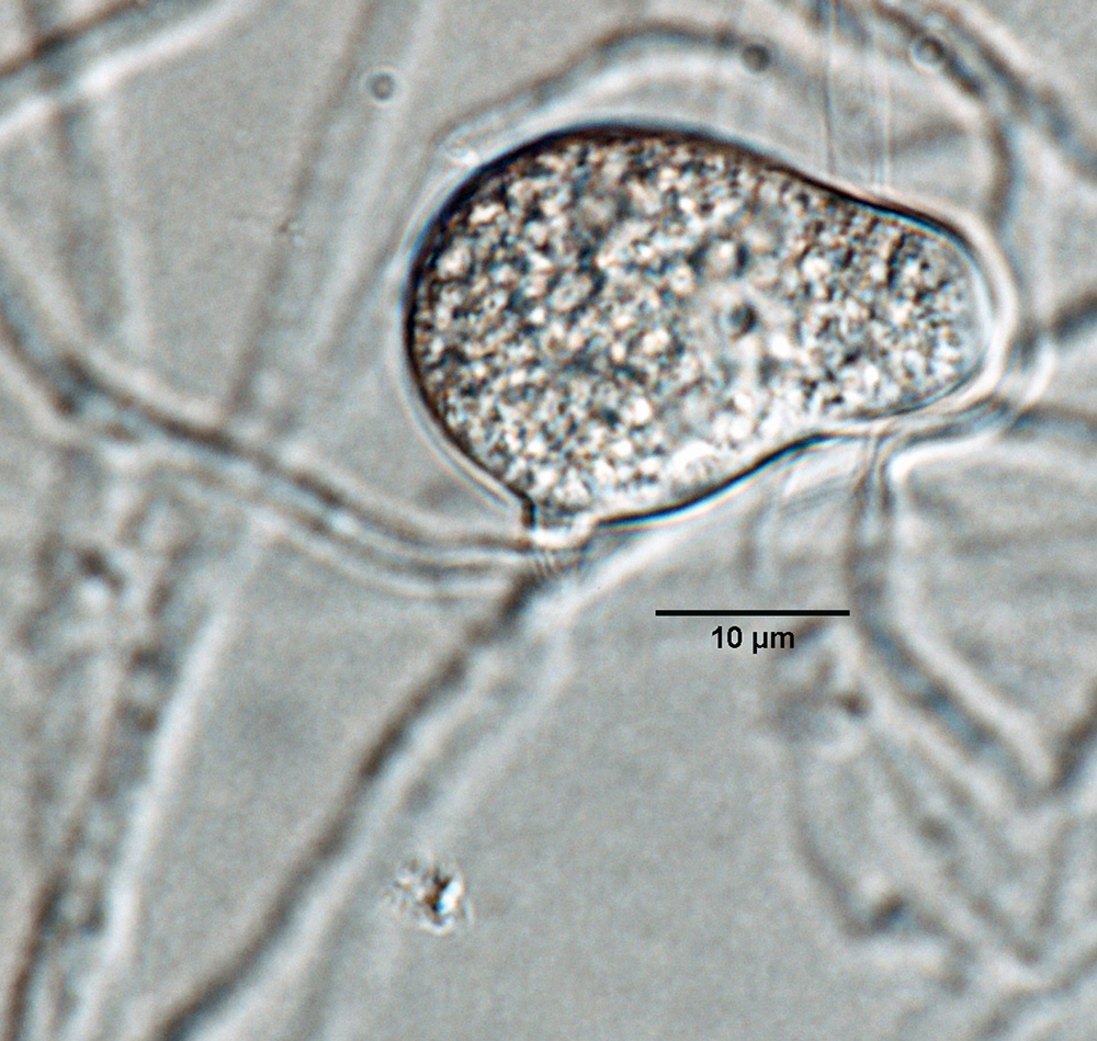
Phytophthora drechsleri (CPHST BL 140, selected specimen) asexual phase: nonpapillate persistent sporangium with tapered base; photo by Gloria Abad, USDA-APHIS-PPQ. |
|
Phytophthora drechsleri (CPHST BL 140, selected specimen) asexual phase: nonpapillate persistent sporangia originated in unbranched sporangiophores; photo by Gloria Abad, USDA-APHIS-PPQ. |
|
Phytophthora drechsleri (CPHST BL 140, selected specimen) asexual phase: nonpapillate persistent sporangium; photo by Gloria Abad, USDA-APHIS-PPQ. |
|
Phytophthora drechsleri (CPHST BL 140, selected specimen) asexual phase: nonpapillate persistent sporangia with tapered bases; photo by Gloria Abad, USDA-APHIS-PPQ. |
|
Phytophthora drechsleri (CPHST BL 140, selected specimen) asexual phase: nonpapillate persistent sporangia; photo by Gloria Abad, USDA-APHIS-PPQ. |
|
Phytophthora drechsleri (CPHST BL 140, selected specimen) asexual phase: nonpapillate persistent sporangia and hyphal swellings in sporangiophore; photo by Gloria Abad, USDA-APHIS-PPQ. |
|
Phytophthora drechsleri (CPHST BL 140, selected specimen) asexual phase: nonpapillate persistent sporangia; photo by Gloria Abad, USDA-APHIS-PPQ. |
|
Phytophthora drechsleri (CPHST BL 140, selected specimen) asexual phase: different shapes of nonpapillate persistent sporangia and hyphal swellings; photo by Gloria Abad, USDA-APHIS-PPQ. |
.JPG)
.JPG)


Name and publication
Phytophthora drechsleri Tucker (1931)
Tucker CM. 1931. Taxonomy of the genus Phytophthora de Bary. Research Bulletin of the Missouri Agricultural Experiment Station 153: 1–208 (pg. 188).
Nomenclature
Mycobank
Synonymy
≡ Phytophthora erythroseptica var. drechsleri (Tucker) Sarej., Annales de l'Institut Phytopathologique Benaki 2: 48 (1936) [MB353067]
Typification
from Tucker (1931)
Type: UNITED STATES OF AMERICA, Idaho, known only from the type isolated by Charles Drechsler from rotting tubers of Solanum tuberosum L, Tucker No 206 (according to Tucker 1931, original publication of the species)
Ex-type: LOST
Well-authenticated specimen(s) selected by Gloria Abad: CPHST BL 17 (A2) = P11637 (WPC) = Tucker, authentic isolate: UNITED STATES OF AMERICA, California, from Beta vulgaris var. altissima, collected by C.M. Tompkins, N0 114 = CBS 292.35, identified by C.M.Tucker, deposited by C.M. Tucker, Apr 1935
CPHST BL 140 (A1) = P1691 (WPC) from Chrysanthemum cinerariifolium, ARGENTINA
Selected specimen in other collections
(SE) CBS 292.35 (A2), NRRL 64328, ATCC 46724 (MCI), WPC P1087 P11638 (Tucker authentic), S&T BL 17 (Abad), 23J5 (Hong), P538 Cooke (SCRI), P41 (Gallegly)
Molecular identification
Voucher sequences for barcoding genes (ITS rDNA and COI) of the selected specimen (see Molecular protocols page)
Phytophthora drechsleri isolate CPHST BL 17 (= P1087 WPC) = ITS rDNA MG865484, COI MH136879
Voucher sequences for Molecular Toolbox with seven genes (ITS, β-tub, COI, EF1α, HSP90, L10, and YPT1
(see Molecular protocols page) (In Progress)
Voucher sequences for Metabarcoding High-throughput Sequencing (HTS) Technologies [Molecular Operational Taxonomic Unit (MOTU)]
(see Molecular protocols page) (In Progress)
Sequences with multiple genes for selected specimen in other sources
- NCBI: Phytophthora drechsleri CPHST BL 17
- NCBI: Phytophthora drechsleri P1087
- NCBI: Phytophthora drechsleri P11638
- NCBI: Phytophthora drechsleri CBS 292.35
- EPPO-Q-bank: Phytophthora drechsleri CBS 292.35
- BOLDSYSTEMS: Phytophthora drechsleri PHYTO07810 = P1087 WOC/WPC, PHYTO08710 = P11638 (barcoding COI & ITS)
Position in multigenic phylogeny with 7 genes (ITS, β-tub, COI, EF1α, HSP90, L10, and YPT1)
Clade 8a
Morphological identification
Colonies and cardinal temperatures
Colonies on V8-A, PDA, and MEA with no distinctive pattern, on PDA and MEA with very light chrysanthemum pattern. Minimum temperature for growth 3°C, optimum 27–33°C, and maximum 36°C.
Asexual phase
Sporangia nonpapillate, persistentpersistent:
pertaining to sporangia that remain attached to the sporangiophore and do not separate or detach easily (cf. caducous)
, ellipsoidellipsoid:
refers to a solid body that forms an ellipse in the longitudinal plane and a circle in cross section; many fungal spores are ellipsoidal or elliptic
, ovoidovoid:
egg-shaped, with the widest part at the base of the sporangium and the narrow part at the apex
, irregular shapes, and some with tapered bases (23–34 × 36–59 µm), showing internal and external proliferationexternal proliferation:
formation of a sporangium after a sporangiophore has emerged from beneath and external to an empty sporangium that has previously emitted its zoospores (cf. internal proliferation)
, and originated in unbranched sporangiophores. Hyphal swellings globose, catenulatecatenulate:
having a chain-like form
(rosary shape), and frequently clustered. ChlamydosporesChlamydospores:
an asexual spore with a thickened inner wall that is delimited from the mycelium by a septum; may be terminal or intercalary, and survives for long periods in soil
absent.
Sexual phase
Heterothallic. OogoniaOogonia:
the female gametangium in which the oospore forms after fertilization by the antheridium
with smooth wall, globoseglobose:
having a rounded form resembling that of a sphere
, some with slightly tapered bases (within antheridiumantheridium:
the male gametangium; a multinucleate, swollen hyphal tip affixed firmly to the wall of the female gametangium (the oogonium)
), 25–40 µm diam.; antheridiaantheridia:
the male gametangium; a multinucleate, swollen hyphal tip affixed firmly to the wall of the female gametangium (the oogonium)
amphigynousamphigynous:
pertaining to the sexual stage in which the antheridium completely surrounds the stalk of the oogonium (cf. paragynous)
, oval (9–16 µm); oosporesoospores:
zygote or thick-walled spore that forms within the oogonium after fertilization by the antheridium; may be long-lived
almost pleroticplerotic:
pertaining to an oospore that fills the oogonium (cf. aplerotic)
to apleroticaplerotic:
pertaining to a mature oospore that does not fill the oogonium; i.e. there is room left between the oospore wall and oogonium wall (cf. plerotic)
(22–35 µm diam) with thin walls (2.5 µm).
Most typical characters
Phytophthora drechsleri is characterized by the typical hyphal swellings that are globoseglobose:
having a rounded form resembling that of a sphere
with rosary shape, frequently clustered, and produced in water cultures.
Specimen(s) evaluated
Phytophthora drechsleri CPHST BL 17 (A2), duplicate of P1087 (World Phytophthora Collection) (G. Abad selected specimen #1)
Phytophthora drechsleri selected specimen CPHST BL 140 (A1), duplicate of P1691 (World Phytophthora Collection) (G. Abad selected specimen #2)
Hosts and distribution
Distribution: cosmopolitan
Substrate: roots, stems, leaves, buds, flowers, fruits, tubers, bark of trunks
Disease note: primarily a root pathogen but also attacks ripening fruit for various crops; root rot, bark canker, fruit rot, stem rot, seedling blight
Hosts: multiple hosts. 113 genera in 40 families
Retrieved January 29, 2018 from U.S. National Fungus Collections Nomenclature Database.
Additional references and links
- SMML USDA-ARS: Phytophthora drechsleri
- EPPO Global Database: Phytophthora drechsleri
- Forest Phytophthoras of the world: Phytophthora drechsleri
- CABI Digital Library: Phytophthora dreschsleri
- Encyclopedia of Life (EOL): Phytophthora drechsleri
- Index Fungorum (IF): Phytophthora drechsleri
- Google All Phytophthora drechsleri
- Google Images Phytophthora drechsleri
- Google Scholar Phytophthora drechsleri
Fact sheet author:
Z. Gloria Abad, Ph.D., USDA-APHIS-PPQ-S&T Plant Pathogen Confirmatory Diagnostics Laboratory (PPCDL), United States of America.
