
Phytophthora cinnamomi
|
Phytophthora spp. in subclade 7c: portion of the seven-loci ML phylogeny featuring the type cultures of 212 described species (by T. Bourret). Notice the position of P. cinnamomi Ex-type CBS 144.22 (A2) Ex-isotype = S&T BL 12. Gloria Abad, USDA S&T.
|
|
Phytophthora spp. in subclade 7c: Morphological Tabular key (PDF) and Tabular key legends (PDF) in IDphy2 KEY SECTION. Notice the data of P. cinnamomi Ex-type CBS 144.22 (A2) Ex-isotype = S&T BL 12. Gloria Abad, USDA S&T.
|
|
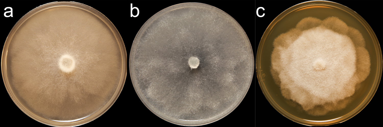
Phytophthora cinnamomi (CPHST BL 12) colonies of the ex-type grown for 7 days on (a) V8® Agar, (b) potato dextrose agar, and (c) malt extract agar; photo by Clinton Greub, Krysta Jennings, and Leandra Knight, USDA-APHIS-PPQ |
|
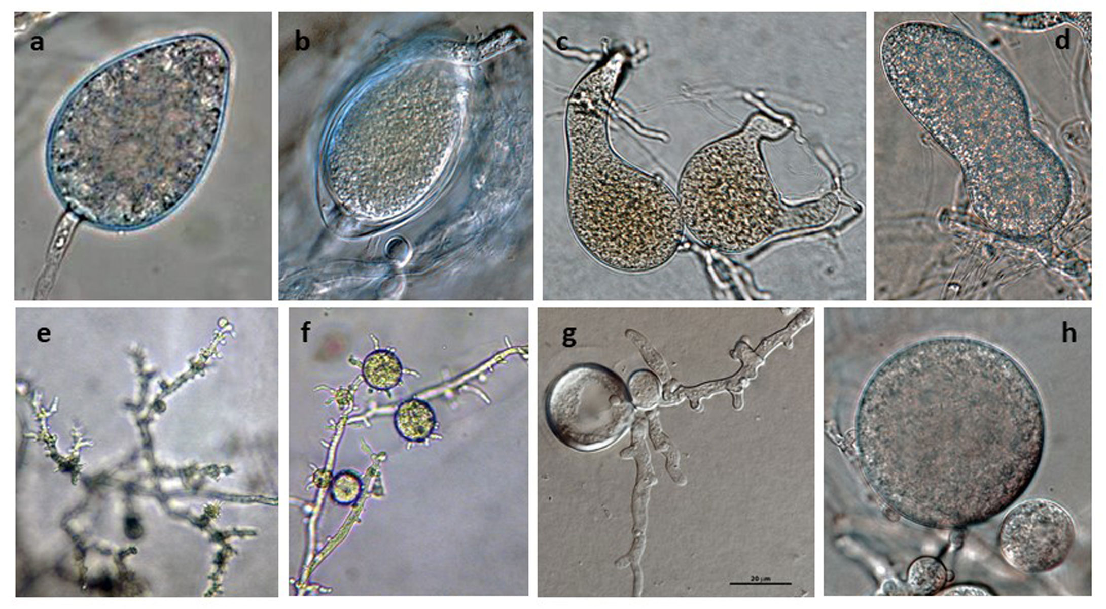
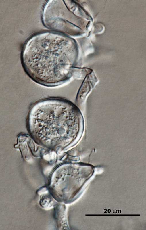
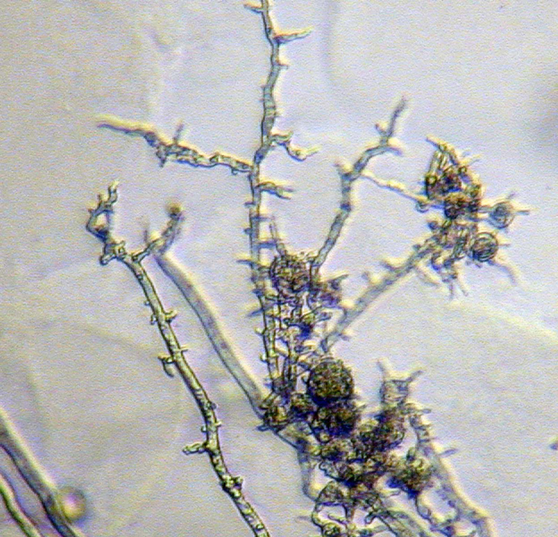
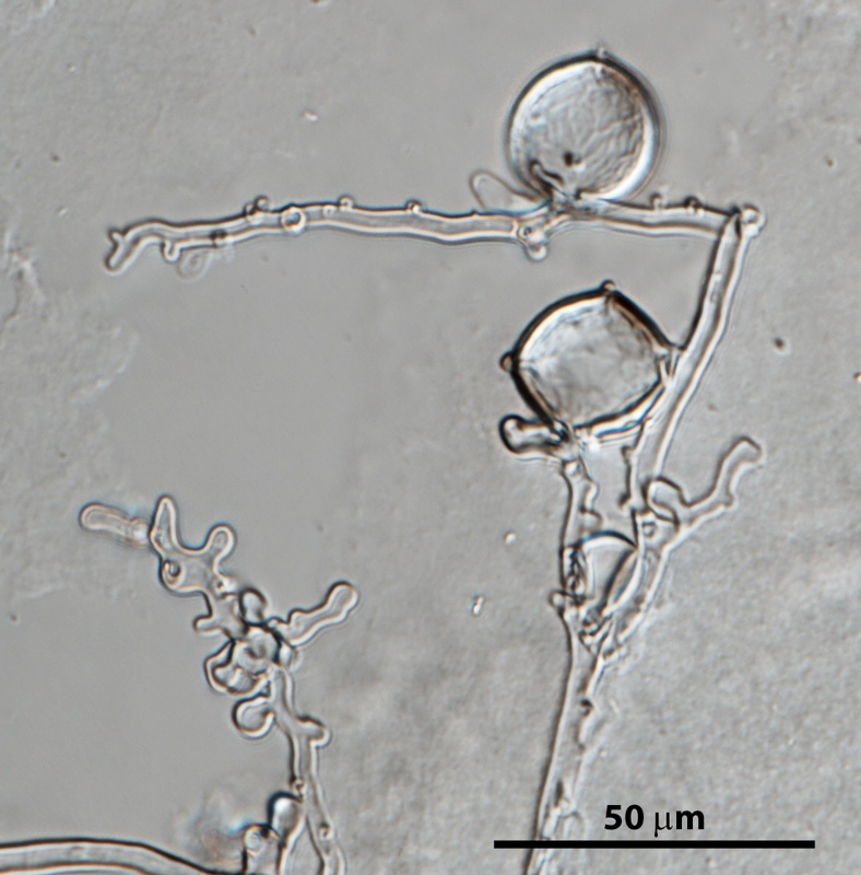
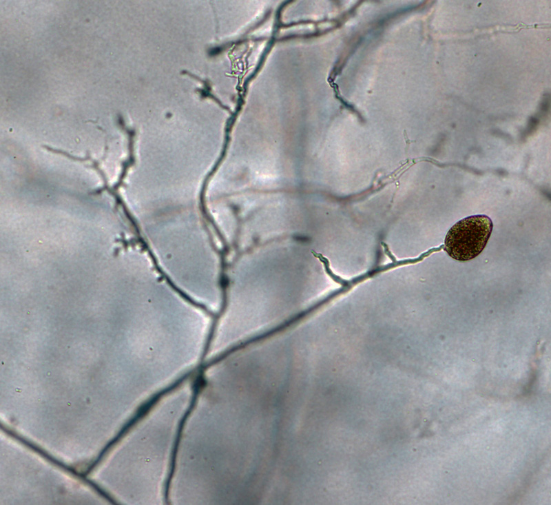
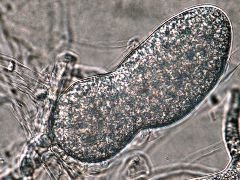
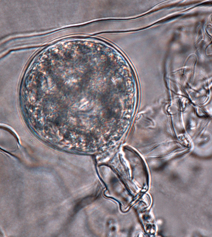
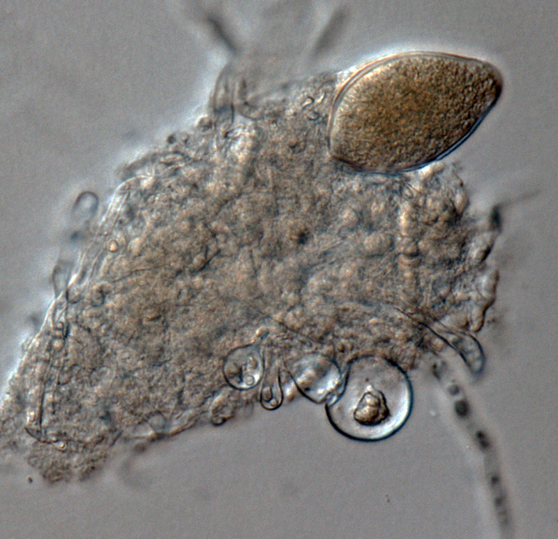
Phytophthora cinnamomi selected specimen P6490 asexual phase (a-h): (a-d) different shapes of persistent sporangia, (e) typical coralloid mycelia, (f, g) hyphal swellings, (h) chlamydospore and hyphal swellings; photos by Gloria Abad, USDA-APHIS-PPQ. |
|
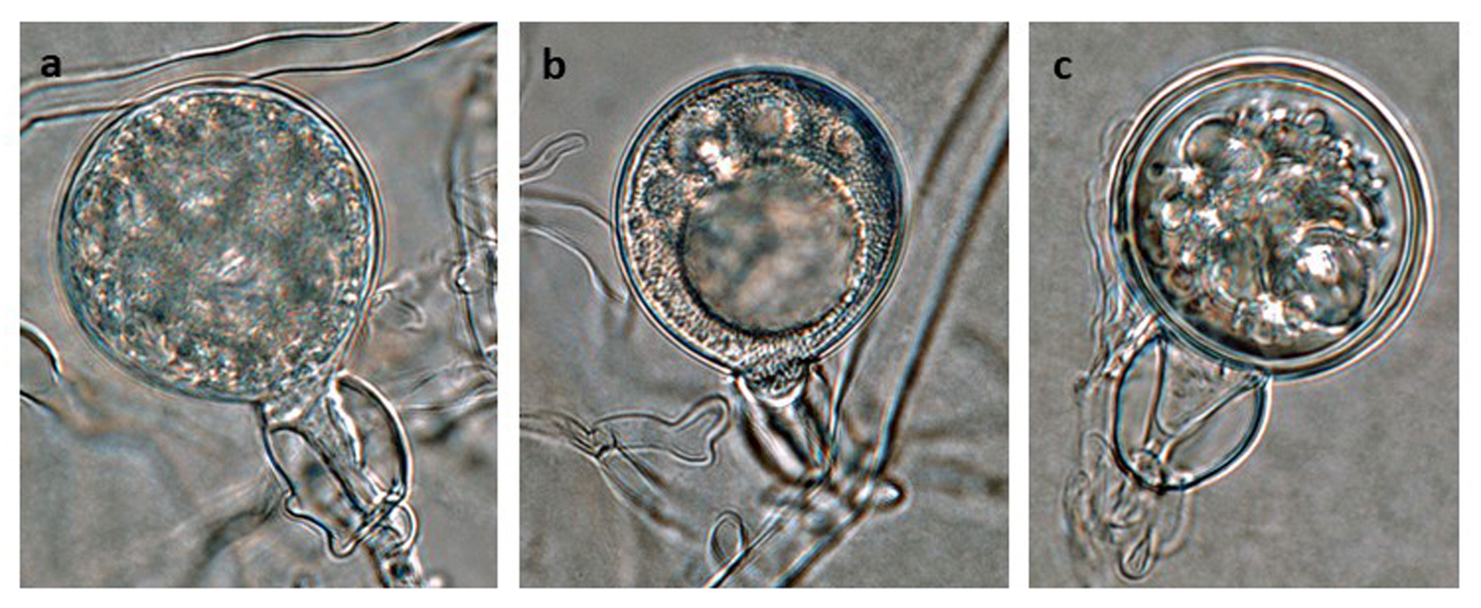
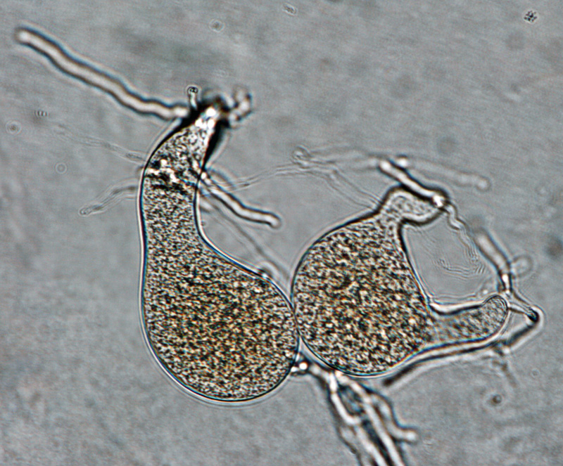
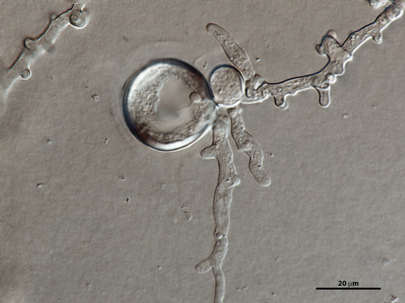
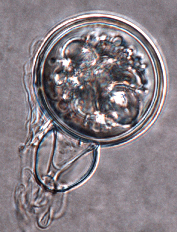
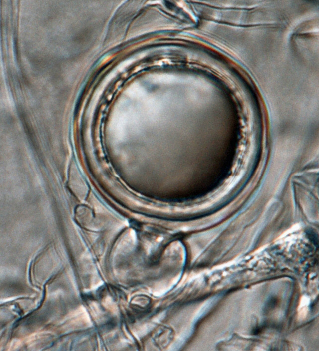
Phytophthora cinnamomi selected specimen P6490 sexual phase (a-c): (a, b) young oogonia with amphigynous antheridia, (c) abortive oogonium; photos by Gloria Abad, USDA-APHIS-PPQ. |
|
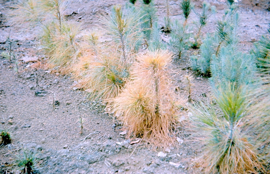
Dieback and mortality of Castanea sativa caused by Phytophthora cinnamomi in Sardinia, Italy; photo by Bruno Scanu and Antonio Franceschini. Università degli Studi di Sassari, Italy |
|
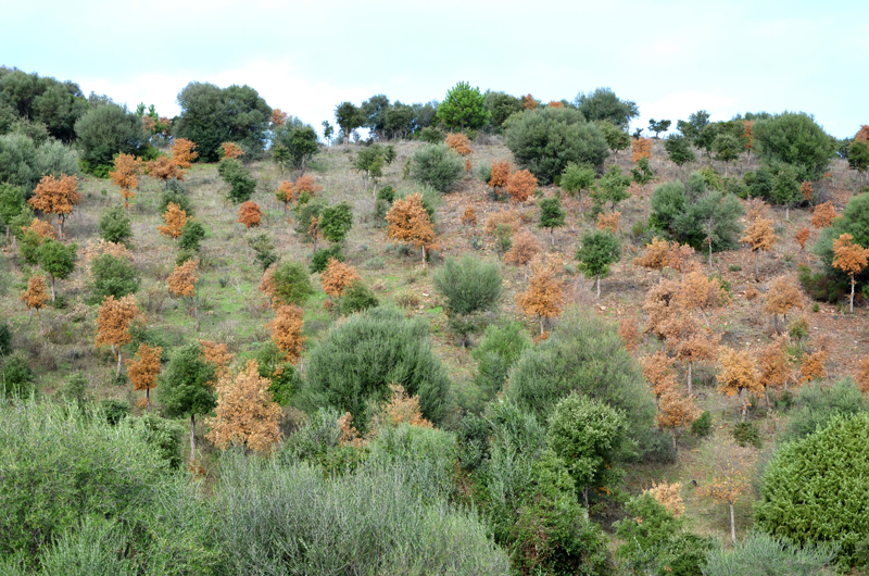
Extensive dieback and mortality of Quercus suber trees caused by Phytophthora cinnamomi in a plantation in Sardinia, Italy; photo by Bruno Scanu and Antonio Franceschini. Università degli di Sassari, Italy |
|
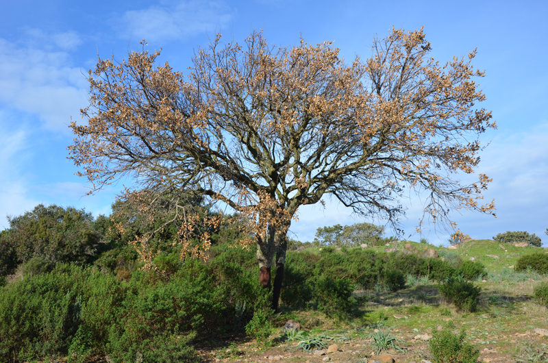
Sudden wilting and dieback of a mature Quercus suber tree caused by Phytophthora cinnamomi in Sardinia, Italy; photo by Bruno Scanu and Antonio Franceschini. Università degli Studi di Sassari, Italy |
|
Girdling collar rot lesion and extensive losses of lateral and fine roots caused by Phytophthora cinnamomi on Quercus suber in Sardinia, Italy; photo by Bruno Scanu and Antonio Franceschini. Università degli Studi di Sassari, Italy |
|
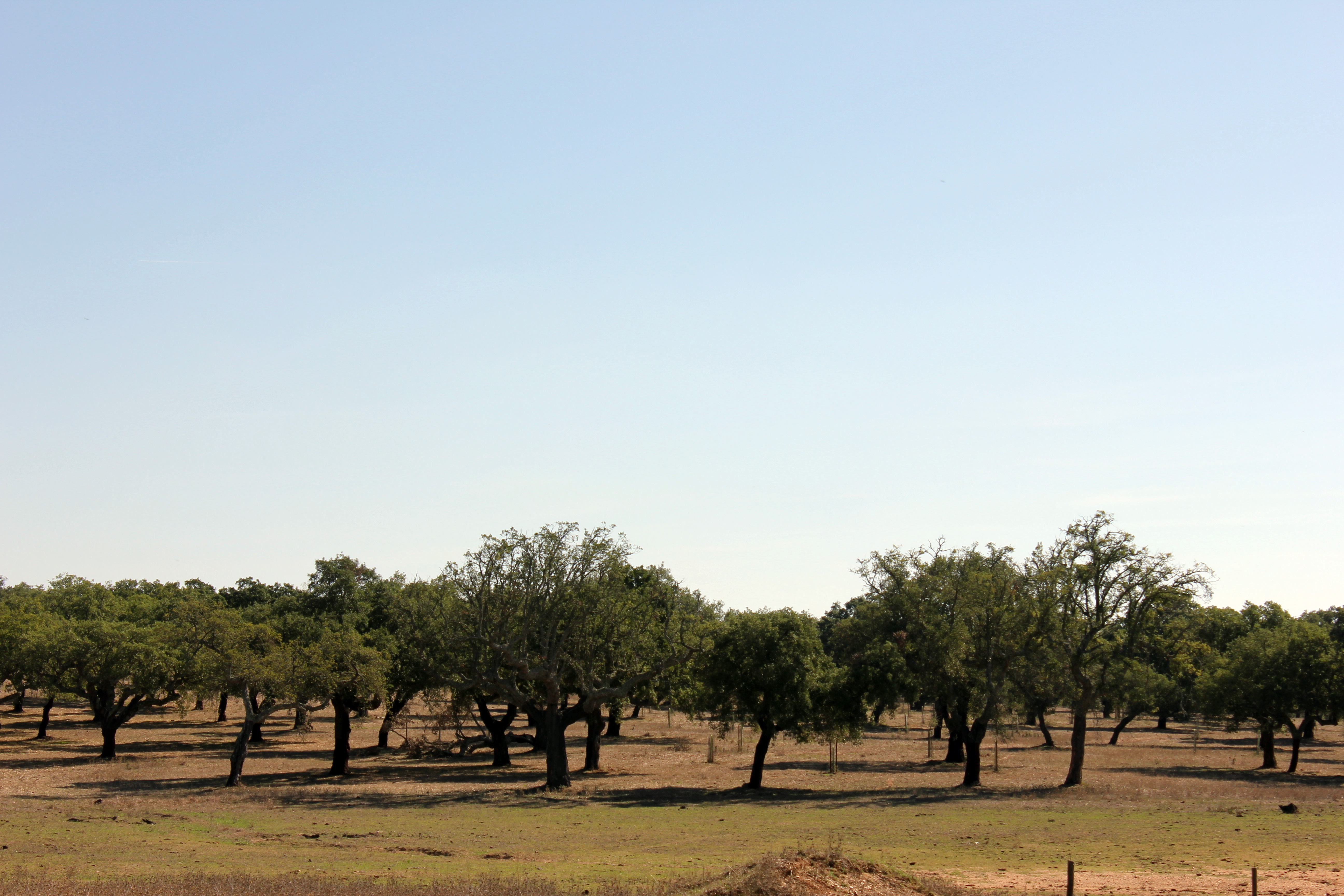
Quercus suber (cork oak) declining symptoms caused by Phytophthora cinnamomi in Alentejo region, Portugal; photo by Alfredo Cravador and Dina Neves, Universidade do Algarve-FCT, Portugal |
|
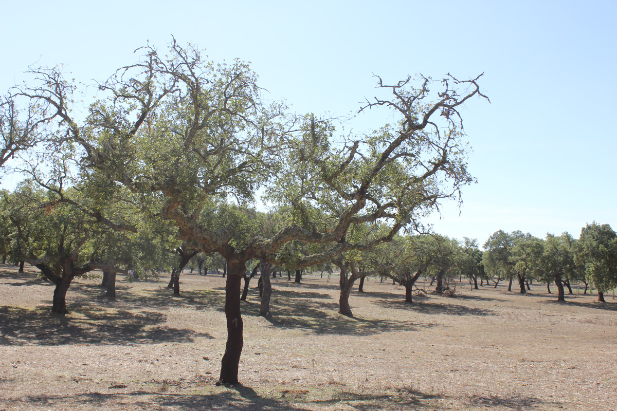
Quercus suber (cork oak) declining symptoms caused by Phytophthora cinnamomi in Alentejo region, Portugal; photo by Alfredo Cravador and Dina Neves, Universidade do Algarve-FCT, Portugal |
|
Quercus suber (cork oak) declining symptoms caused by Phytophthora cinnamomi in Alentejo region, Portugal; photo by Alfredo Cravador and Dina Neves, Universidade do Algarve-FCT, Portugal |
|
Heart rot of pineapple (Ananas comosus) caused by Phytophthora cinnamomi; photo by Scot Nelson, University of Hawaii at Manoa |
|
Root rot of pineapple (Ananas comosus) caused by Phytophthora cinnamomi; photo by Scot Nelson, University of Hawaii at Manoa |
|
Pine root rot caused by Phytophthora cinnamomi (1961); photo by Eduardo Trujillo, courtesy of Scot Nelson, University of Hawaii at Manoa
|
|
Phytophthora cinnamomi selected specimen P6490 asexual phase: different shapes of persistent sporangia and typical coralloid mycelia; photo by Gloria Abad, USDA-APHIS-PPQ. |
|
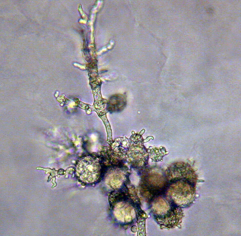
Phytophthora cinnamomi selected specimen P6490 asexual phase: botryose hyphal swellings; photo by Gloria Abad, USDA-APHIS-PPQ. |
|
Phytophthora cinnamomi selected specimen P6490 asexual phase: irregular shaped sporangia; photo by Gloria Abad, USDA-APHIS-PPQ. |
|
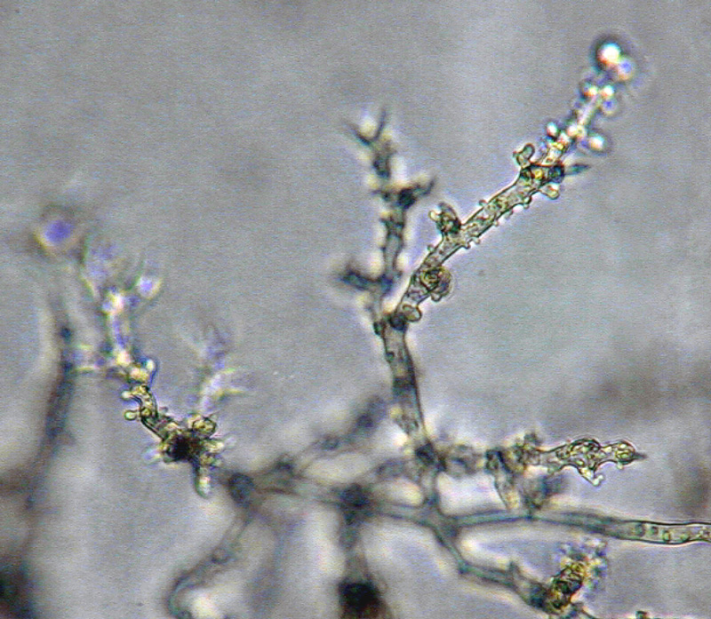
Phytophthora cinnamomi selected specimen P6490 asexual phase: typical coralloid mycelia; photo by Gloria Abad, USDA-APHIS-PPQ. |
|
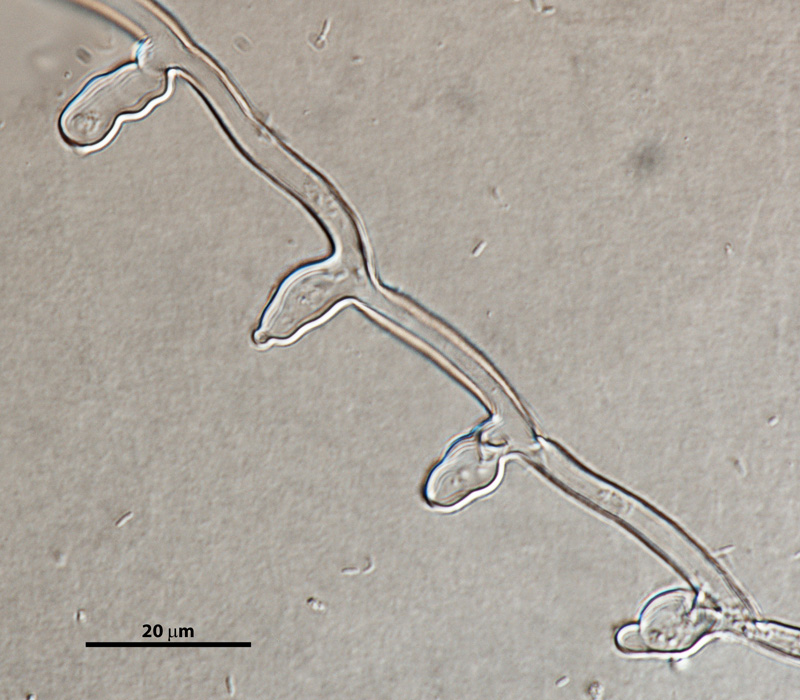
Phytophthora cinnamomi asexual phase: typical coralloid mycelia and botryose hyphal swellings; photo by Gloria Abad, USDA-APHIS-PPQ. |
|
Phytophthora cinnamomi selected specimen P6490 asexual phase: coralloid mycelia; photo by Gloria Abad, USDA-APHIS-PPQ. |
|
Phytophthora cinnamomi selected specimen P6490 asexual phase: hyphal swellings and typical coralloid mycelia; photo by Gloria Abad, USDA-APHIS-PPQ. |
|
Phytophthora cinnamomi selected specimen P6490 asexual phase: hyphal swellings and typical coralloid mycelia; photo by Gloria Abad, USDA-APHIS-PPQ. |
|
Phytophthora cinnamomi selected specimen P6490 asexual phase: hyphal swellings and typical coralloid mycelia; photo by Gloria Abad, USDA-APHIS-PPQ. |
|
Phytophthora cinnamomi selected specimen P6490 asexual phase: nonpapillate sporangia borne on simple sympodial sporangiophore; photo by Gloria Abad, USDA-APHIS-PPQ. |
|
Phytophthora cinnamomi selected specimen P6490 asexual phase: nonpapillate sporangium; photo by Gloria Abad, USDA-APHIS-PPQ. |
|
Phytophthora cinnamomi selected specimen P6490 sexual phase: abortive oogonium; photo by Gloria Abad, USDA-APHIS-PPQ. |
|
Phytophthora cinnamomi sexual phase: young oogonium with amphigynous antheridium; photo by Gloria Abad, USDA-APHIS-PPQ. |
|
Phytophthora cinnamomi selected specimen P6490 sexual phase: young oogonium with amphigynous antheridium; photo by Gloria Abad, USDA-APHIS-PPQ. |
|
Phytophthora cinnamomi selected specimen P6490 sexual phase: young oogonium with amphigynous antheridium; photo by Gloria Abad, USDA-APHIS-PPQ. |
|
Phytophthora cinnamomi selected specimen P6490 asexual phase: nonpapillate sporangium; photo by Gloria Abad, USDA-APHIS-PPQ. |
|
Phytophthora cinnamomi selected specimen P6490 asexual phase: typical coralloid mycelia; photo by Gloria Abad, USDA-APHIS-PPQ. |
|
Phytophthora cinnamomi asexual phase: typical coralloid mycelia and botryose hyphal swellings; photo by Gloria Abad, USDA-APHIS-PPQ. |
.JPG)
.JPG)


Name and publication
Phytophthora cinnamomi Rands (1922)
Rands RD. 1922. Streepkanker van Kaneel, veroorzaakt door Phytophthora cinnamomi n. sp. Meded. Inst. voor Plantenziekt 54: 1–54.
Nomenclature
Mycobank
Typification
from Rands (1922)
Type: INDONESIA, West Sumatra, from Indonesian cinnamon in cortice of Cinnamomum burmannii IMI 22938 deposited in Herb IMI ex-type collections by Randes, R.D. 1928-121
Note: IMI 22938 states that a culture is available from CABI Collections. However, this isolate is not available at CABI.
Isotype: INDONESIA, (West Sumatra) Matoer near Padang Indonesian cinnamon (Cinnamomum burmannii) Herbarium: CBS H-7638 collected by C. Hartley, isolated by R.D. Rands, No. 13, deposited by C. Hartley Jan. 1922 to CBS
Ex-isotype: CBS 144.22
NOTE: according to Mycologia: duplicates of the holotype collection are isotypes when deposited elsewhere or are otherwise separate from the holotype.
Ex-isotype in other collections
(ET) CBS 144.22 (A2) Ex-isotype, NRRL 64213, ATCC 46671 MCI, CABI IMI22938 (PA), WPC P2110, S&T BL 12 (Abad), 61J1 (Hong), Pc 110 (Zentmyer)
Selected specimen (A1) (G. Abad): P cinnamomi P2121 (WPC) Persea americana, Madagascar; other codes: ATCC 46672, Pc 121 (Zentmyer), CPHST BL 137 (Abad)
Molecular identification
Voucher sequences for barcoding genes (ITS rDNA and COI) of the ex-type (see Molecular protocols page)
Phytophthora cinnamomi isolate CPHST BL 12 (A2) (= P2110 WPC) = ITS rDNA MG865473, COI MH136869
Sequences of selected specimens
Phytophthora cinnamomi isolate CPHST BL 137 (A1) (= P2121 WPC) = ITS rDNA MG865474, COI MH136870
Voucher sequences for Molecular Toolbox with seven genes (ITS, β-tub, COI, EF1α, HSP90, L10, and YPT1
(see Molecular protocols page) (In Progress)
Voucher sequences for Metabarcoding High-throughput Sequencing (HTS) Technologies [Molecular Operational Taxonomic Unit (MOTU)]
(see Molecular protocols page) (In Progress)
Sequences with multiple genes for ex-type in other sources
- NCBI: Phytophthora cinnamomi CPHST BL 12
- NCBI: Phytophthora cinnamomi CBS 144.22
- NCBI: Phytophthora cinnamomi P2110
- EPPO-Q-bank: Phytophthora cinnamomi CBS 144.22
- BOLDSYSTEMS: Phytophthora cinnamomi (barcoding COI & ITS)
Position in multigenic phylogeny with 7 genes (ITS, β-tub, COI, EF1α, HSP90, L10, and YPT1)
Clade 7c
Genome sequence
Phytophthora cinnamomi strain ex-type CBS 144.22. Accession genome ASM2028349v1, BioProject PRJNA68241, University of Liverpool (2021)
Morphological identification
Colonies and cardinal temperatures
Colonies on V8-A with no distinctive colonycolony:
assemblage of hyphae which usually develops form a single source and grows in a coordinated way
pattern, on PDA and MEA with rosette (petaloid) pattern. Minimum temperature for growth 9°C, optimum 27°C, and maximum 30°C.
Asexual phase
SporangiaSporangia:
sac within which zoospores form, especially when water is cooled to about 10°C below ambient temperature; in solid substrates, sporangia usually germinate by germ tubes
nonpapillatenonpapillate:
pertaining to the production of a non-distinct, or inconspicuous, papilla at the distal end of the sporangium (cf. papillate and semipapillate)
; persistentpersistent:
pertaining to sporangia that remain attached to the sporangiophore and do not separate or detach easily (cf. caducous)
with internal proliferationinternal proliferation:
internal proliferation occurs when the sporangiophore continues to grow through an empty sporangium
, sometimes nested; ovoidovoid:
egg-shaped, with the widest part at the base of the sporangium and the narrow part at the apex
, ellipsoidellipsoid:
refers to a solid body that forms an ellipse in the longitudinal plane and a circle in cross section; many fungal spores are ellipsoidal or elliptic
, obpyriformobpyriform:
inversely pear-shaped, i.e. with the widest part at the point of attachment (cf. pyriform)
or rarely irregularly shaped (14–83 x 13-43 μm); produced in unbranched or lax simple sympodial sporangiophores. Hyphal swellings abundant; globoseglobose:
having a rounded form resembling that of a sphere
to sub-globose, coralloid, clustered, or aggregated. ChlamydosporesChlamydospores:
an asexual spore with a thickened inner wall that is delimited from the mycelium by a septum; may be terminal or intercalary, and survives for long periods in soil
abundant; globoseglobose:
having a rounded form resembling that of a sphere
, terminal, intercalaryintercalary:
positioned within a hypha (cf. terminal)
, rarely lateral (16–43 μm).
Sexual phase
Heterothallicheterothallic:
pertaining to sexual reproduction in which conjugation is possible only through interaction of different thalli (i.e. different mating types) (cf. homothallic)
. OogoniaOogonia:
the female gametangium in which the oospore forms after fertilization by the antheridium
smooth-walled; spherical (23–41 μm); antheridiaantheridia:
the male gametangium; a multinucleate, swollen hyphal tip affixed firmly to the wall of the female gametangium (the oogonium)
amphigynousamphigynous:
pertaining to the sexual stage in which the antheridium completely surrounds the stalk of the oogonium (cf. paragynous)
spherical to broadly ellipsoidellipsoid:
refers to a solid body that forms an ellipse in the longitudinal plane and a circle in cross section; many fungal spores are ellipsoidal or elliptic
; oosporesoospores:
zygote or thick-walled spore that forms within the oogonium after fertilization by the antheridium; may be long-lived
pleroticplerotic:
pertaining to an oospore that fills the oogonium (cf. aplerotic)
with a moderately thick wall (22–36 μm).
Most typical characters
Phytophthora cinnamomi is characterized by the presence of typical botryose hyphal swellings and thick-walled chlamydosporeschlamydospores:
an asexual spore with a thickened inner wall that is delimited from the mycelium by a septum; may be terminal or intercalary, and survives for long periods in soil
which are common and useful for the morphological diagnostics.
Specimen(s) evaluated
Phytophthora cinnamomi CPHST BL 12 (A2) duplicate of P2110 (World Phytophthora Collection)
Phytophthora cinnamomi CPHST BL 137 (A1) duplicate of P2121 (World Phytophthora Collection)
Phytophthora cinnamomi P6940 (WPC) from avocado, USA
Hosts and distribution
Distribution: primarily Southern Hemisphere
Substrate: roots, stems, branches, heartwood
Disease note: root rot, heart rot, wilt; causes ink disease of chestnut in conjunction with Phytophthora cambivora; a serious pathogen of hardwood forests and various crop species
Hosts: 266 genera in 90 families, commonly hardwood trees
Retrieved January 29, 2018 from U.S. National Fungus Collections Nomenclature Database.
Additional references and links
Zentmyer GA, et al. 1976. Variability in growth of Phytophthora cinnamomi in relation to temperature. Phytopathology 66: 982-986. Cites ATCC 46671.
- SMML USDA-ARS: Phytophthora cinnamomi
- EPPO Global Database: Phytophthora cinnamomi
- Forest Phytophthoras of the world: Phytophthora cinnamomi
- CABI Digital Library: Phytophthora cinnamomi
- Encyclopedia of Life (EOL): Phytophthora cinnamomi
- Index Fungorum (IF): Phytophthora cinnamomi
- Google All Phytophthora cinnamomi
- Google Images Phytophthora cinnamomi
- Google Scholar Phytophthora cinnamomi
Fact sheet author
Z. Gloria Abad, Ph.D., USDA-APHIS-PPQ-S&T Plant Pathogen Confirmatory Diagnostics Laboratory (PPCDL), United States of America.
