
Phytophthora chlamydospora
|
Phytophthora spp. in subclade 6b: portion of the seven-loci ML phylogeny featuring the type cultures of 212 described species (by T. Bourret). Notice the position of P. chlamydospora Ex-type CBS 149403 = S&T BL 156. Gloria Abad, USDA S&T.
|
|
Phytophthora spp. in subclade 6b: Morphological Tabular key (PDF) and Tabular key legends (PDF) in IDphy2 KEY SECTION. Notice the data of P. chlamydospora Ex-type CBS 149403 = S&T BL 156. Gloria Abad, USDA S&T.
|
|
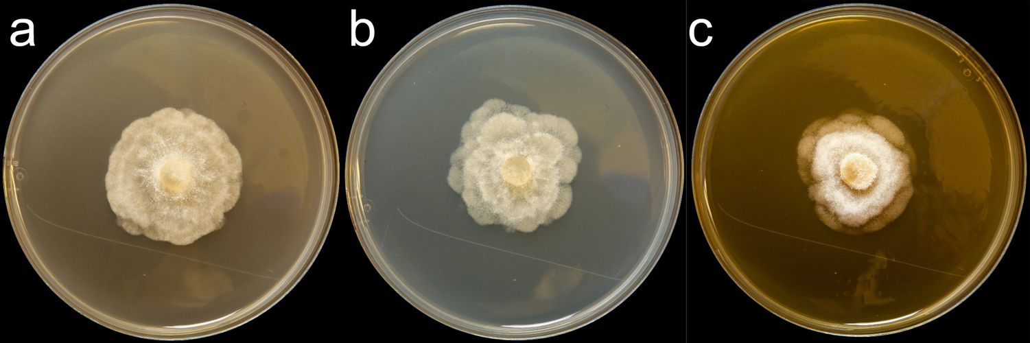
Phytophthora chlamydospora (CPHST BL 156) colonies of the ex-type grown for 7 days on (a) V8® Agar, (b) potato dextrose agar, and (c) malt extract agar; photo by Krysta Jennings and Leandra Knight, USDA-APHIS-PPQ. |
|
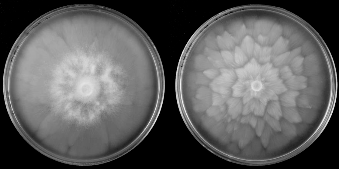
Phytophthora chlamydospora asexual phase: (a) cultures of the ex-type isolate P236 on V8 agar (left) and carrot agar (right), (b) hyphal swellings, (c) chlamydospore, (d) persistent nonpapillate sporangium, (e) sporangia in water, showing subsporangial elongation; Scale bar = 20μm; photos by Paul Reeser. |
|
Phytophthora chlamydospora cultures of the ex-type isolate P236: V8 agar (left) and carrot agar (right); Scale bar = 20μm; photos by Paul Reeser. |
|
Phytophthora chlamydospora asexual phase: hyphal swellings; Scale bar = 20μm; photo by Paul Reeser. |
|
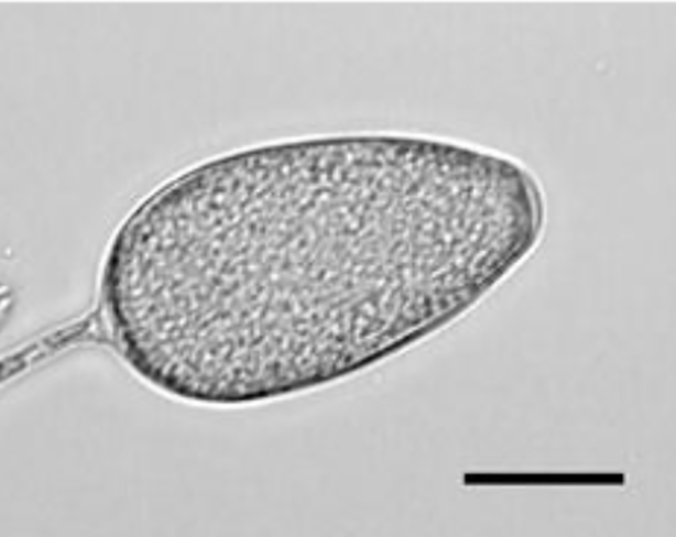
Phytophthora chlamydospora asexual phase: persistent nonpapillate sporangium; Scale bar = 20μm; photos by Paul Reeser. |
|
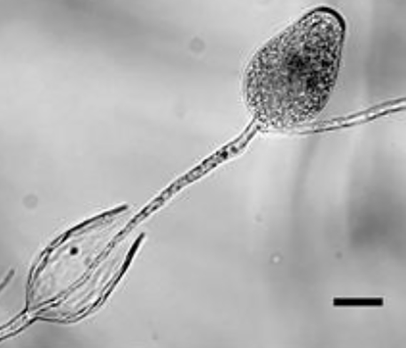
Phytophthora chlamydospora asexual phase: sporangia in water, showing subsporangial elongation; Scale bar = 20μm; photos by Paul Reeser. |
|
Phytophthora chlamydospora asexual phase:chlamydospore; Scale bar = 20μm; photos by Paul Reeser. |
.JPG)
.JPG)


Name and publication
Phytophthora chlamydospora Brasier & E.M. Hansen (2015)
Hansen EM, Reeser P, Sutton W, and Brasier CM. 2015. Redesignation of Phytophthora taxon Pgchlamydo as Phytophthora chlamydospora sp. nov. North American Fungi 10 (2): 1–14.
Corresponding author: hansene@science.oregonstate.edu
Nomenclature
from Hansen et al. (2015)
Mycobank
Etymology
refers to the distinguishing chlamydosporeschlamydospores:
an asexual spore with a thickened inner wall that is delimited from the mycelium by a septum; may be terminal or intercalary, and survives for long periods in soil
formed especially at higher temperatures
Typification
Type: UNITED KINGDOM, isolated from ornamental Prunus in Cheltenham, Gloucestershire in 1971 by C.M Brasier and R.G Strouts OSC # 153153 (Forest Research Advisory Service Record 72/445/82 - isolate P236)
Ex-type: P6133 (World Phytophthora Collection) duplicate of isolate P236
Sequences for ex-type in original manuscript: Phytophthora taxon Pgchlamydo isolate P236 = COX spacer KF750598, β-tubulin KF750602
NOTES:
Although sequence ITS AF541890 is cited as the holotype, it corresponds to Phytophthora gonapodyides strain P501 isolated from ivy roots in UK.
The correct code for the ITS rDNA for the type of P. chlamydospora is: Phytophthora sp. P236 = ITS rDNA AF541900 (provided by H. Hansen to G. Abad on 7.22.15).
Specimen P6133 ITS rDNA GU258890 (under P. gonapodyides)
Ex-type in other collections
(ET) CBS 149403, NRRL 64368, WPC P6133, P236, S&T BL 156 (Abad)
Molecular identification
Voucher sequences for barcoding genes (ITS rDNA and COI) of the ex-type (see Molecular protocols page)
Phytophthora chlamydospora isolate CPHST BL 156 (= P6133 WPC) = ITS rDNA MG865471, COI MH136867
Voucher sequences for Molecular Toolbox with seven genes (ITS, β-tub, COI, EF1α, HSP90, L10, and YPT1
(see Molecular protocols page) (In Progress)
Voucher sequences for Metabarcoding High-throughput Sequencing (HTS) Technologies [Molecular Operational Taxonomic Unit (MOTU)]
(see Molecular protocols page) (In Progress)
Sequences with multiple genes for ex-type in other sources
- NCBI: Phytophthora chlamydospora CPHST BL 156
- NCBI: Phytophthora pgchlamydo isolate P236
- EPPO-Q-bank: Phytophthora chlamydospora
- BOLDSYSTEMS: Phytophthora chlamydospora (barcoding COI & ITS)
Position in multigenic phylogeny with 7 genes (ITS, β-tub, COI, EF1α, HSP90, L10, and YPT1)
Clade clade:
a taxonomic group of organisms classified together on the basis of homologous features traced to a common ancestor
6b
Morphological identification
adapted from Hansen et al. (2015)
Colonies and cardinal temperatures
Colonies on V8-A, PDA, and MEA with rosette (petaloid) pattern. Optimum 25–28°C, and maximum 36–37°C.
Conditions for growth and sporulation
Sporangia and hyphal swellings formed in water. ChlamydosporesChlamydospores:
an asexual spore with a thickened inner wall that is delimited from the mycelium by a septum; may be terminal or intercalary, and survives for long periods in soil
formed in agar media, often scarce at 22°C but usually abundant at 28°C.
Asexual phase
SporangiaSporangia:
sac within which zoospores form, especially when water is cooled to about 10°C below ambient temperature; in solid substrates, sporangia usually germinate by germ tubes
nonpapillatenonpapillate:
pertaining to the production of a non-distinct, or inconspicuous, papilla at the distal end of the sporangium (cf. papillate and semipapillate)
, persistentpersistent:
pertaining to sporangia that remain attached to the sporangiophore and do not separate or detach easily (cf. caducous)
(noncaducous), obpyriformobpyriform:
inversely pear-shaped, i.e. with the widest part at the point of attachment (cf. pyriform)
or ovoidovoid:
egg-shaped, with the widest part at the base of the sporangium and the narrow part at the apex
, often somewhat elongated, with internal proliferationinternal proliferation:
internal proliferation occurs when the sporangiophore continues to grow through an empty sporangium
; average 56 μm x 36 μm; sporangiasporangia:
sac within which zoospores form, especially when water is cooled to about 10°C below ambient temperature; in solid substrates, sporangia usually germinate by germ tubes
produced in unbranched or occasionally sympodial sporangiophores. Hyphal swellings large globoseglobose:
having a rounded form resembling that of a sphere
, subglobose to elongate, produced in clumps and in branched chains (catenulate). ChlamydosporesChlamydospores:
an asexual spore with a thickened inner wall that is delimited from the mycelium by a septum; may be terminal or intercalary, and survives for long periods in soil
are globoseglobose:
having a rounded form resembling that of a sphere
, mostly intercalaryintercalary:
positioned within a hypha (cf. terminal)
but also lateral, terminal, and sessile.
Sexual phase
Self-sterile.
Additional specimen(s) evaluated
Phytophthora chlamydospora ex-type CPHST BL 156 = P6133 (World Phytophthora Collection)
Hosts and distribution
Distribution: Europe, North America
Substrate: roots, leaves, water and riparian areas, soil
Disease note: canker, root rot
Host: wide host range of hard and soft woods
Retrieved from U.S. National Fungus Collections Nomenclature Database.
Additional references and links
- SMML USDA-ARS: Phytophthora chlamydospora
- EPPO Global Database: Phytophthora chlamydospora
- Forest Phytophthoras of the world: Phytophthora chlamydospora
- CABI Digital Library: Phytophthora chlamydospora
- Encyclopedia of Life (EOL): Phytophthora chlamydospora
- Index Fungorum (IF): Phytophthora chlamydospora
- Google All Phytophthora chlamydospora
- Google Images Phytophthora chlamydospora
- Google Scholar Phytophthora chlamydospora
Fact sheet author
Z. Gloria Abad, Ph.D., USDA-APHIS-PPQ-S&T Plant Pathogen Confirmatory Diagnostics Laboratory (PPCDL), United States of America.
