
Phytophthora cactorum (in progress - Abad et al. 2023b)
|
Phytophthora spp. in subclade 1a: portion of the seven-loci ML phylogeny featuring the type cultures of 212 described species (by T. Bourret). Notice the position of P. cactorum selected specimen CBS 231.30 = S&T BL 9. Gloria Abad, USDA S&T.
|
|
Phytophthora spp. in subclade 1a: Morphological Tabular key (PDF) and Tabular key legends (PDF) in IDphy2 KEY SECTION. Notice the data of P. cactorum selected specimen CBS 231.30 = S&T BL 9. Gloria Abad, USDA S&T.
|
|
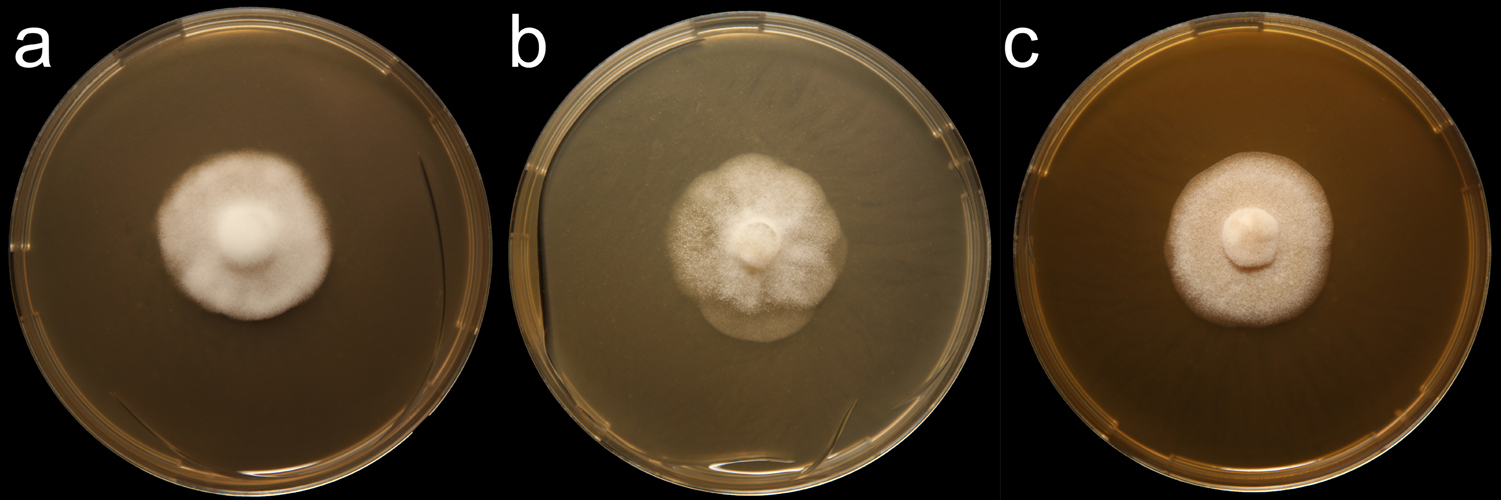
Phytophthora cactorum (CPHST BL 9) colony of a selected specimen grown for 7 days on (a) V8® agar (b) potato dextrose agar (c) malt extract agar; photos by Krysta Jennings and Leandra Knight, USDA-APHIS-PPQ |
|
Phytophthora cactorum (CPHST BL 178) colony of a selected specimen grown for 7 days on (a) V8® agar (b) potato dextrose agar (c) malt extract agar; photos by Krysta Jennings and Leandra Knight, USDA-APHIS-PPQ |
|
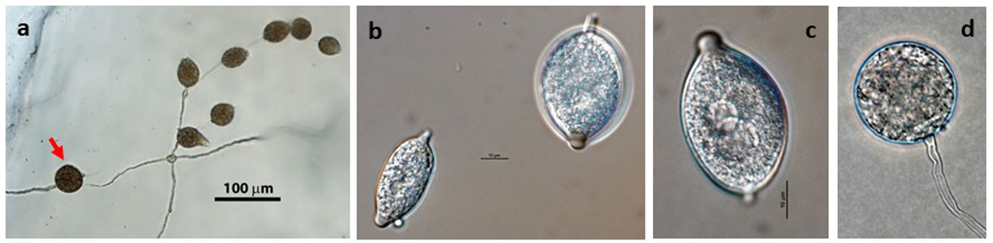
Phytophthora cactorum (CPHST BL 9) asexual phase formed on V8 agar flooded with soil extract (a-c): (a) sporangia originated in lax sympodial sporangiophore, chlamydospore in bottom left (arrow), (b) papillate caducous sporangia with short pedicel, (c) papillate caducous sporangium with short pedicel, (d) chlamydospore; photos by G. Abad, USDA-APHIS-PPQ. |
|
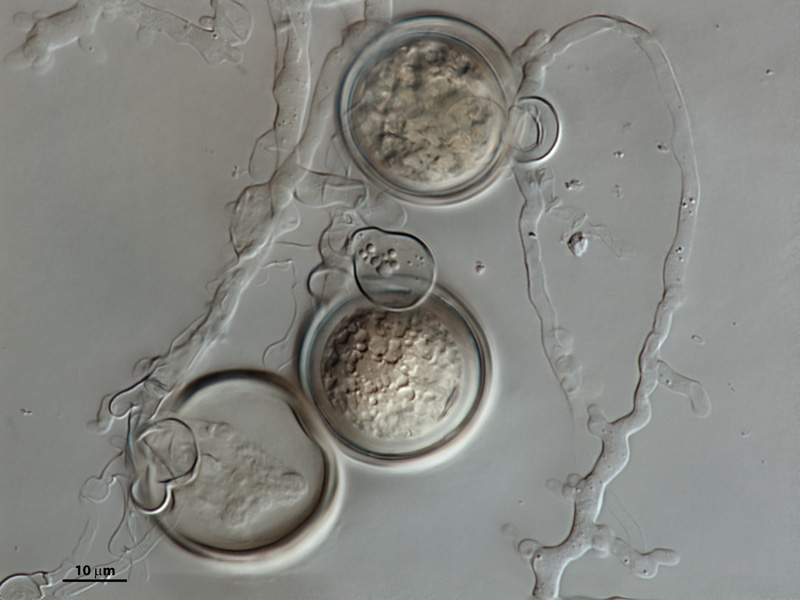
Phytophthora cactorum (CPHST BL 9) sexual phase formed on V8 agar flooded with soil extract: (a) three oogonia with paragynous antheridia, in bottom abortive oogonium, (b) aplerotic oospore in smooth wall oogonium with paragynous antheridium, (c) plerotic oospore in smooth wall oogonium with tapered base and paragynous antheridium; photos (a) and (b) by V. Brewster, USDA-APHIS-PPQ and (c) by G. Abad, USDA-APHIS-PPQ |
|
Phytophthora cactorum (CPHST BL 9) asexual phase formed on V8 agar flooded with soil extract: sporangia originated in lax sympodial sporangiophore, chlamydospore in bottom left. Photo by G. Abad, USDA-APHIS-PPQ. |
|
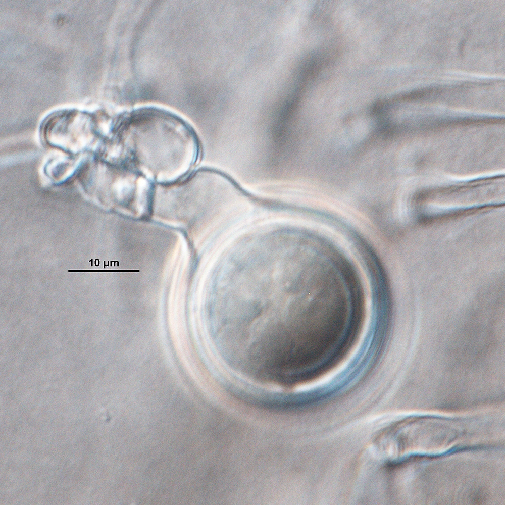
Phytophthora cactorum (CPHST BL 9) sexual phase formed on V8 agar flooded with soil extract: aplerotic oospore in smooth wall oogonium with paragynous antheridium. Photo by V. Brewster, USDA-APHIS-PPQ. |
|
Phytophthora cactorum (CPHST BL 9) sexual phase formed on V8 agar flooded with soil extract: three oogonia with paragynous antheridia, in bottom abortive oogonium. Photo by V. Brewster, USDA-APHIS-PPQ. |
|
Phytophthora cactorum (CPHST BL 9) asexual phase: chlamydospore. Photo by G. Abad, USDA-APHIS-PPQ. |
|
Phytophthora cactorum (CPHST BL 9) sexual phase formed on V8 agar flooded with soil extract: plerotic oospore in smooth wall oogonium with tapered base and paragynous antheridium. Photo by G. Abad, USDA-APHIS-PPQ. |
|
Phytophthora cactorum (CPHST BL 9) asexual phase formed on V8 agar flooded with soil extract: papillate caducous sporangium with short pedicel. Photo by G. Abad, USDA-APHIS-PPQ. |
.JPG)
.JPG)


Name and publication
Phytophthora cactorum (Lebert & Cohn) J. Schröt. (1886)
Schröter J. 1886. pp. 129–256 (236). In: F. Cohn, Kryptogamen-Flora von Schlesien. Band 3, Heft 3, Pilze J.U. Kern's Verlag, Breslau, 1889, pp. 1–814.
Nomenclature
from Schroeter (1886)
Mycobank
Synonym(s)
≡ Peronospora cactorum Lebert & Cohn, Beiträge zur Biologie der Pflanzen 1: 182 (1872) [MB166699]
≡ Nozemia cactorum (Lebert & Cohn) Pethybr., Scientific Proceedings of the Royal Dublin Society 13: 566 (1913) [MB562134]
≡ Phloeophthora cactorum (Lebert & Cohn) G.W. Wilson, Mycologia 6 (2): 80 (1914) [MB453641]
= Phytophthora paeoniae D.C. Cooper & Porter, Phytopathology 18: 881 (1928) [MB273265]
Typification
Type: SLOVAKIA, Bratislava, in the garden of Jacobi, Type Figures: Peronospora cactorum was first identified on cacti in 1870 by Lebert and Cohn. Habitat, intercellular in the parequima of various diseased rotten cacti. Observed in the host 1868/9.
Ex-type: LOST
Well-authenticated specimen selected by Gloria Abad: CPHST BL 9 = P0714 (WPC) (Collection MUCL 9638)
Selected specimens in other collections
(SE) CBS 231.30, NRRL 64109, MUCL 9638, WPC P0714, ATCC 10091, 200785 MCI, MUCL 9638, S&T BL 9 (Abad), N93 (Gallegly)
Molecular identification
Voucher sequences for barcoding genes (ITS rDNA and COI) of the selected specimen (see Molecular protocols page)
Phytophthora cactorum isolate CPHST BL 9 (= P0714 WPC) = ITS rDNA MG783385, COI MH136858
Additional sequences for molecular identification
ITS rDNA HQ261514 (isolate P0714), COI HQ261261 (isolate P0714)
Voucher sequences for Molecular Toolbox with seven genes (ITS, β-tub, COI, EF1α, HSP90, L10, and YPT1
(see Molecular protocols page) (In Progress)
Voucher sequences for Metabarcoding High-throughput Sequencing (HTS) Technologies [Molecular Operational Taxonomic Unit (MOTU)] (see Molecular protocols page)
(In Progress)
Sequences with multiple genes for selected specimen in other sources
- NCBI: Phytophthora cactorum CPHST BL 9
- NCBI: Phytophthora cactorum P0714
- NCBI: Phytophthora cactorum CBS 231.30
- EPPO-Q-bank: Phytophthora cactorum CBS 231.30
- BOLDSYSTEMS: Phytophthora cactorum OOMYA2078-10 = CBS 231.30, PHYTO008-10 = P0714 (barcoding COI & ITS)
Position in multigenic phylogeny with 7 genes (ITS, β-tub, COI, EF1α, HSP90, L10, and YPT1)
Clade clade:
a taxonomic group of organisms classified together on the basis of homologous features traced to a common ancestor
1a
Morphological identification
Colonies and cardinal temperatures
Colonies in V8 agar, potato dextrose agar, and malt extract agar with no distinct pattern. The minimum temperature for growth is 4°C, the optimum 24°C, and the maximum 30°C.
Asexual phase
SporangiaSporangia:
sac within which zoospores form, especially when water is cooled to about 10°C below ambient temperature; in solid substrates, sporangia usually germinate by germ tubes
papillatepapillate:
pertaining to the production of a distinct papilla at the distal end of the sporangium (cf. nonpapillate and semipapillate)
, caducouscaducous:
pertaining to sporangia that become dislodged readily (i.e. deciduous) and separate from the sporangiophore (cf. persistent)
with short pedicels (less than 4 μm in length), ellipsoidal, obpyriformobpyriform:
inversely pear-shaped, i.e. with the widest part at the point of attachment (cf. pyriform)
, ovoidovoid:
egg-shaped, with the widest part at the base of the sporangium and the narrow part at the apex
, or globoseglobose:
having a rounded form resembling that of a sphere
(24–50 L x 19–36 W μm) borne in simple or in close or lax sympodial sporangiophores. Hyphal swellings absent. ChlamydosporesChlamydospores:
an asexual spore with a thickened inner wall that is delimited from the mycelium by a septum; may be terminal or intercalary, and survives for long periods in soil
terminal and intercalaryintercalary:
positioned within a hypha (cf. terminal)
, globoseglobose:
having a rounded form resembling that of a sphere
(17–55 μm diam).
Sexual phase
Homothallic. OogoniaOogonia:
the female gametangium in which the oospore forms after fertilization by the antheridium
smooth-walled and usually hyaline (19–38 µm diam); antheridiaantheridia:
the male gametangium; a multinucleate, swollen hyphal tip affixed firmly to the wall of the female gametangium (the oogonium)
paragynousparagynous:
pertaining to the sexual stage in which the antheridium is attached to the side of the oogonium (cf. amphigynous)
, nearly spherical to club-shaped, and nearly always applied close to the oogonial stalk; oospores plerotic plerotic:
pertaining to an oospore that fills the oogonium (cf. aplerotic)
and apleroticaplerotic:
pertaining to a mature oospore that does not fill the oogonium; i.e. there is room left between the oospore wall and oogonium wall (cf. plerotic)
(20–26 µm diam), with an average wall thickness of 2 μm.
Most typical characters
Phytophthora cactorum is characterized by its production of caducouscaducous:
pertaining to sporangia that become dislodged readily (i.e. deciduous) and separate from the sporangiophore (cf. persistent)
sporangia with short pedicels and paragynousparagynous:
pertaining to the sexual stage in which the antheridium is attached to the side of the oogonium (cf. amphigynous)
antheridiaantheridia:
the male gametangium; a multinucleate, swollen hyphal tip affixed firmly to the wall of the female gametangium (the oogonium)
.
Specimen(s) evaluated
Phytophthora cactorum CPHST BL 9 (Abad) = P0714 [World Phytophthora Collection (WPC) California, USA]
Hosts and distribution
Distribution: cosmopolitan
Substrate: leaves, stems, fruits, roots
Disease note: damping off of seedlings, fruit rot, leaf and stem rot, collar and crown rot, stem canker, root rot (Waterhouse & Waterston 1966)
Host: at least 154 genera of vascular plants in 54 families (Waterhouse & Waterston 1966)
Retrieved May 18, 2018 from U.S. National Fungus Collections Nomenclature Database.
Quarantine status
There is no quarantine for Phytophthora cactorum, as it is very prevalent and widely distributed around the world.
Additional references and links
Phytophthora cactorum in OSU Phytophthora Online course: Training for Nursery Growers. Oregon State University.
- SMML USDA-ARS: Phytophthora cactorum
- EPPO Global Database: Phytophthora cactorum
- Forest Phytophthoras of the world: Phytophthora cactorum
- CABI Digital Library: Phytophthora cactorum
- Encyclopedia of Life (EOL): Phytophthora cactorum
- Index Fungorum (IF): Phytophthora cactorum
- Google All Phytophthora cactorum
- Google Images Phytophthora cactorum
- Google Scholar Phytophthora cactorum
Fact sheet author
Z. Gloria Abad, Ph.D., USDA-APHIS-PPQ-S&T Plant Pathogen Confirmatory Diagnostics Laboratory (PPCDL), United States of America.
