
Ganoderma Butt Rot
|
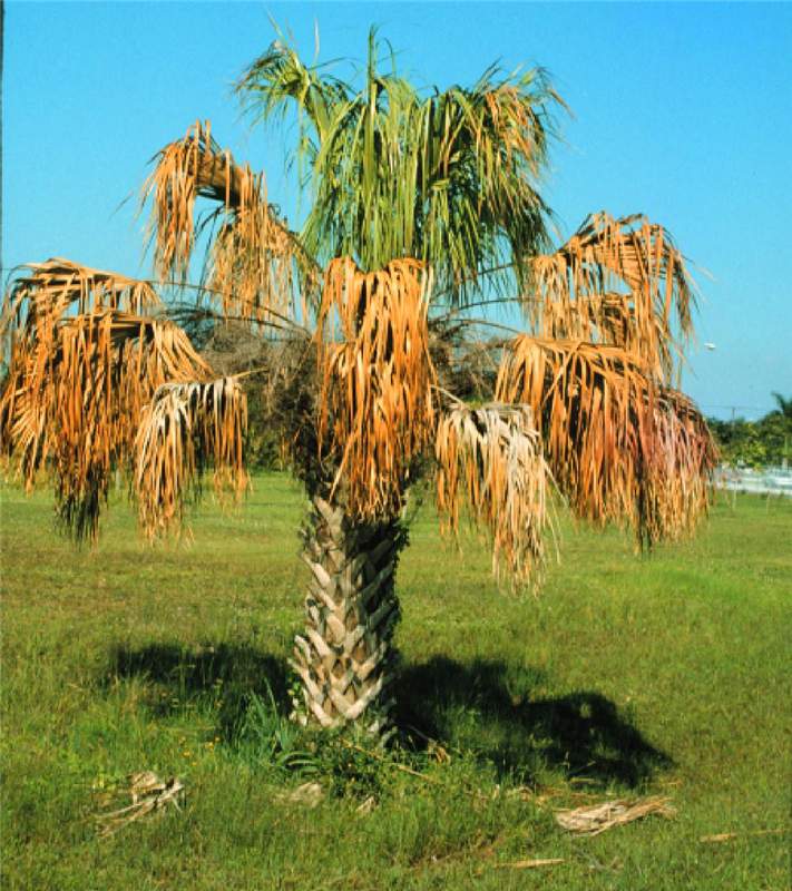
Figure 1. Sabal palmetto exhibiting an overall wilted canopy and premature death of the oldest leaves due to Ganoderma butt rot. Photo by M. L. Elliott.
|
|
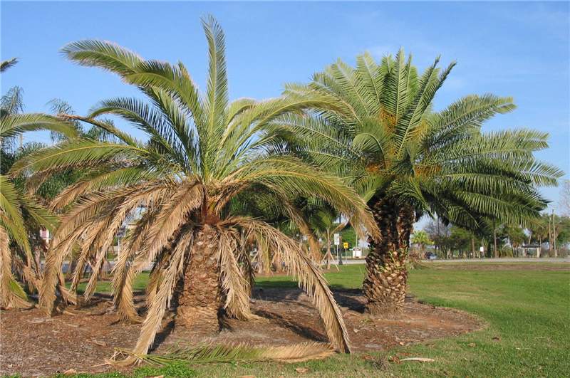
Figure 2. The Phoenix canariensis on the left is exhibiting premature death of the oldest leaves due to Ganoderma butt rot. This is in contrast to the healthy palm on the right. Photo by B. Dick, City of Lakeland, Florida.
|
|
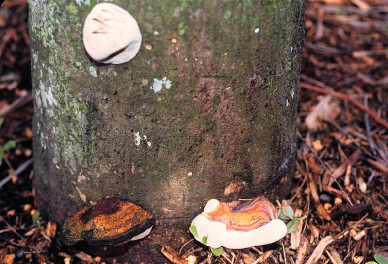
Figure 3. The white mass in the upper-left corner is an early stage of basidiocarp formation of Ganoderma zonatum. The lower-right corner illustrates the shelf or bracket structure of a developing basidiocarp. Because it is still white at the edges and underneath, it has not yet matured and has not released the basidiospores. The lower-left corner illustrates an old, decaying basidiocarp. Photo by M. L. Elliott.
|
|
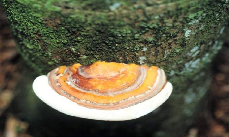
Figure 4. A developing basidiocarp of Ganoderma zonatum. Note the "zones" on the top portion of the basidiocarp, and that the fungus is directly attached to the palm trunk. Photo by M. L. Elliott.
|
|
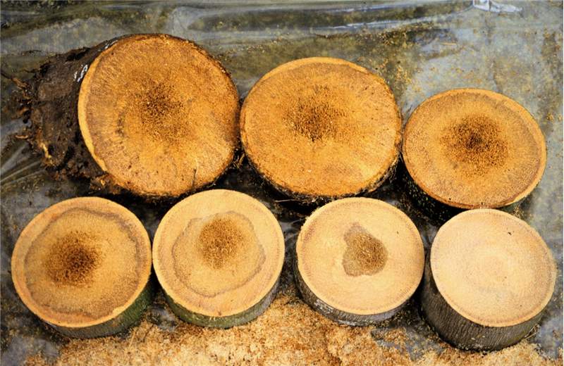
Figure 5. A series of cross-sections of a Ganoderma butt rot affected palm trunk. The section in the upper-left corner is the lowest section (at the soil line). The section in the lower-right corner is the furthest section from the soil line. The remaining sections are in the correct sequence from the soil line upwards. Note how the discoloration due to degrading tissue remains centralized within the trunk, but decreases as the cross-section ascends from the soil line. Photo by M. L. Elliott.
|
|
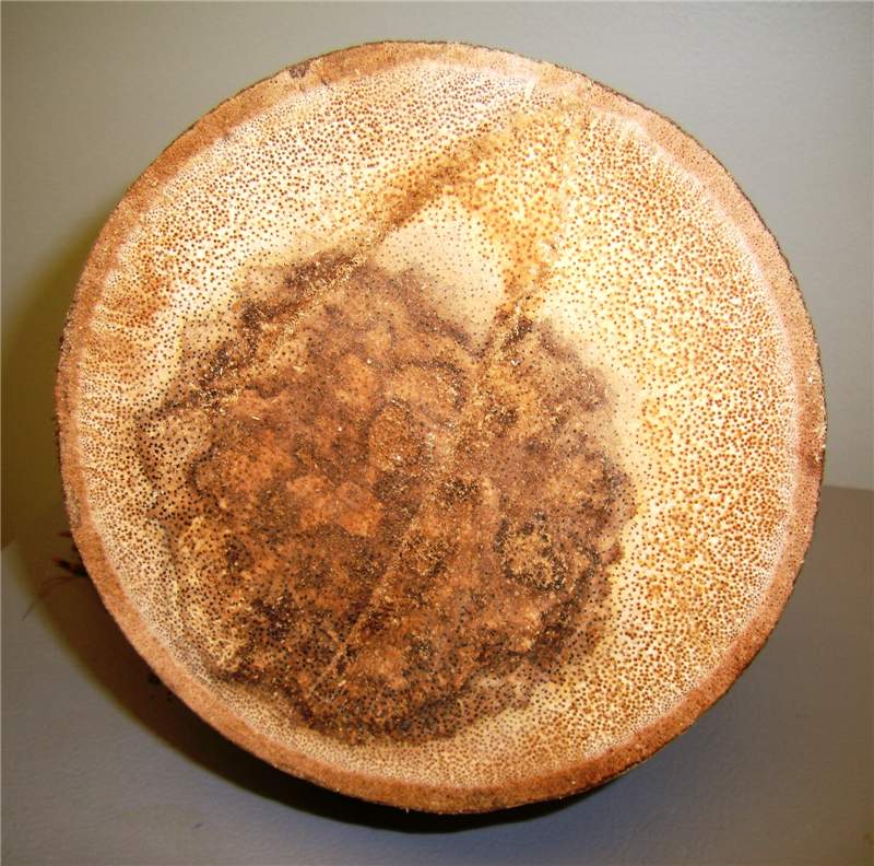
Figure 6. A cross-section through a Cocothrinax sp. trunk with Ganoderma butt rot. The darkened area in the center is a symptom of the trunk rot. Photo by M. L. Elliott.
|
|
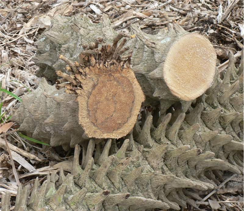
Figure 7. The section of Phoenix roebelenii on the left is the end of the trunk at the soil line and illustrates the typical trunk discoloration associated with Ganoderma butt rot. The section on the right was located further up the trunk and illustrates a trunk section unaffected by Ganoderma zonatum. Photo by M. L. Elliott.
|
Scientific name of pathogen
Ganoderma zonatum: Kingdom Fungi, Phylum Basidiomycota
Hosts
All palm species are considered hosts of this pathogen.
Distribution
Southeastern USA, primarily Florida, but also known to occur in Georgia and South Carolina. While the fungus may be present in Mexico or the Caribbean basin, it has not been documented thus far.
Symptoms/signs
The primary symptoms that may be observed include: wilting, mild to severe, of either all leaves or just the lowest leaves in the canopycanopy:
the cluster of leaves borne at the tip of the stem
(Fig. 1); premature death of the oldest leaves (Figs. 1 and 2); or a general decline of the palm (slower growth and off-color foliage, despite good fertilization).
The basidiocarp or conk is the most easily identifiable structure associated with the fungus. The basidiocarp originates from fungal growth inside the palm trunk. When the basidiocarp first starts to form on the side of a palm trunk or palm stump, it is a solid white mass that is relatively soft when touched (Fig. 3). It will have an irregular to circular shape and is relatively flat on the trunk or stump. As the basidiocarp matures, a small shelf or bracket will start to form as the basidiocarp begins to extend or protrude from the trunk (Fig. 3). It will still be white, both on the top and bottom surfaces. Eventually, the fungus will form a very distinct shelf-like structure that is quite hard with a glazed reddish-brown top surface and a white undersurface (Fig. 4).
A mature basidiocarp will have distinct zones, hence the name Ganoderma zonatum (Fig. 4). The basidiocarp will have a half-moon shape that is directly attached to the trunk. There is no stalk that attaches the basidiocarp to the trunk. Once the basidiocarp is mature, it will release rust-colored spores that will cover the basidiocarp and the surrounding area. Basidiocarps of Ganoderma zonatum can be up to 8 inches at their widest point and 2 inches thick. However, basidiocarps will take on the shape and size of the area in which they are forced to grow.
If a basidiocarp is present on the trunk at the same time the wilt or decline symptoms appear, then it is safe to diagnose Ganoderma butt rot. However, it is quite common for basidiocarps not to appear prior to severe decline and death of a palm. In that situation, the only way to determine if Ganoderma butt rot is the cause is to cut cross-sections through the lower four feet of trunk after the palm is felled and examine the cross-sections for the typical pattern of rot - greatest near the soil line, decreasing in sections further from the soil line (Fig. 5). Basidiocarps may form on the palm stump after the diseased trunk is felled.
The tissue degradation (rot) that occurs does not result in a soft, mushy trunk, nor does it smell "rotten". Instead, it is a dry rot of lignified tissue found in the vascular bundles (Figs. 6 and 7).
May be confused with
Ganoderma butt rot may be confused with the initial symptoms caused by the phytoplasma diseases, lethal yellowing or Texas Phoenix palm decline - i.e., a wilt or premature death of the lowest leaves in the canopycanopy:
the cluster of leaves borne at the tip of the stem
. Thielaviopsis trunk rot normally causes a trunk rot in the upper half of the trunk, but Thielaviopsis paradoxa rots the trunk from the outside to the inside of the trunk, beginning at a precise infection point. Drought stress and deep planting would also cause wilt symptoms.
Additional comments
The fungus is spread primarily by the spores produced by the basidiocarp (conk). It is believed that the fungus uses the roots as a means of moving from the soil to the woody trunk tissue at or below the soil line. Once a palm is infected with Ganoderma zonatum, it may remain symptomless for months or years. However, the fungus will move with that infected palm to the location in which it is transplanted. It is also possible that soil associated with transplanted palms is infested with the fungus. Another Ganoderma species, G. boninense, is the cause of a similar disease of Elaeis guineensis in southeast Asia.
