

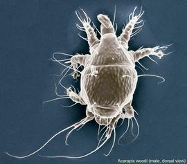
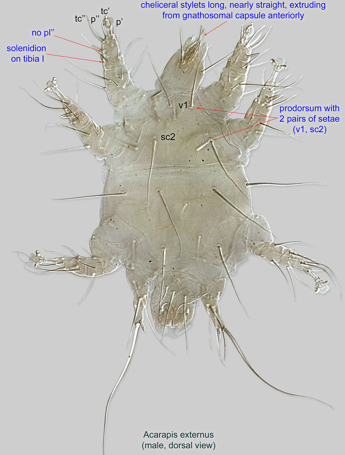
Fig. 5. Acarapis woodi male, dorsal view; LT-SEM (digitally colorized) photo by Ron Ochoa & Gary Bauchan, USDA-ARS.
Fig. 5. Acarapis woodi male, dorsal view; LT-SEM (digitally colorized) photo by Ron Ochoa & Gary Bauchan, USDA-ARS.
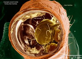
Fig. 6. Acarapis woodi female inside honey bee trachea; LT-SEM (digitally colorized) photo by Ron Ochoa & Gary Bauchan, USDA-ARS.
Fig. 6. Acarapis woodi female inside honey bee trachea; LT-SEM (digitally colorized) photo by Ron Ochoa & Gary Bauchan, USDA-ARS.

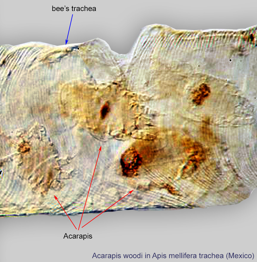
Fig. 7. Acarapis woodi in a trachea of the European honey bee, Apis mellifera; photo by Barry OConnor, University of Michigan.
Fig. 7. Acarapis woodi in a trachea of the European honey bee, Apis mellifera; photo by Barry OConnor, University of Michigan.
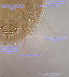
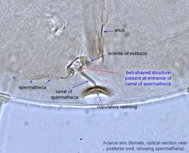
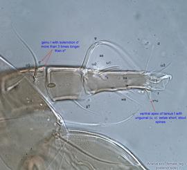
Fig. 11. Acarus siro female optical section at the posterior end of the body, showing spermatheca.
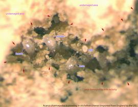
Fig. 15. Acarus chaetoxysilos burrowing in Stichelton cheese imported from England to the US.
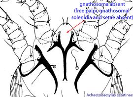
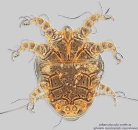
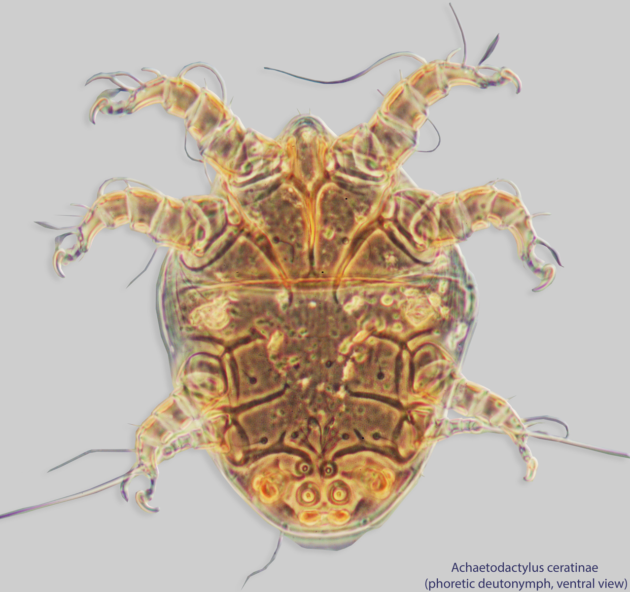
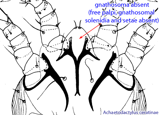
Fig. 2. Anteroventral region of the phoretic deutonymph of Achaetodactylus ceratinae, showing the absence of the gnathosoma; drawing courtesy of Belgian GTI Focal Point 2009, http://www.taxonomy.be.
Fig. 2. Anteroventral region of the phoretic deutonymph of Achaetodactylus ceratinae, showing the absence of the gnathosoma; drawing courtesy of Belgian GTI Focal Point 2009, http://www.taxonomy.be.
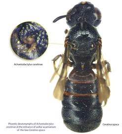
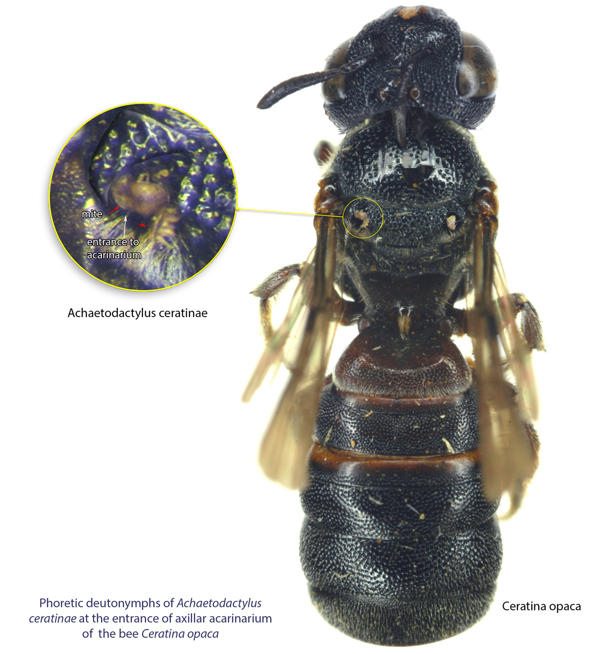
Fig. 3. Phoretic deutonymphs of Achaetodactylus ceratinae at the entrance of axillar acarinarium of the bee Ceratina opaca.
Fig. 3. Phoretic deutonymphs of Achaetodactylus ceratinae at the entrance of axillar acarinarium of the bee Ceratina opaca.
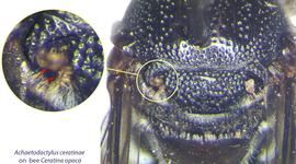
Fig. 4. Closeup of phoretic deutonymphs of Achaetodactylus ceratinae at the entrances of axillar acarinaria of Ceratina opaca.
Fig. 4. Closeup of phoretic deutonymphs of Achaetodactylus ceratinae at the entrances of axillar acarinaria of Ceratina opaca.
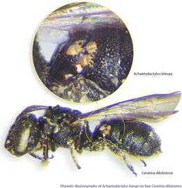
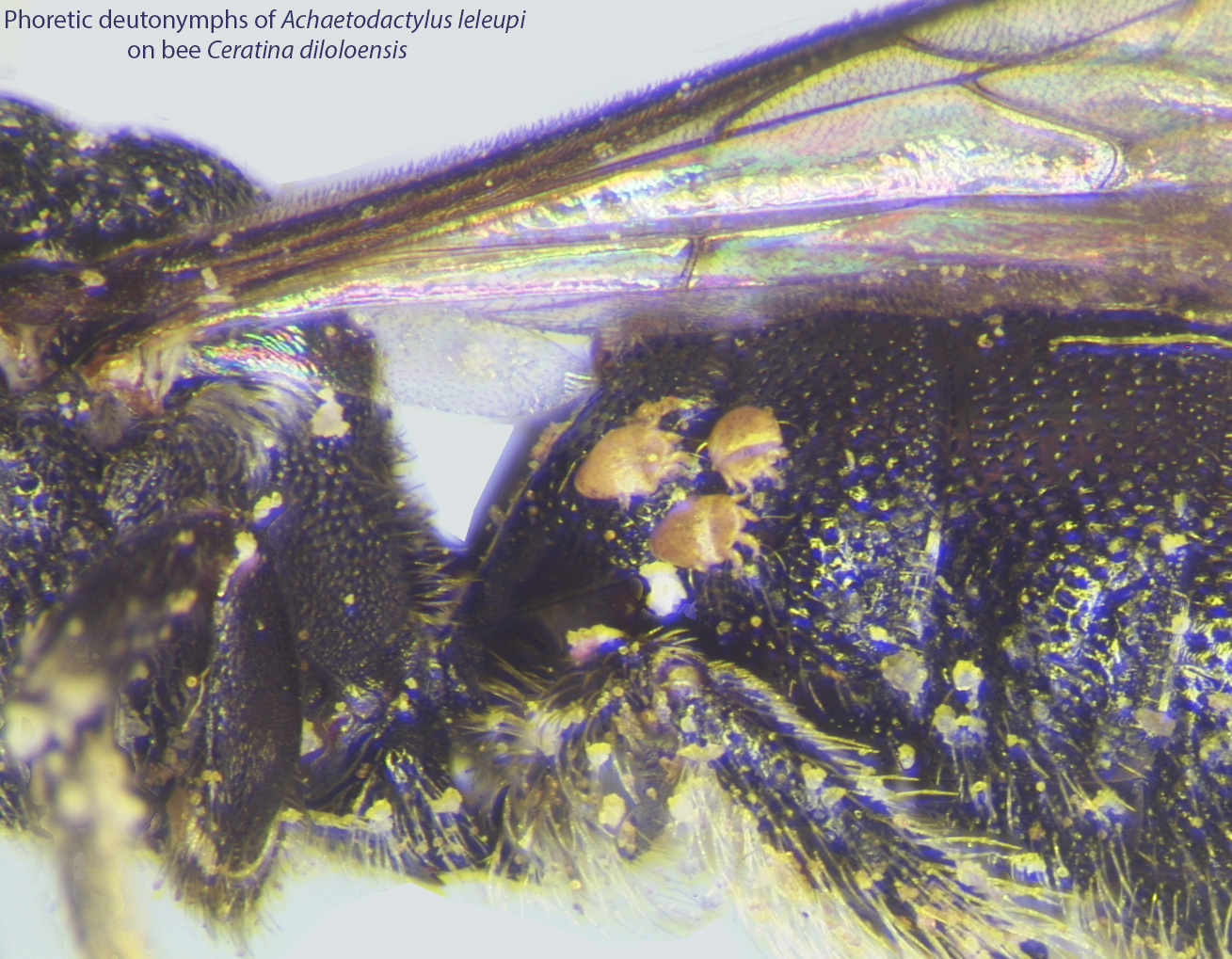
Fig. 5. Phoretic deutonymphs of Achaetodactylus leleupi on bee Ceratina diloloensis.
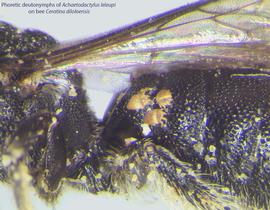
Fig. 6. Closeup of phoretic deutonymphs of Achaetodactylus leleupi on bee Ceratina diloloensis.
Authors: P. Klimov, B. OConnor, R. Ochoa, G. Bauchan, A. Redford, J. Scher
Last updated October 2016
tool images at ITP Node
idtools.org

