

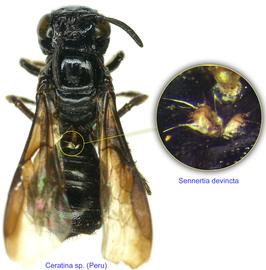
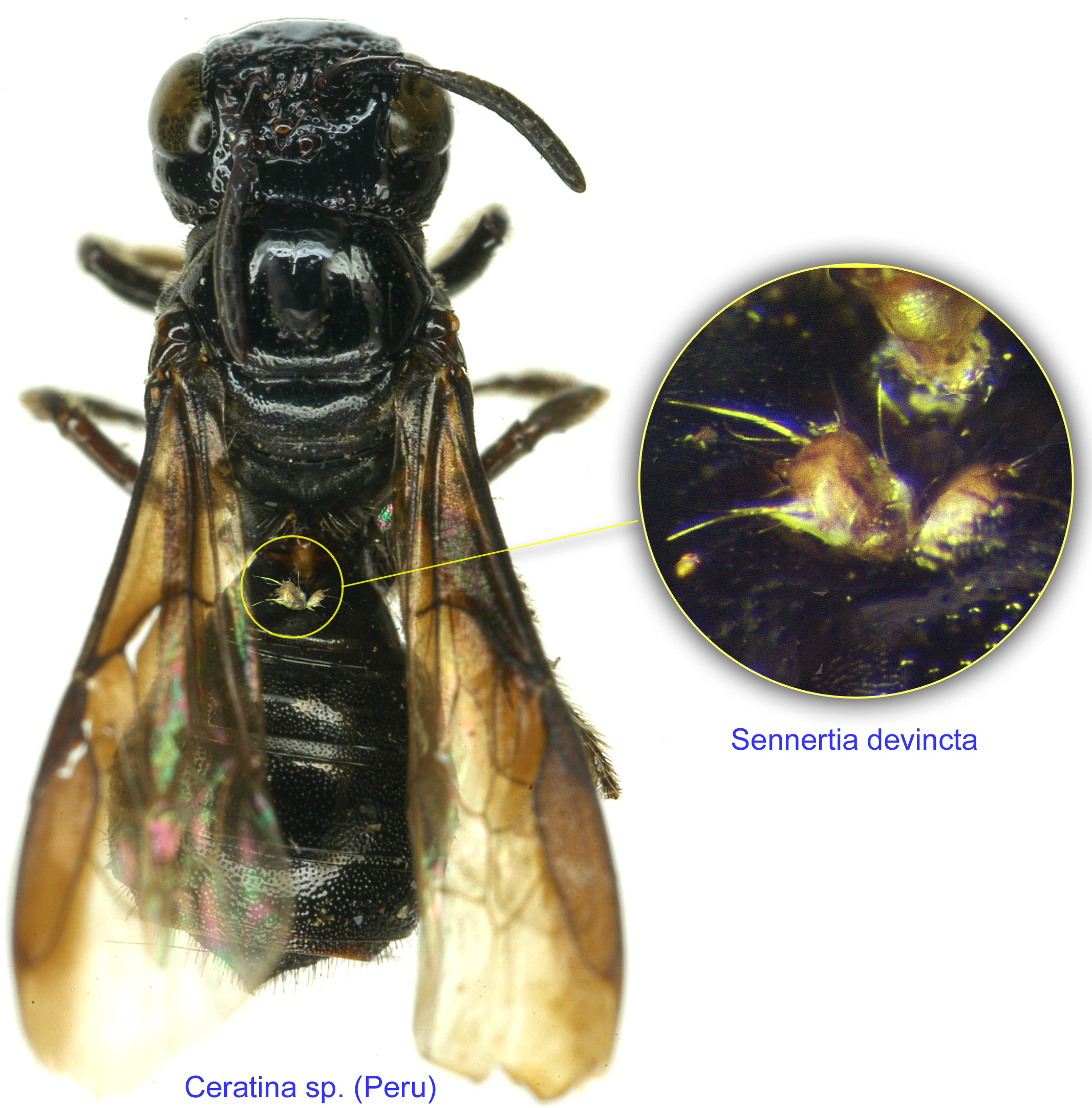
Fig. 14. Phoretic deutonymphs of Sennertia devincta at the entrance of the metasomal acarinarium of bee Ceratina sp. from Peru.
Fig. 14. Phoretic deutonymphs of Sennertia devincta at the entrance of the metasomal acarinarium of bee Ceratina sp. from Peru.

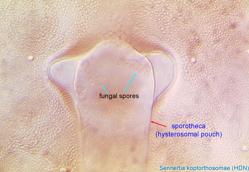
Fig. 15. Sporotheca (hysterosomal pouch) of the phoretic deutonymph of Sennertia koptorthosomae. Situated on the hysterosomal shield.
Fig. 15. Sporotheca (hysterosomal pouch) of the phoretic deutonymph of Sennertia koptorthosomae. Situated on the hysterosomal shield.
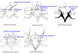
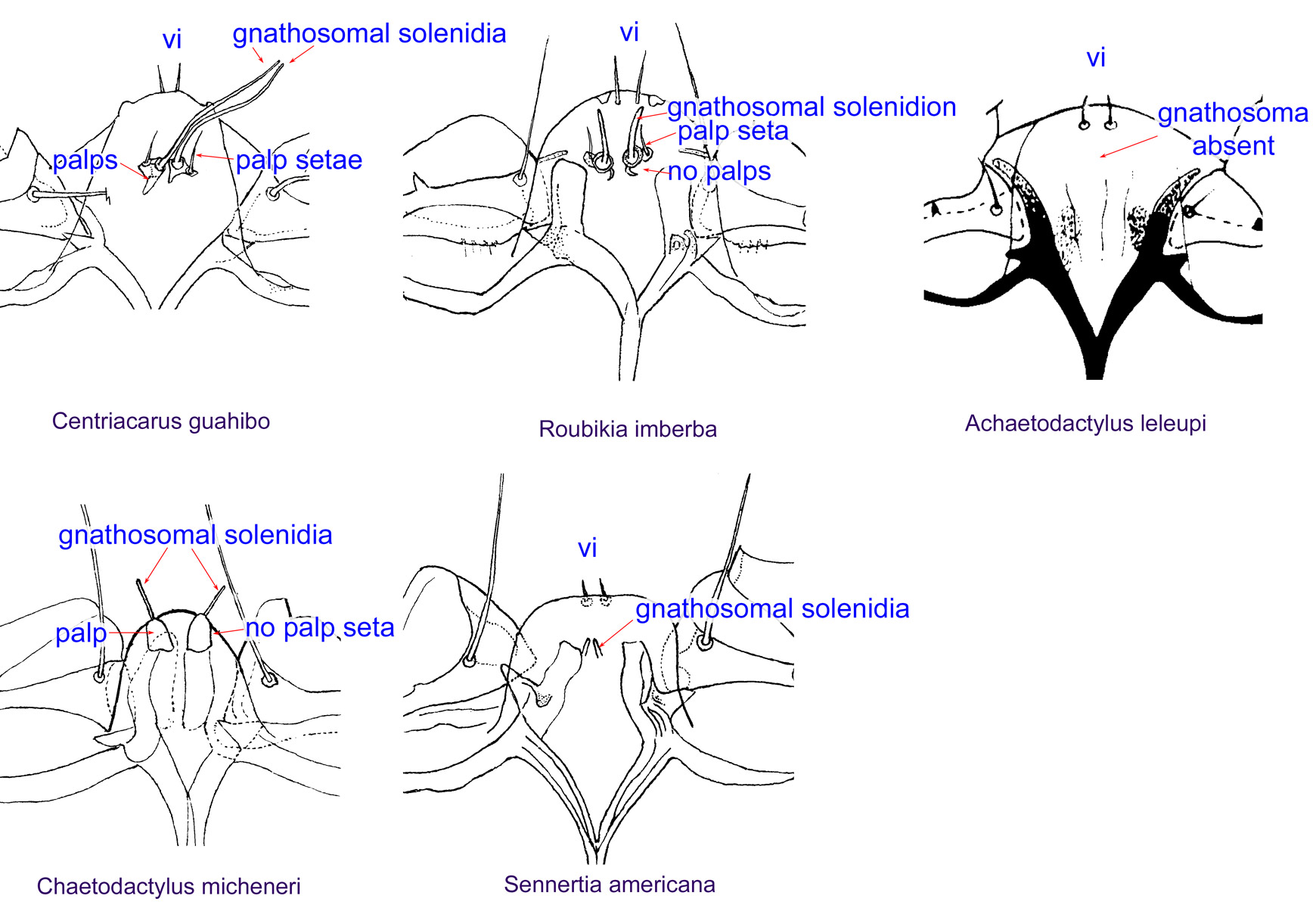
Fig. 16. Gnathosomal morphology is a key character separating genera of the family Chaetodactylidae (Centriacarus, Roubikia, Achaetodactylus, Chaetodactylus, and Sennertia) based on phoretic deutonymphs. In particular, Sennertia can be distinguished by its gnathosomal solenidion present and free palpi and setae on free palpi absent; drawing of Achaetodactylus leleupi courtesy of Belgian GTI Focal Point 2009, http://www.taxonomy.be.
Fig. 16. Gnathosomal morphology is a key character separating genera of the family Chaetodactylidae (Centriacarus, Roubikia, Achaetodactylus, Chaetodactylus, and Sennertia) based on phoretic deutonymphs. In particular, Sennertia can be distinguished by its gnathosomal solenidion present and free palpi and setae on free palpi absent; drawing of Achaetodactylus leleupi courtesy of Belgian GTI Focal Point 2009, http://www.taxonomy.be.
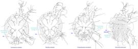
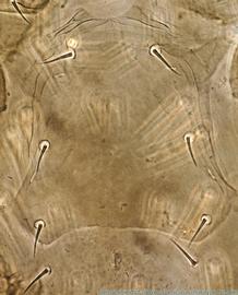
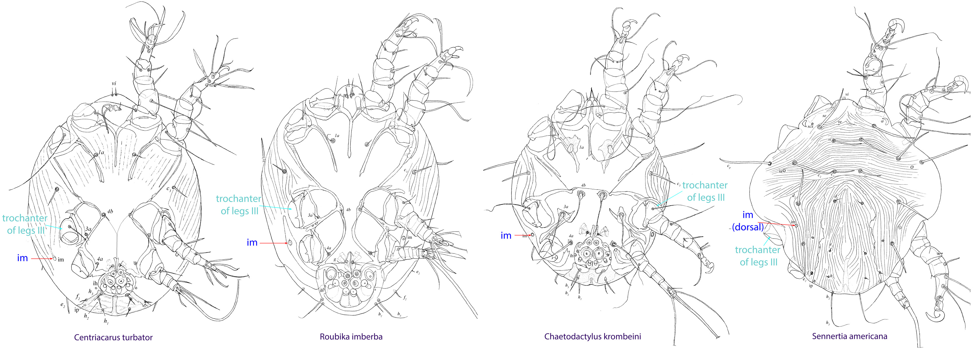
Fig. 17. Cupules im of deutonymphs of Chaetodactylidae. The genus Sennertia is unique in having cupules im situated at the level of trochanters III, while in other genera these cupules are situated posterior to the level of trochanters III. Also, cupules im are dorsal in Sennertia and ventral in all other genera (but may appear dorsal on slide-mounted specimens of Chaetodactylus, due to flattening).
Fig. 17. Cupules im of deutonymphs of Chaetodactylidae. The genus Sennertia is unique in having cupules im situated at the level of trochanters III, while in other genera these cupules are situated posterior to the level of trochanters III. Also, cupules im are dorsal in Sennertia and ventral in all other genera (but may appear dorsal on slide-mounted specimens of Chaetodactylus, due to flattening).
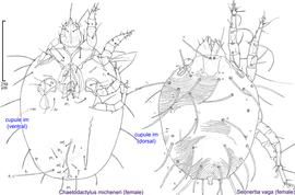
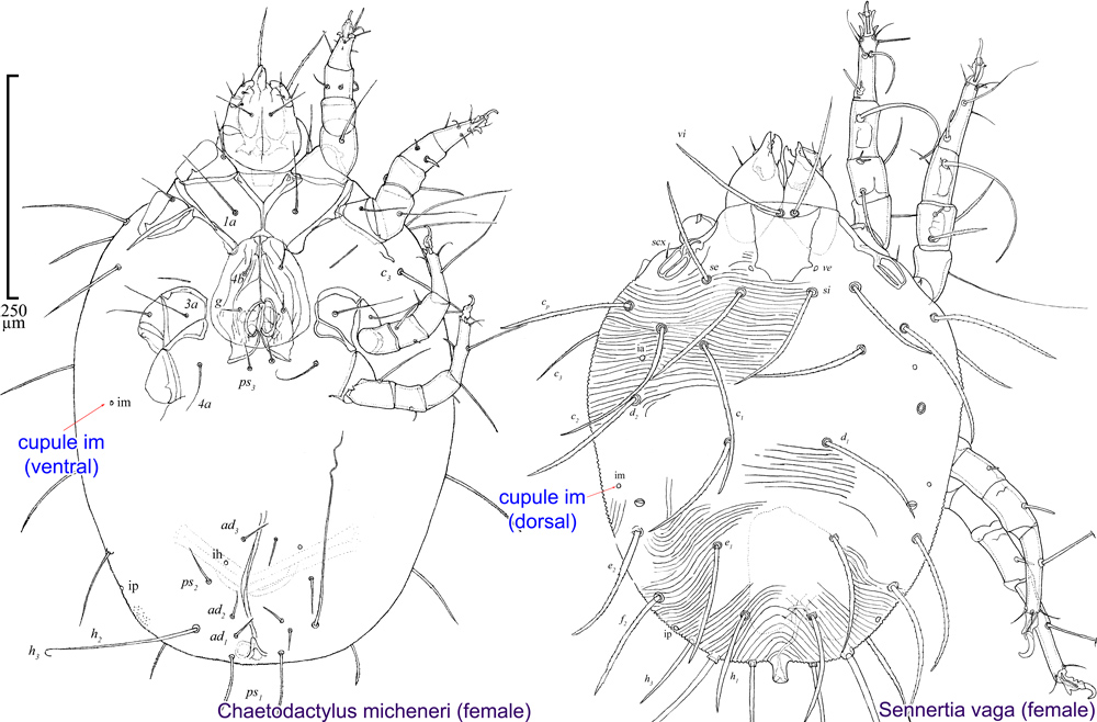
Fig. 18. Adults of Sennertia differ from those of other chaetodactylid genera with known adults (Chaetodactylus and Roubikia) by the dorsal position of cupules im. In Chaetodactylus and Roubikia these cupules im are ventral or ventro-lateral.
Fig. 18. Adults of Sennertia differ from those of other chaetodactylid genera with known adults (Chaetodactylus and Roubikia) by the dorsal position of cupules im. In Chaetodactylus and Roubikia these cupules im are ventral or ventro-lateral.
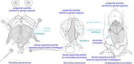
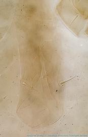
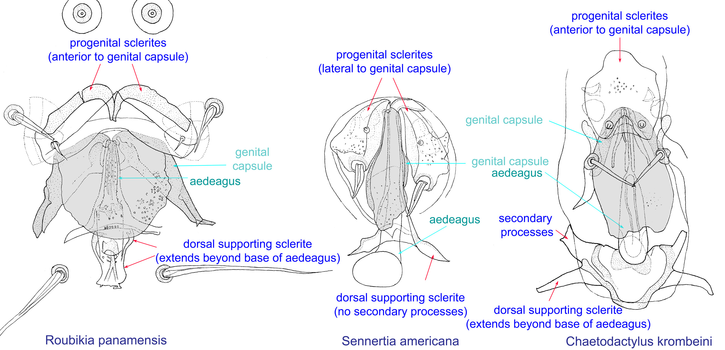
Fig. 19. External genital apparatus of males of Roubikia, Sennertia, and Chaetodactylus, showing key diagnostic feature of Sennertia males: progenital sclerites are lateral to genital capsule, while in other genera they are anterior to genital capsule.
Fig. 19. External genital apparatus of males of Roubikia, Sennertia, and Chaetodactylus, showing key diagnostic feature of Sennertia males: progenital sclerites are lateral to genital capsule, while in other genera they are anterior to genital capsule.
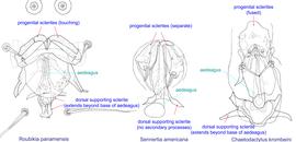
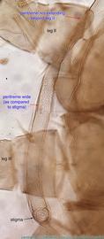
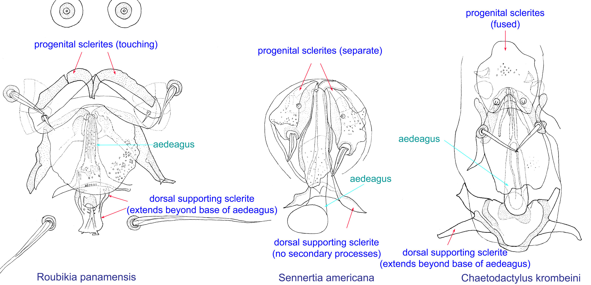
Fig. 20. External genital apparatus of males of Roubikia, Sennertia, and Chaetodactylus, showing key diagnostic features of Sennertia males: progenital sclerites are separate (not touching each other or fused) and dorsal supporting sclerite does not extend posterior to base of aedeagus.
Fig. 20. External genital apparatus of males of Roubikia, Sennertia, and Chaetodactylus, showing key diagnostic features of Sennertia males: progenital sclerites are separate (not touching each other or fused) and dorsal supporting sclerite does not extend posterior to base of aedeagus.
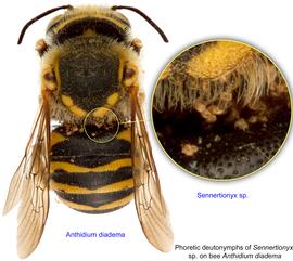
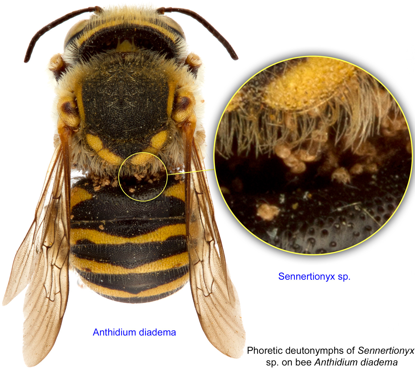
Fig. 3. Phoretic deutonymphs of Sennertionyx sp. on bee Anthidium diadema; photo by Lindsey Seastone & Laura Hartmann, ITP.
Fig. 3. Phoretic deutonymphs of Sennertionyx sp. on bee Anthidium diadema; photo by Lindsey Seastone & Laura Hartmann, ITP.
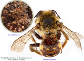
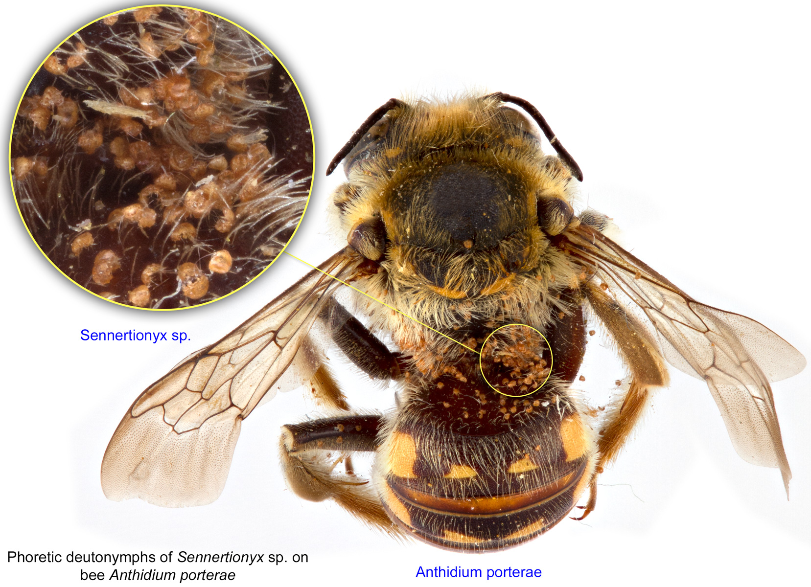
Fig. 4. Phoretic deutonymphs of Sennertionyx sp. on bee Anthidium porterae; photo by Lindsey Seastone & Laura Hartmann, ITP.
Fig. 4. Phoretic deutonymphs of Sennertionyx sp. on bee Anthidium porterae; photo by Lindsey Seastone & Laura Hartmann, ITP.
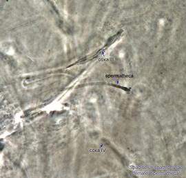
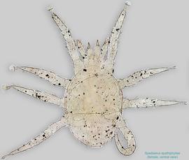
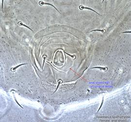
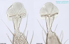
Fig. 16. Spadiseius spathiphyllae paratype female spermatheca, optical section of hysterosoma in region of insertion of leg coxae III-IV.
Fig. 16. Spadiseius spathiphyllae paratype female spermatheca, optical section of hysterosoma in region of insertion of leg coxae III-IV.
Authors: P. Klimov, B. OConnor, R. Ochoa, G. Bauchan, A. Redford, J. Scher
Last updated October 2016
tool images at ITP Node
idtools.org
