

generalist predators that are expected to be neutral to beneficial, but may be harmful if they can enter brood cells; preys on microarthropods in bee nests
Blattisocius Keegan, 1944
Superorder Parasitiformes » Order Mesostigmata » Suborder Monogynaspida » Hyporder Dermanyssiae » Family Blattisociidae » Genus Blattisocius
Blattisocius triodons Keegan, 1944 (=Lasioseius tarsalis Berlese, 1918)
Paragarmania Nesbitt, 1951 (this synonymy has not been accepted by all authors; in these references (Bregetova, 1977cBregetova, 1977c:
Bregetova, N. G. 1977c. [Family Aceosejidae]. In [Opredelitel' obytayshchikh v pochve kleshchey Mesostigmata = Identification key to soil-inhabiting mites Mesostigmata], eds. M. S. Gilarov amp; N. G. Bregetova, 169-226. Leningrad: Nauka.; Karg, 1971Karg, 1971:
Karg, W. 1971. Acari (Acarina), Milben. Unterordnung Anactinochaeta (Parasitiformes). Die freilebenden Gamasina (Gamasides), Raubmilben. Die Tierwelt Deutschlands. 59. 475 pp.), Paragarmania is treated as a separate genus).
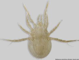
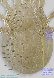
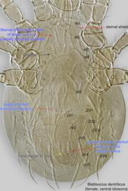
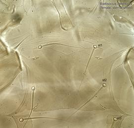
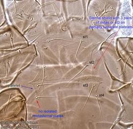
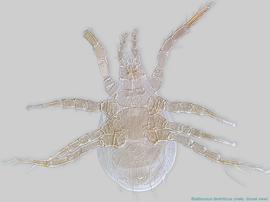
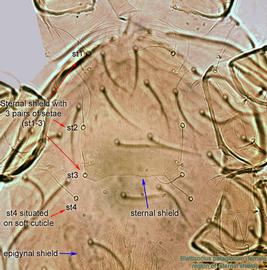
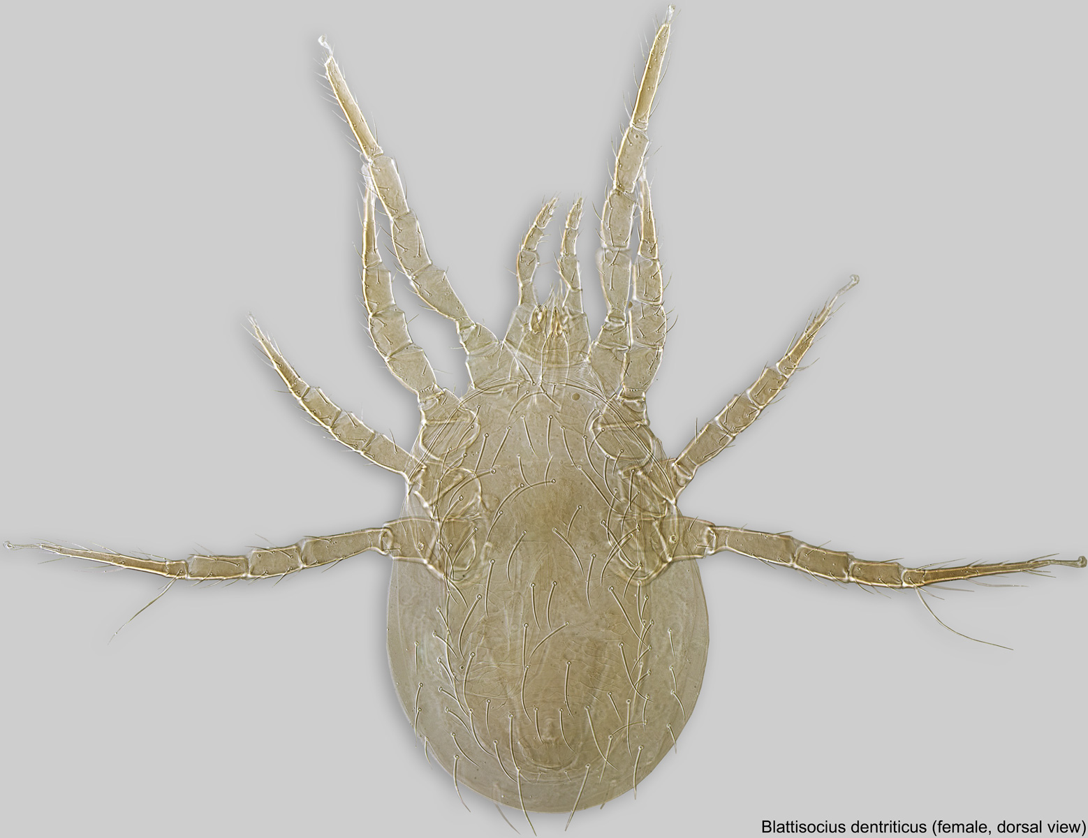
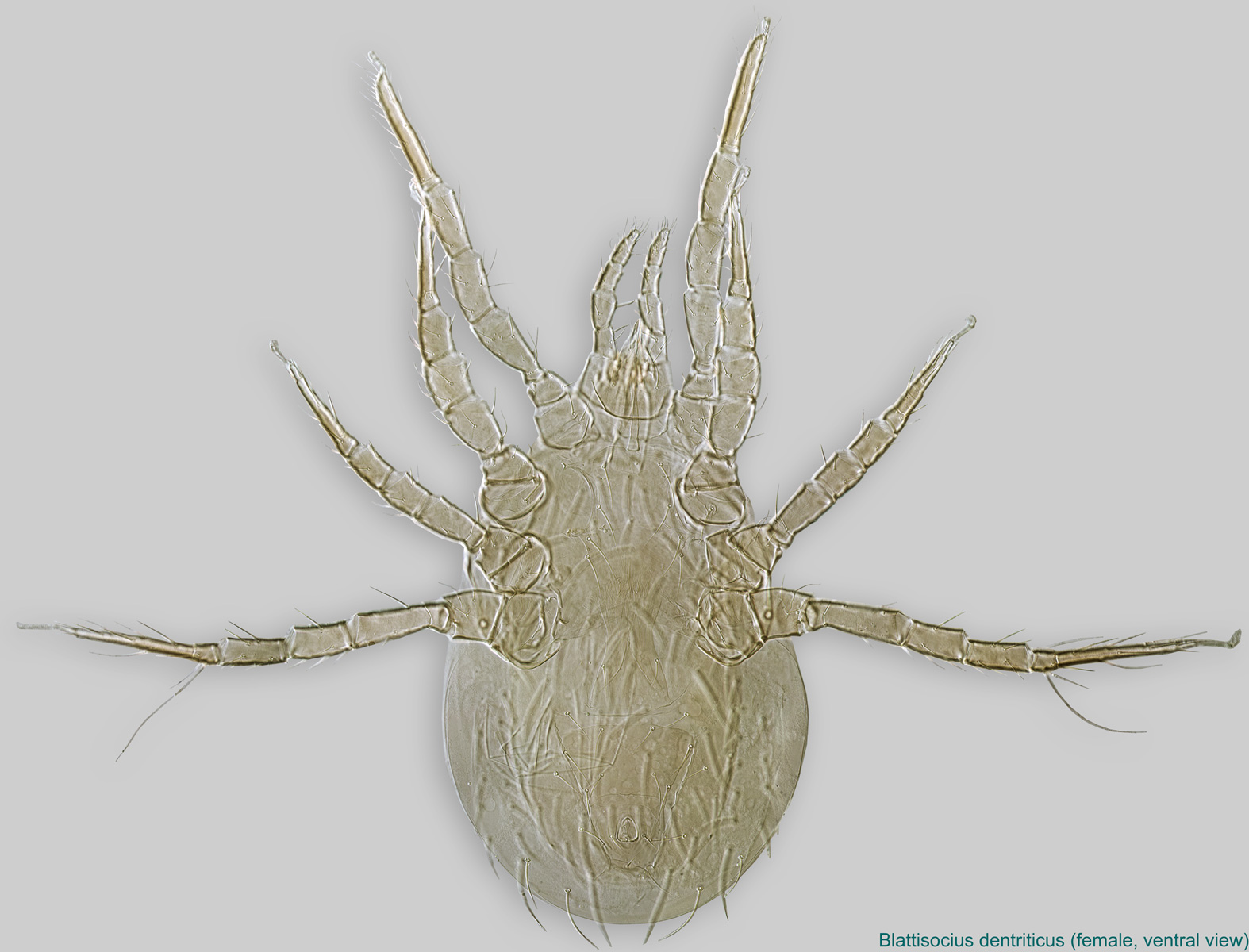
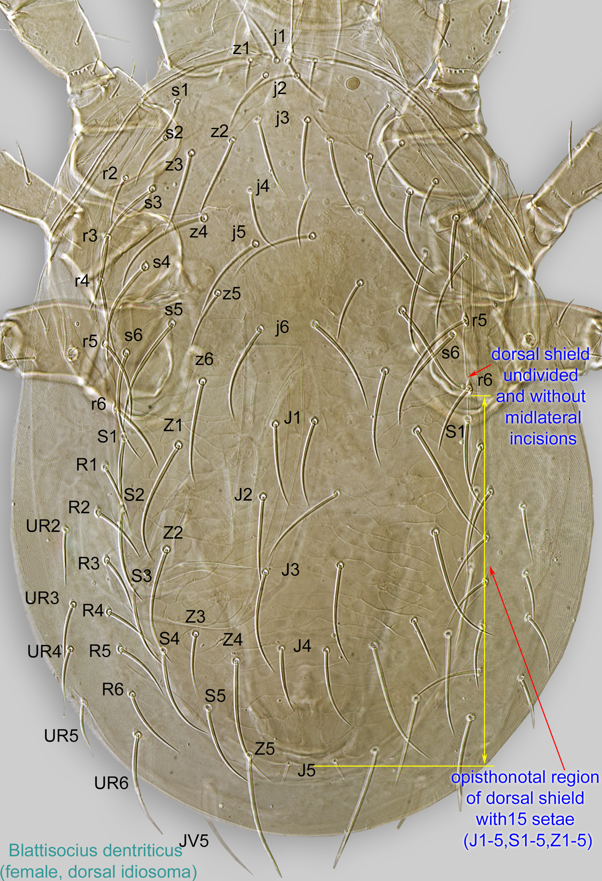
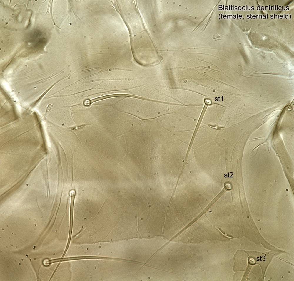
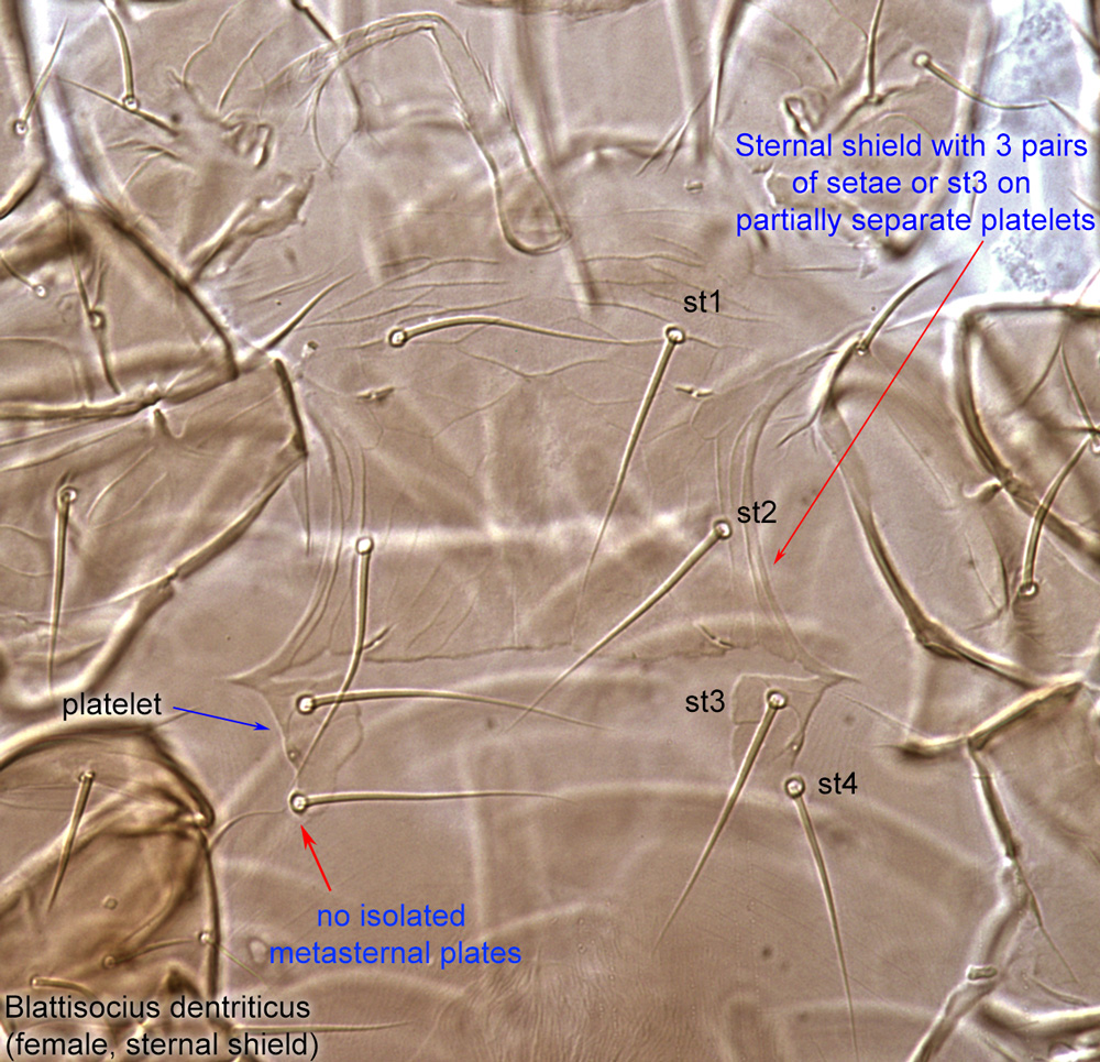
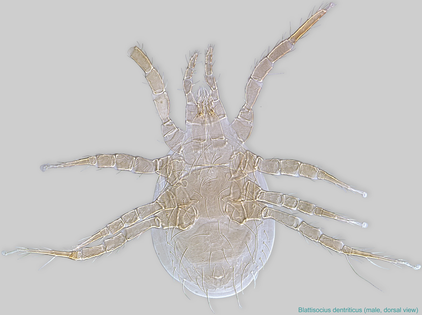
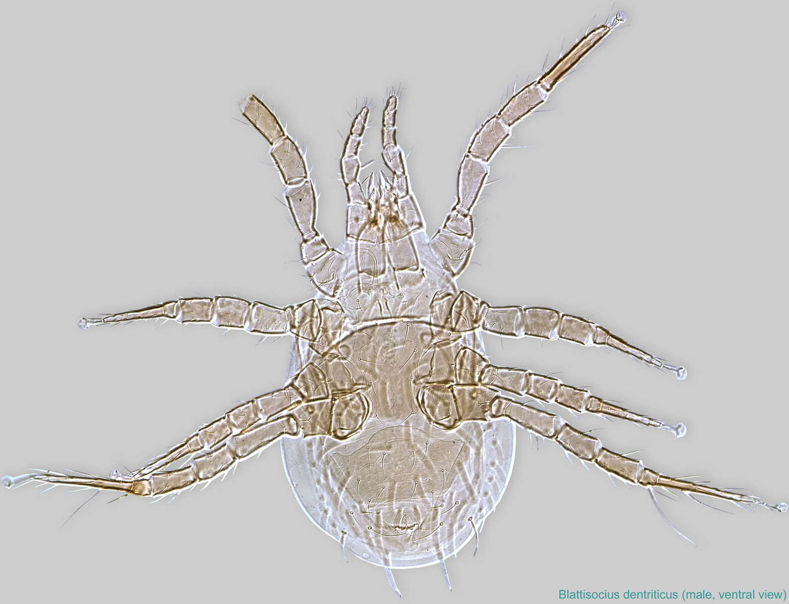
Female: Dorsal shield undivided and without midlateral incisions (Figs. 1, 3). Sternal shieldsternal shield:
A shield in the anterior intercoxal region of parasitiform mites that bears one or more pairs of sternal setae.
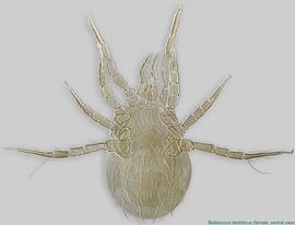
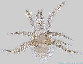
 with 3 pairs of setae (Fig. 20) or st3 on partially separate platelets (Figs. 4, 5, 6, 7); st4 situated on soft cuticle (Fig. 20) or on corners of sternal shieldsternal shield:
A shield in the anterior intercoxal region of parasitiform mites that bears one or more pairs of sternal setae.
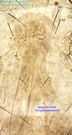
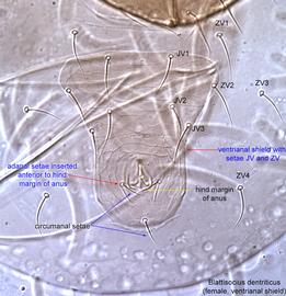
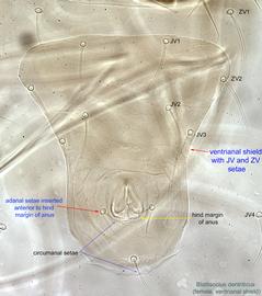
platelets (Figs. 4, 6, 7). Metsternal shields absent (Figs. 4, 6, 7, 20). Ventrianal shieldventrianal shield:
In Mesostigmata, a ventral shield bearing the anal opening, circumanal (postanal and adanal) setae, and one or more pairs of ventral setae or pores (lyrifissures) (see anal shield); may be narrow or very broad and covering most of the gaster.
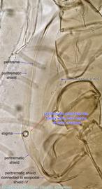
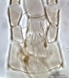
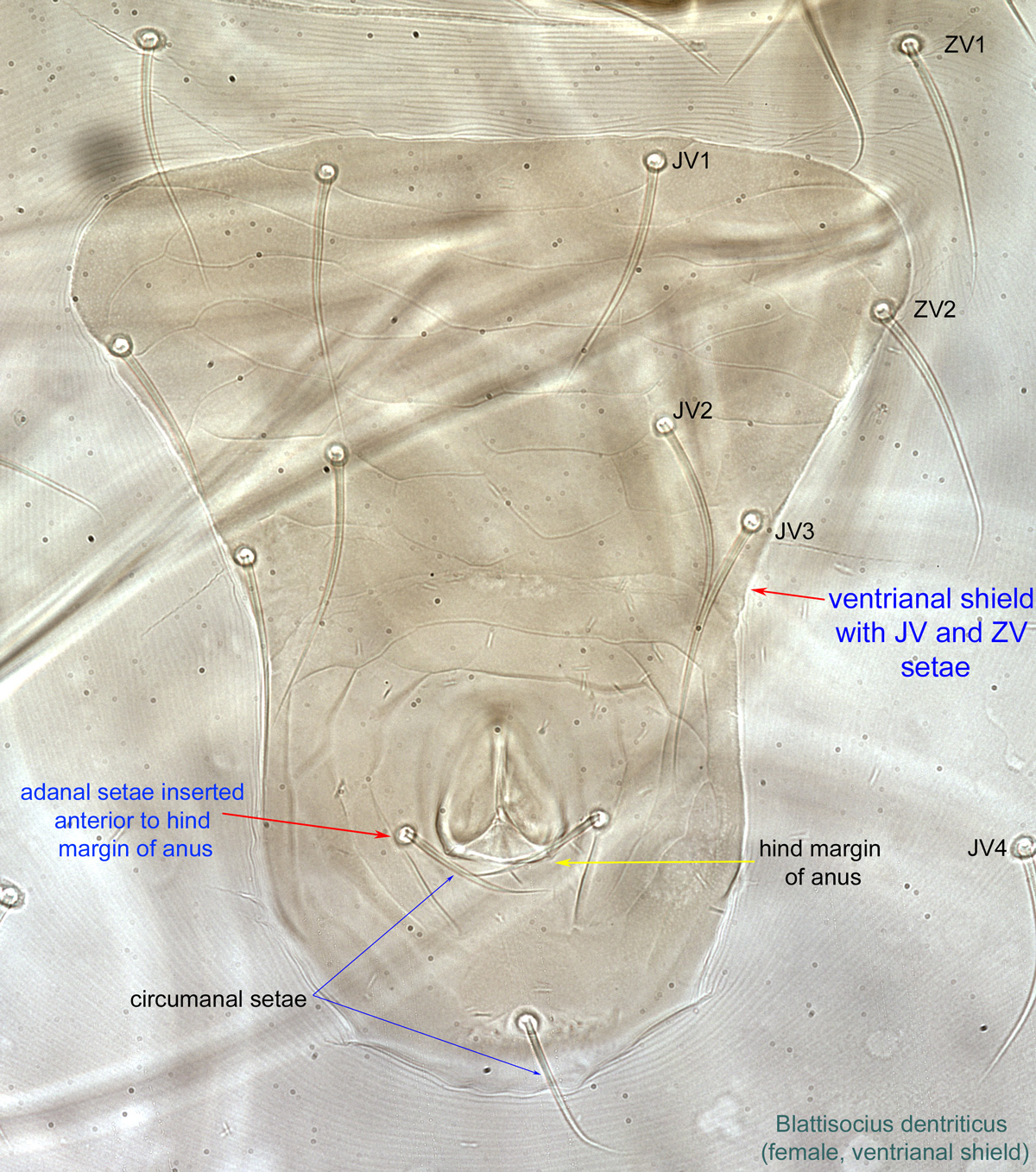
 elongated, subtriangular to bullet-shaped, bearing 3-4 pairs of ventral (preanal) setae (JV, ZV), in addition to the three circumanal setae (Figs. 8, 9). Adanal setae inserted anterior to hind margin of anus (Figs. 8, 9). Peritrematic shield slender, barely wider than stigma at level of stigma (Fig. 10). Corniculicorniculus:
Paired, horn-like process (sometimes toothed, bifurcate, trifurcate, spine-like, spatulate, or membranous) on the subcapitulum of parasitiform mites. These processes usually support the salivary styli. If toothed could be confused with a rutellum, a possibly homologous structure in Acariformes and Opilioacarida.
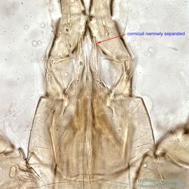
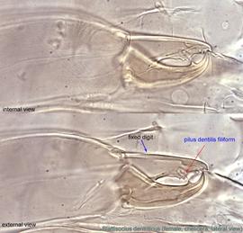
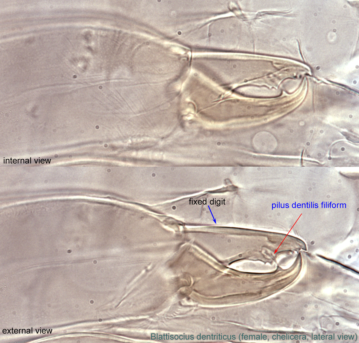
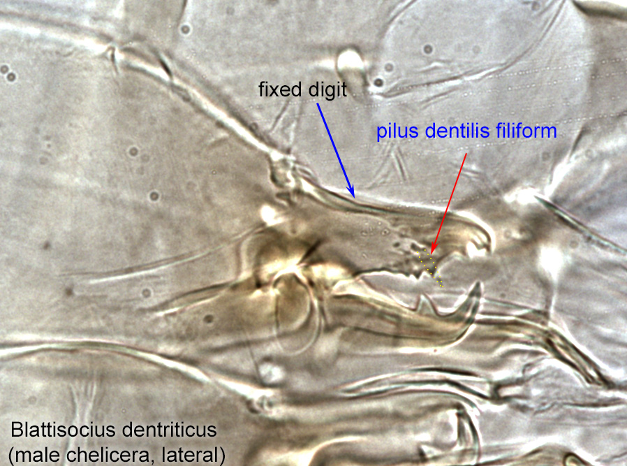
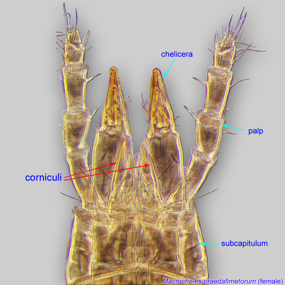 narrowly separated, slender (Fig. 11). Fixed digit with filiform pilus dentilispilus dentilis:
A seta-like or membranous sensory organ inserted ventrolaterally on the fixed digit of the chelicera of many Mesostigmata.
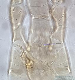
(Figs. 12, 17). Tectumtectum:
The leading dorsal, shelf-like projection of the basis capitulum in Mesostigmata. Also known as gnathotectum or epistome.
convex (Fig 16).
A dichotomous key is available in Britto et al., 2012Britto et al., 2012:
Britto, E. P. J., P. C. Lopes amp; G. J. De Moraes. 2012. Blattisocius (Acari, Blattisociidae) species from Brazil, with description of a new species, redescription of Blattisocius keegani and a key for the separation of the world species of the genus. Zootaxa.3479: 33-51..
Lasioseius. Blattisocius can be separated from Lasioseius by narrow ventrianal shieldventrianal shield:
In Mesostigmata, a ventral shield bearing the anal opening, circumanal (postanal and adanal) setae, and one or more pairs of ventral setae or pores (lyrifissures) (see anal shield); may be narrow or very broad and covering most of the gaster.
(broader in Lasioseius), metasternal shieldsmetasternal shield:
Small, usually teardrop to subtriangular paired shields bearing metasternal setae st4; sometimes fused to the sternal shield or the endopodal shields. Present in Mesostigmata.
 absent (present in Lasioseius); corniculicorniculus:
Paired, horn-like process (sometimes toothed, bifurcate, trifurcate, spine-like, spatulate, or membranous) on the subcapitulum of parasitiform mites. These processes usually support the salivary styli. If toothed could be confused with a rutellum, a possibly homologous structure in Acariformes and Opilioacarida.
narrowly separated (widely separated in Lasioseius), and peritrematic shield barely wider than diameter of stigma at level of stigma (clearly wider in Lasioseius).
Worldwide. Species associated with bees have been found in the Nearctic (Canada), Palaearctic (Czech Republic, Poland, Russia, and Ukraine), Neotropical (Argentina), Oriental (India), and Australasian (New Zealand and Australia) regions.
Members of this genus have been found in honey bee (Apis) hives (e.g., B. tarsalis) and on adult honey bees (e.g., B. apisassociae and B. apis). Blattisocius tarsalis has also been found in nests of leafcutter bees Megachile gomphrenae and M. pallefacta.
facultativefacultative:
can complete entire life cycle without bees or their close relative, wasps
Six species (Blattisocius apis, B. apisassociae, B. dentriticus, B. keegani, B. mali, and B. tarsalis) have been recorded from hives of honey bees or on adult bees.
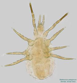
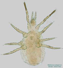
Species of Blattisocius are also predators in soil, stored food, the nests of small mammals and birds, ripe, dried, and rotting vegetables, on plants (e. g., roses, apple trees, and Citrus trees), and in insect cultures. Prey may include acarid mites. One species, Blattisocius patagiorum (Figs. 18-20), is parasitic on adult noctuid moths.
Authors: P. Klimov, B. OConnor, R. Ochoa, G. Bauchan, A. Redford, J. Scher
Last updated October 2016
tool images at ITP Node
idtools.org

