

mostly neutral generalists that may live in hives and disperse on bees; feed on honey and pollen stored in honeycombs
Glycyphagus Hering, 1838
Superorder Acariformes » Order Sarcoptiformes » Suborder Oribatida » Infraorder Desmonomata » Hyporder Astigmata » Family Glycyphagidae » Genus Glycyphagus
Glycyphagus prunorum Hering, 1838 (=Acarus domesticus De Geer, 1778, synonymized in Samšiňák, 1965Samšiňák, 1965:
Samšiňák, K. 1965. A new species of the genus Diamesoglyphus (Acari) from Angola. Publicacoes Culturais da Companhia de Diamantes de Angola. No. 72: 81-86., but this synonymy has not been accepted by all).
Lepidoglyphus Zachvatkin, 1936 (as a genus; here considered a subgenus of Glycyphagus)
Glycyphagus domesticus: furniture mite, grocer's itch mite, hairy grain mite, house mite; Glycyphagus destructor: storage mite, hay mite, fodder mite
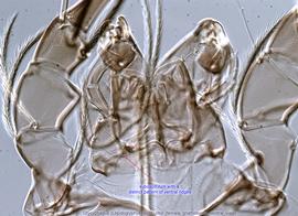
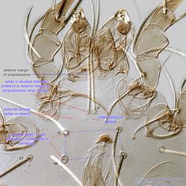
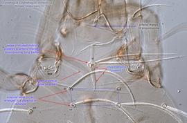
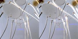
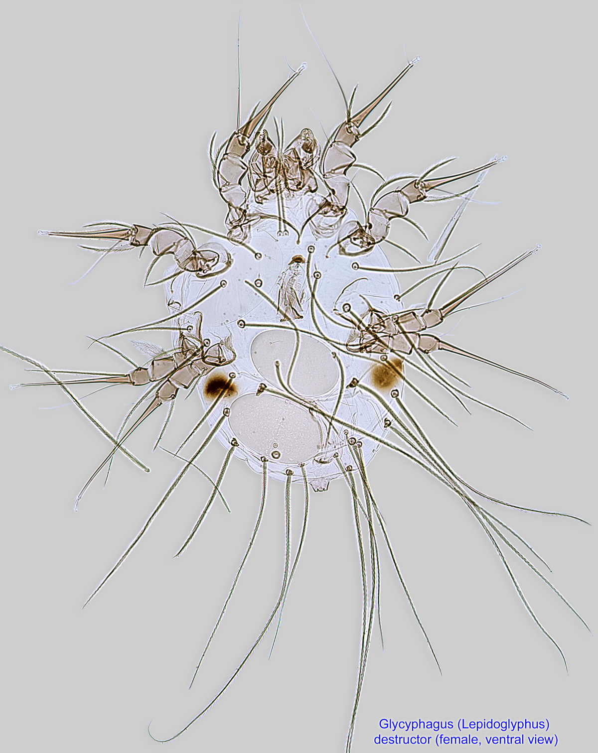
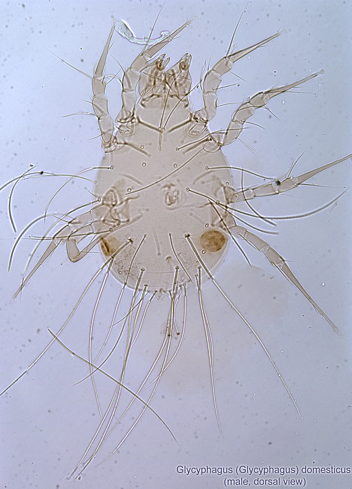
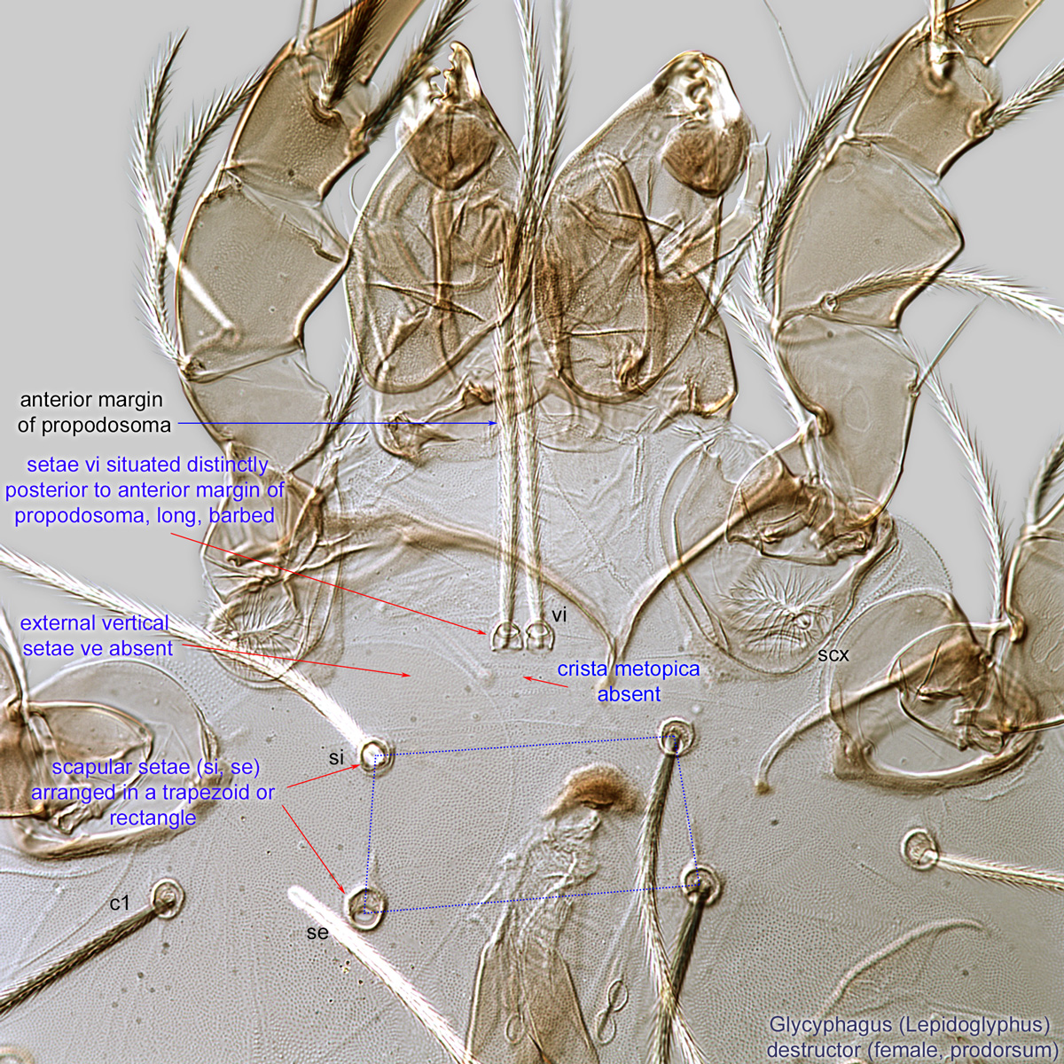
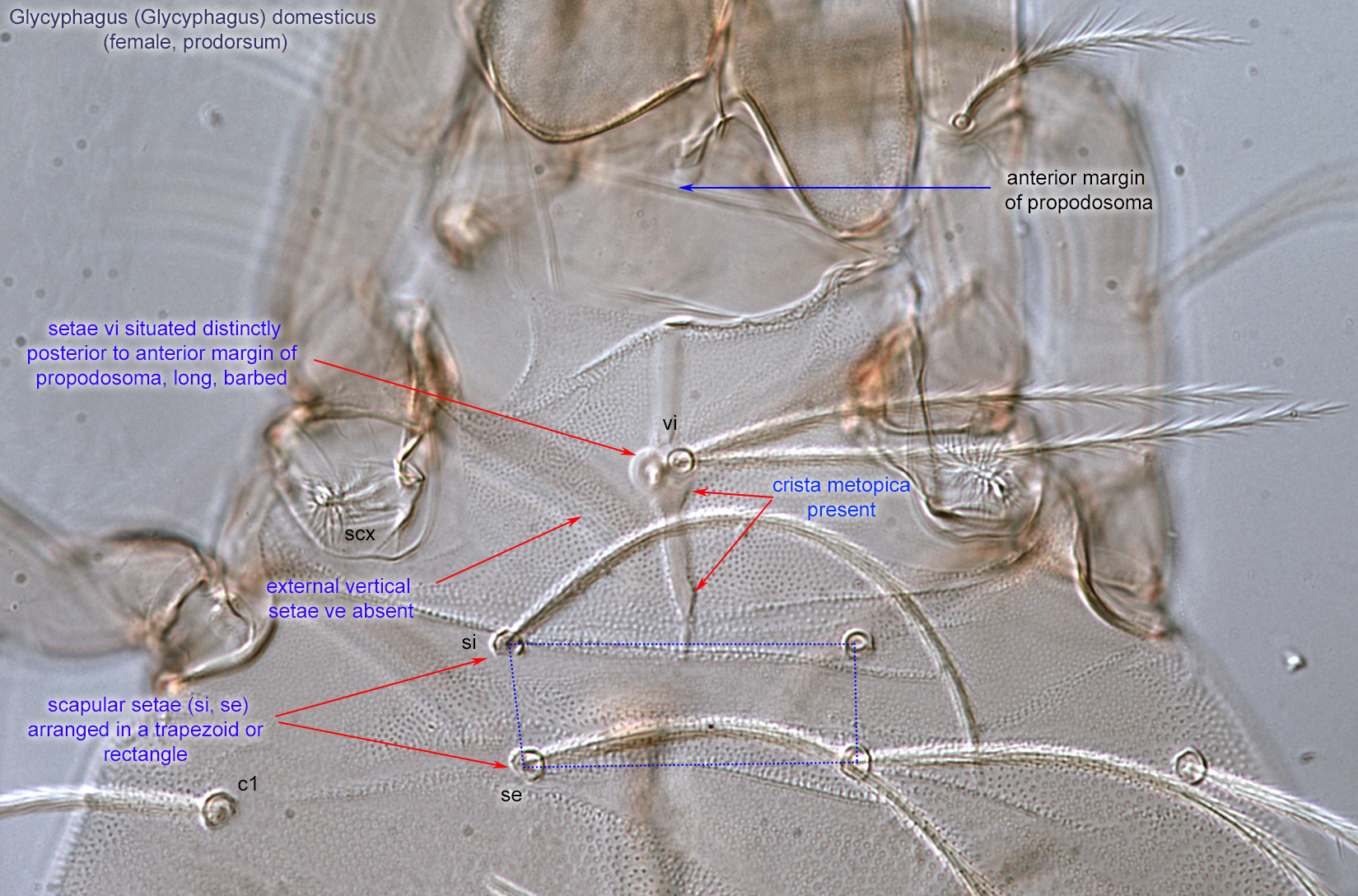
Adult: Prodorsumprodorsum:
Dorsal surface of propodosoma.
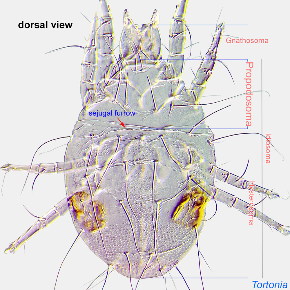 with external vertical setae ve absent (Figs. 6, 7). Internal vertical setae (vi) situated distinctly posterior to the anterior margin of the propodosomapropodosoma:
Anterior part of idiosoma, in front of sejugal furrow.
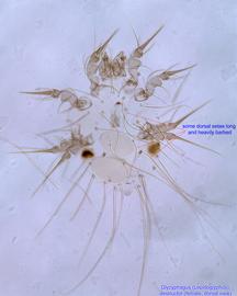
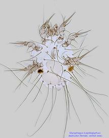
, long, barbed (Figs. 6, 7). Scapular setae (si, se) arranged in a trapezoid or rectangle (Figs. 6, 7). Prodorsalprodorsal:
Pertaining to the prodorsum.
sclerotization called a crista metopica (Fig. 7), a widened scleritesclerite:
A component section of an exoskeleton; a plate forming the skeleton of an arthropod.
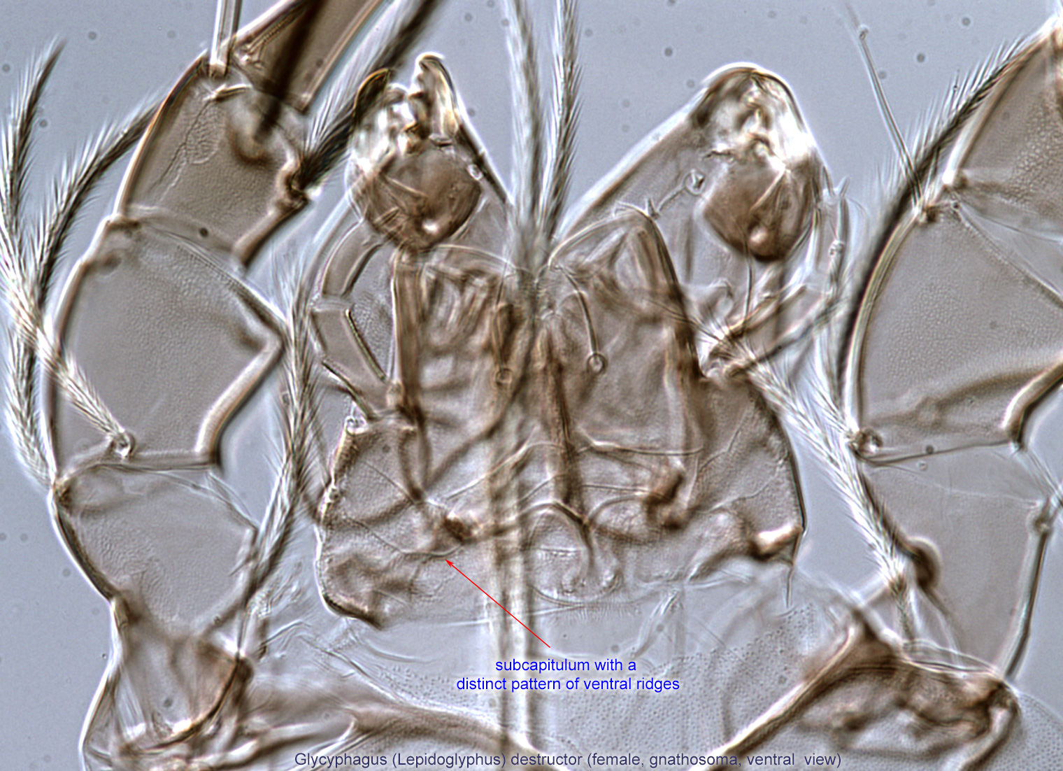
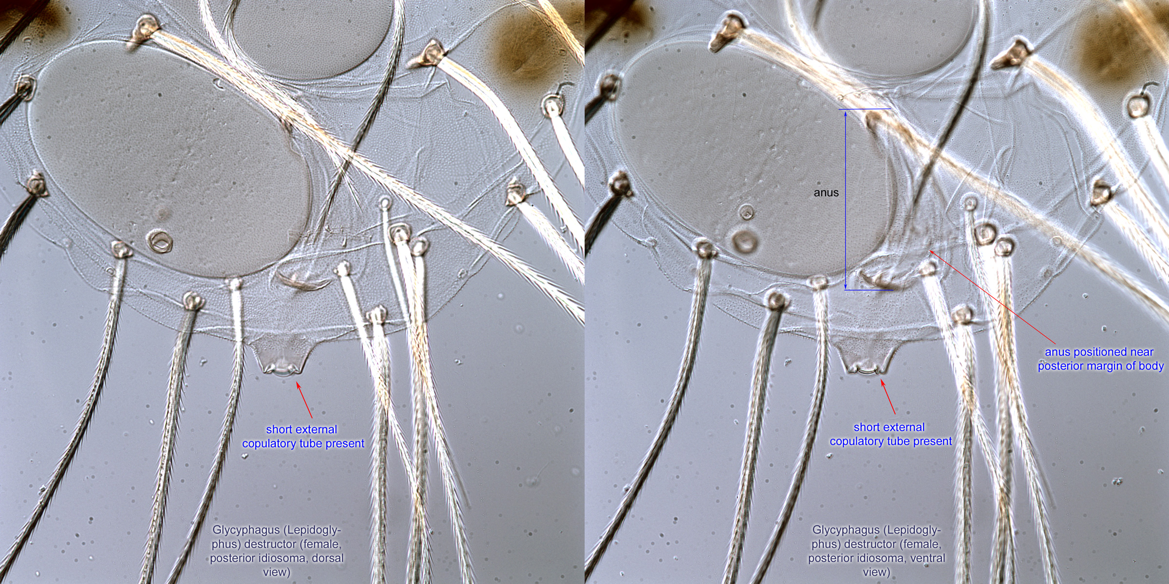
or absent (Fig. 6). Some dorsal setae long and heavily barbed (Fig. 1). Anus positioned near posterior margin of body (Fig. 8). Subcapitulumsubcapitulum:
Ventral faces of the fused palpcoxae.
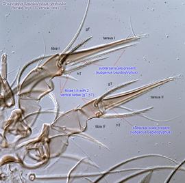
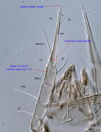
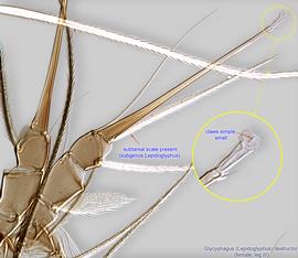
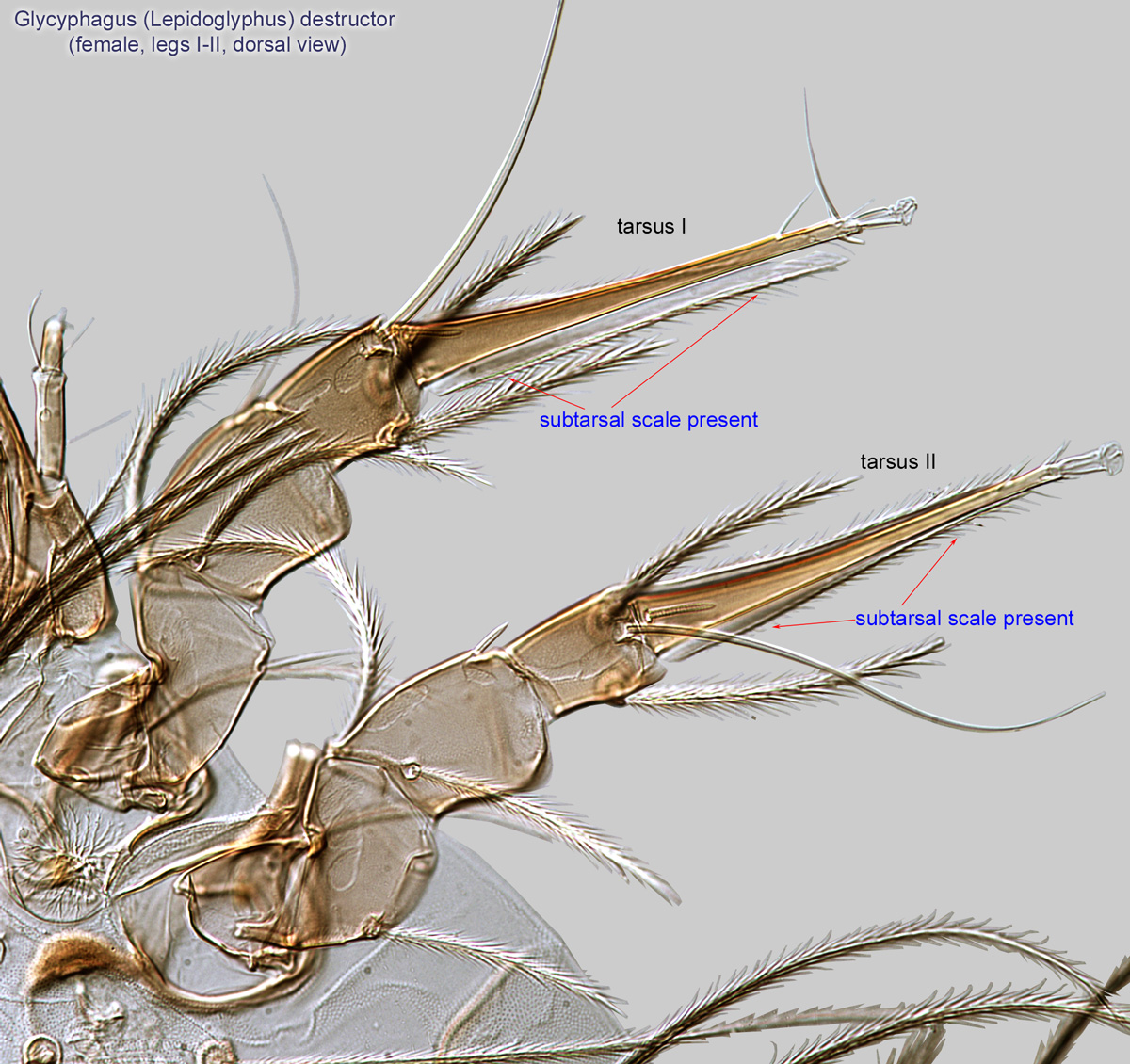
with a distinct pattern of ventral ridges (Fig. 5). Tibiaetibia:
Leg or palp segment (also known as podomere or palpomere) between tarsus and genu.
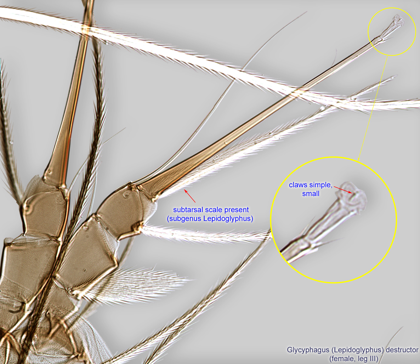
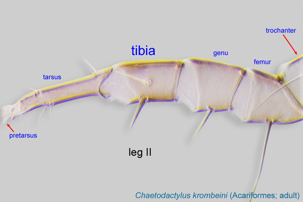 I-II with 2 ventral setae (Figs. 10, 11). Claws simplesimple:
Of claws or setae; not modified or not bi- or trifurcate at tip.
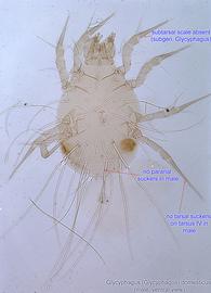
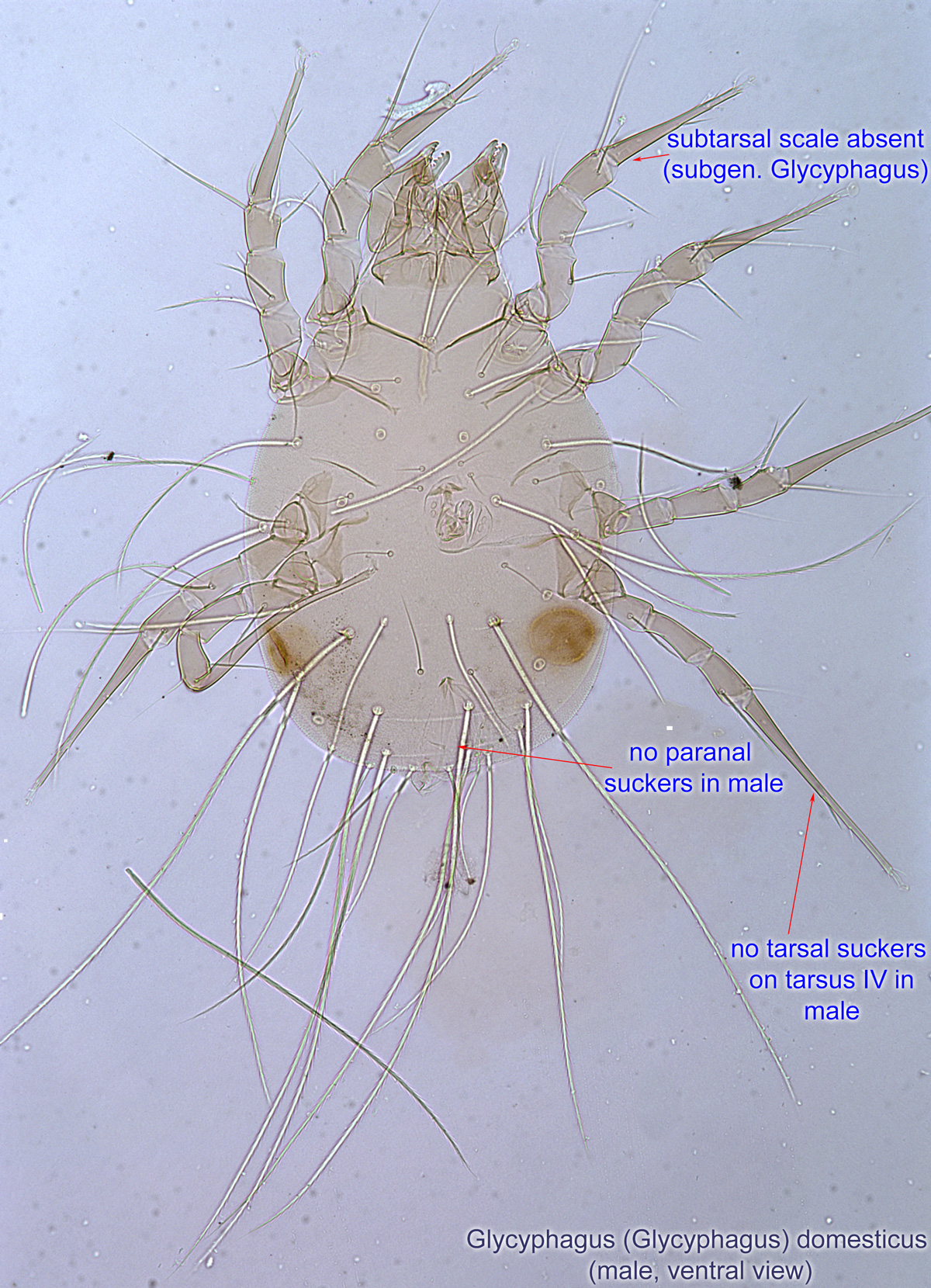
, small (Figs. 11, 12). Female usually with a short external copulatory tube (Fig. 8). Males without paranal suckers and tarsal suckers on tarsustarsus:
Terminal segment (also known as podomere or palpomere) of legs or palps. In Parasitoformes it can be subdivided into telotarsus and basitarsus.
 IV (Fig. 4). Without a subtarsal scale (Glycyphagus (Glycyphagus)) (Figs. 4, 11) or with a subtarsal scale Glycyphagus (Lepidoglyphus) (Fig. 9, 10, 12).
A dichotomous key is available in Hughes, 1976Hughes, 1976:
Hughes, A. M. 1976. The mites of stored food and houses. London: Her Majesty's Stationery Office. 400 pp., which can be used to identify all species recorded from bees and bee nests.
Cosmopolitan. Found in association with bees in the Palaearctic and Oriental regions.
The following bees have been recorded to harbor Glycyphagus in their nests: honey bees (Apis) (a very common host), bumble bees (Bombus), and large carpenter bees (Xylocopa). Phoresyphoresy:
Attaching to or boarding another organism (i.e., a host) for dispersal to new habitats. Can be distinguished from parasitism because feeding typically does not occur.
of feeding stages of mites on adult bees has been reported.
facultativefacultative:
can complete entire life cycle without bees or their close relative, wasps
 are absent. Feeding stages disperse on hosts (including bees) by active movements or by air currents.
These are generalist species occurring in a variety of habitats: rodent and bird nests, stored products (as a pest), house dust, grass and hay, and others. These mites have also been found in nests of social insects, including bees. Glycyphagus causes dermatitis, gastritis, and allergies in humans.
These mites are common in beehives, especially on debris from bottom boards. Infestation rates have been reported ranging from 20% (Grobov, 1978Grobov, 1978:
Grobov, O. F. 1978. Kleshchi medonosnoy pchely ( Apis mellifera L.): ikh znachenie i osnovnue printsypy bor'by s kleschchevymi porazheniyami [=Mites of the honeybee ( Apis mellifera L.): their significance and main principles of control of diseases caused by mites]. Doctor of Sciences (Habilitation) Thesis. 536. Moscow: All-Union Institute of Experimental Veterinary, All-Union Academy of Agricultural Sciences.), to 98.8% (Chmielewski, 1991cChmielewski, 1991c:
Chmielewski, W. 1991c. Stored products mites (Acaroidea) in Polish bee hives. In Modern acarology. Volume I: proceedings of the 8 International Congress of Acarology, held in Ceske Budejovice, Czechoslovakia, 6-11 August 1990. , 615-619. The Hague, The Netherlands: SPB Academic Publishing.), to 100% (Vitzthum, 1936Vitzthum, 1936:
Vitzthum, H. G. 1936. Über das Vorkommen von saprohytischen und parasitaren Milben im Wabenwerk der Honigbiene. Deutscher Imkerführer : Mitteilungen . 10 : 460-461.). Often there are hundreds of thousands of mites in a single beehive. They feed on decomposing organic matter, stored pollen, beehive debris, and honey, but often prefer dead bees. Glycyphagus (Glycyphagus) domesticus and Glycyphagus (Lepidoglyphus) destructor are the most common and abundant species, while other species are much more rare [Glycyphagus (Glycyphagus) ornatus, Glycyphagus (Lepidoglyphus) privatus, and Glycyphagus (Lepidoglyphus) michaeli]. In Poland, Glycyphagus domesticus has been found to be the most common species of all mites, occurring in 98.8% of samples (Chmielewski, 1991cChmielewski, 1991c:
Chmielewski, W. 1991c. Stored products mites (Acaroidea) in Polish bee hives. In Modern acarology. Volume I: proceedings of the 8 International Congress of Acarology, held in Ceske Budejovice, Czechoslovakia, 6-11 August 1990. , 615-619. The Hague, The Netherlands: SPB Academic Publishing.), while G. destructor was found only in 4.9% of samples. In German beehives, these two species occurred at nearly the same frequency (Homann, 1933Homann, 1933:
Homann, H. 1933. Die Milben in gesunden Bienenstöcken. Zeitschrift für Parasitenkunde 6: 350-415.).
Under laboratory conditions, Glycyphagus domesticus preferred bee bread, pollen, mold, and beehive debris, followed by combs and wax, honey, propolispropolis:
A red or brown resinous substance collected by honey bees from tree buds that is used by them to fill crevices and to seal and varnish honeycombs.
, and dead bees; no reproduction occurred on royal jelly. In contrast, G. destructor preferred dead brood bees, mold, and beehive debris (Chmielewski, 1991cChmielewski, 1991c:
Chmielewski, W. 1991c. Stored products mites (Acaroidea) in Polish bee hives. In Modern acarology. Volume I: proceedings of the 8 International Congress of Acarology, held in Ceske Budejovice, Czechoslovakia, 6-11 August 1990. , 615-619. The Hague, The Netherlands: SPB Academic Publishing.).
Authors: P. Klimov, B. OConnor, R. Ochoa, G. Bauchan, A. Redford, J. Scher
Last updated October 2016
tool images at ITP Node
idtools.org

