
Beneficial
none known
Family: Scarabaeidae Subfamily: Scarabaeinae Genus: Onitis Species: Onitis vanderkelleni Lansberge, 1886Lansberge, 1886:
Lansberge J. 1886. Scarabaeides, Buprestides, et Cerembycides de L'Afrique occidentale. Notes from the Leyden Museum. 8: 79-108.
none available
Total body length 18.0–26.0 cm (0.71–1.02 in). Body shape subquadratesubquadrate:
somewhat quadrate in shape
posteriorly; may be caked in dried dung. Color dull black. Clypealclypeal:
of, or referring to, the clypeus
apexapex:
point or edge furthest from the body
rounded to weakly sinuatesinuate:
gently curved (specifically of margins or edges); often in reference to the clypeus
. Fronsfrons:
part of the head generally positioned between the eyes (posterior to the clypeus and anterior to the vertex) and visible dorsally
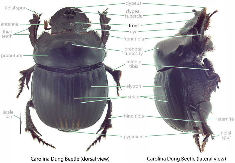 with weak, central tubercletubercle:
a small, conical knob or horn-like protuberance
. Front tibiatibia:
a segment of the leg articulated with the tarsus and femur
 of male elongate, curving ventrally and inward at apexapex:
point or edge furthest from the body
; female tibiatibia:
a segment of the leg articulated with the tarsus and femur
somewhat shorter; tarsitarsi:
the distal part of an insect leg attached to the tibia and consisting of five sub-segments in scarab beetles; bears the tarsal claws
lacking in both sexes. Tibiatibia:
a segment of the leg articulated with the tarsus and femur
of middle leg gradually expanded to a triangulate apexapex:
point or edge furthest from the body
. Hind trochantertrochanter:
a segment of the leg articulated with the femur and coxa
lacking spine on posteriorposterior:
towards the rear end; opposite of anterior
margin in both sexes. Hind femurfemur:
segment of the leg that is articulated to the body by the trochanter and bears the tibia at the distal end
of male with well-developed, posteriorly produced, straight spine; female lacking spine.
Undescribed. For Scarabaeinae (Ritcher, 1966Ritcher, 1966:
Ritcher P. 1966. White grubs and their allies: a study of North American scarabaeoid larvae. Oregon State University Monographs, Studies in Entomology 4: 1-219.): Grub C-shaped, hump-backed, cylindrical, cream-colored. Maxillamaxilla:
set of paired mouthparts located posterior to the mandibles
with galeagalea:
outer branch or lobe of the maxilla
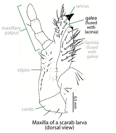 and lacinialacinia:
inner portion of the maxilla distinctly separate. AntennaeAntennae:
paired sensory organ on head, formed from numerous segments
 with 4 or 5 apparent segments. Distaldistal:
situated away from the point of articulation, thus usually furthest from the body
segment of antennaantenna:
paired sensory organ on head, formed from numerous segments
much reduced in size. Epipharnyx with tormaetormae:
in scarab larvae, sclerotized structures on the ends of the clypeolateral suture extending towards the mesal line
united mesallymesally:
at or near midline of body
, anterioranterior:
the front or forward; opposite of posterior
phoba present. Anal opening surrounded by fleshy lobes. Legs 2-segmented.
Africa. This species occurs in the tropical highlands of sub-Saharan Africa, with records from Angola, Burundi, Cameroon, Kenya, Rwanda, Tanzania, Uganda, and the Democratic Republic of the Congo (formerly Zaire) (Krikken, 1977Krikken, 1977:
Krikken J. 1977. Notes on African Onitini, mainly from southeastern Kenya (Coleoptera: Scarabaeidae). Zoologische Mededelingen: 141-170. full text (accessed 2015)). It usually occurs at elevations over 1,800 meters (5,900 ft) where rainfall ranges from 800–2,000 mm (31–79 in) per year (Edwards, 2007Edwards, 2007:
Edwards P. 2007. Introduced dung beetles in Australia 1967-2007. Landcare Australia, Sydeny, New South Wales, Australia. full text (accessed 2015)).
None. Onitis spp. feed on dung as both adults and larvaelarvae:
the immature form of an insect; in scarabs, also called grub or white grub; preceded by the egg stage, followed by the pupal stage
 (Edwards and Aschenborn, 1987Edwards and Aschenborn, 1987:
Edwards P and Aschenborn H. 1987. Patterns of nesting and dung burial in Onitis dung beetles: implications for pasture productivity and fly control. Journal of Applied Ecology 24: 837-851. full text (accessed 2015)).
Poorly known. Related species of Onitis are dung burrowers. A vertical tunnel lined with dung is created under the initial dung source. Adult male and female beetles cooperate to transport dung pieces down the burrow where the dung is shaped into sausage-like masses. An egg (or eggs) is deposited into each of the dung sausages, and larval development occurs within the dung mass.
None. This species recycles dung and is beneficial for ranching and farming in Hawaii. Being a dung feeder, this species poses no threat to crop or ornamental plants. Additionally, this species is not a threat to native dung beetles because none are known from Hawaii or Guam.
Established. Onitis vanderkelleni was released in 1976 at Parker Ranch on Big Island (Nakao and Funasaki, 1979Nakao and Funasaki, 1979:
Nakao H and Funasaki G. 1979. Introductions for biological control in Hawaii: 1975 amp; 1976. Proceedings of the Hawaiian Entomological Society 23: 125-128. full text (accessed 2015)) where it is now established (Nishida, 2002Nishida, 2002:
Nishida G (editor). 2002. Hawaiian terrestrial arthropod checklist, fourth edition. Bishop Museum Technical Report 22: 1-313.). Like most of Hawaii's dung beetles, this species was introduced to help control populations of the horn fly (Haematobia irritans), a biting pest of livestock.
Not established or recorded. There are no records of this species from Guam.
In Hawaii, this species was intentionally introduced.
Three species of Onitis are recorded from Hawaii (none are known from Guam). Onitis vanderkelleni can be separated from the other Onitis species by examination of the middle tibiatibia:
a segment of the leg articulated with the tarsus and femur
(O. vanderkelleni with gradually expanded middle tibiatibia:
a segment of the leg articulated with the tarsus and femur
with a triangulate apexapex:
point or edge furthest from the body
versus O. phartopuswith an abruptly expanded tibiatibia:
a segment of the leg articulated with the tarsus and femur
with trapezoidal apexapex:
point or edge furthest from the body
), the hind trochantertrochanter:
a segment of the leg articulated with the femur and coxa
(O. vanderkelleni hind trochantertrochanter:
a segment of the leg articulated with the femur and coxa
lacking spine-like process versus trochanter trochanter:
a segment of the leg articulated with the femur and coxa
with a well-developed spine in male O. phartopus), hind femurfemur:
segment of the leg that is articulated to the body by the trochanter and bears the tibia at the distal end
of male (O. vanderkelleni with a posteriorly produced, straight spine versus O. alexiswith a curved spine and O. phartopus that lacks a spine), and color (O. vanderkelleni dull black versus greenish-black with brown elytraelytra:
the hardened and chitinous wing-cover of a beetle that protect and overlie the flight wing
in O. alexis).
none known
Report your observation of this beneficial species at our iNaturalist project.
Authors: J.B. Dunlap, M.L. Jameson, E.L. Engasser, P.E. Skelley, A.J. Redford
Content last updated February 2016
idtools.org
Lucid mobile app for Android and iOS | tool images at ITP Node


