
Family: Megachilidae
Subfamily: Megachilinae
Tribe: Anthidiini
Genus: Epanthidium Moure, 1947
Subgenera: Ananthidium, Carloticola, Epanthidium
Common name: none
Epanthidium range from robust to moderately elongate body form. Small Epanthidium range in body length from 6.5–7.5 mm, while large Epanthidium range in length from 9–12 mm. They have black integumentintegument:
a tough, protective outer layer
, sometimes tinged with red, and have yellow to cream-colored maculations on their head, thorax, and abdomen (Michener 2007Michener 2007:
Michener, C.D. 2007. The Bees of the World (2nd ed.). Johns Hopkins University Press, Baltimore and London, 953 pp.).
Epanthidium contains approximately 23 species within 3 subgenera (Michener 2007Michener 2007:
Michener, C.D. 2007. The Bees of the World (2nd ed.). Johns Hopkins University Press, Baltimore and London, 953 pp.); none are known to occur in the U.S. or Canada.
(modified from Michener 2007Michener 2007:
Michener, C.D. 2007. The Bees of the World (2nd ed.). Johns Hopkins University Press, Baltimore and London, 953 pp.)
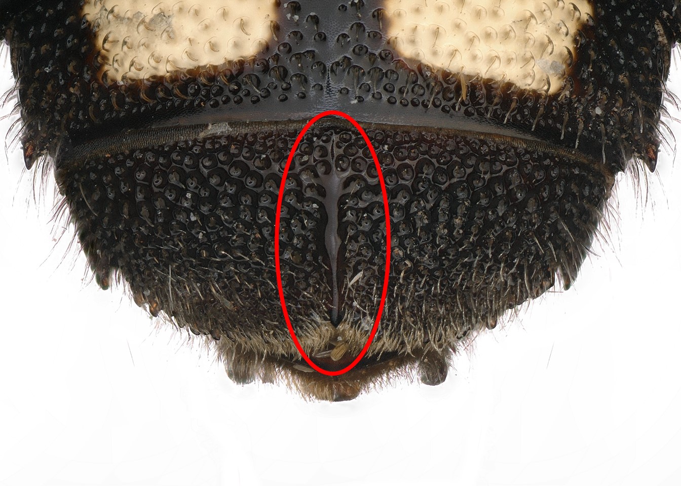
 carinatecarinate:
 rounded.
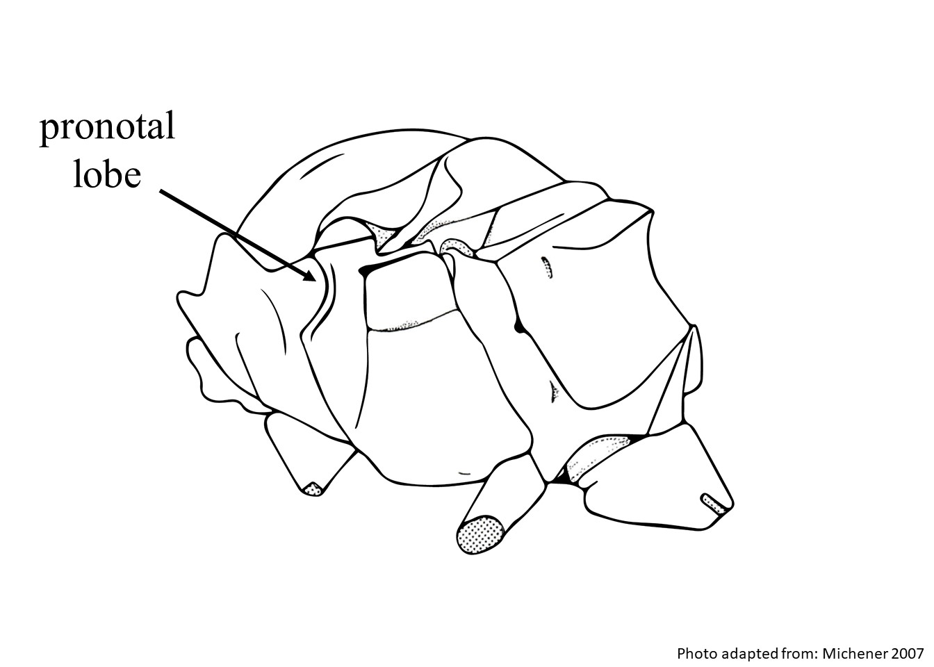 with pronounced lamellalamella:
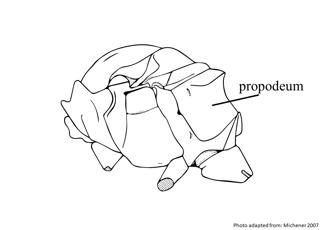 with foveafovea:
 straight, or nearly so.
 with laterallateral:
 with preapicalpreapical:
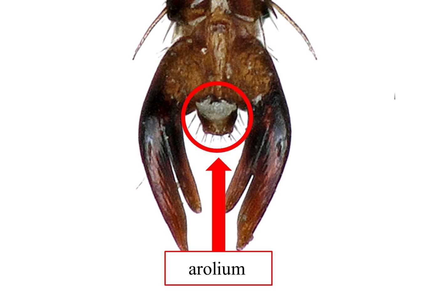 present, can be present or absent in females, although usually present.
Epanthidium may be confused with Dianthidium due to a similar sloping lamellalamella:
thin, plate-like, often somewhat translucent structure
on the pronotal lobepronotal lobe:
a part of the pronotum located dorsally on the posterior margin of the pronotum and overlaps the anterior thoracic spiracle; however, Epanthidium can be distinguished by the characteristics listed above (Michener 2007Michener 2007:
Michener, C.D. 2007. The Bees of the World (2nd ed.). Johns Hopkins University Press, Baltimore and London, 953 pp.).
There are no known invasives.
Little is known about the floral resources used by Epanthidium. In the subgenus Epanthidium, many species appear to be generalists, visiting a wide variety of plants including Zygophyllaceae, Fabaceae, and Asteraceae (Parizotto and Melo 2015Parizotto and Melo 2015:
Parizotto, D.R. and G.A.R. Melo. 2015. Nests of the bees of the anthidiine genus Ananthidium Urban (Hymenoptera, Apidae, Megachilinae). Journal of Hymenoptera Research 47: 115ndash;122.).
Epanthidium are solitary bees that build nests primarily from plant resins. Epanthidium tigrinum builds nests consisting of groups of cells in preexisting cavities (Michener 2007Michener 2007:
Michener, C.D. 2007. The Bees of the World (2nd ed.). Johns Hopkins University Press, Baltimore and London, 953 pp.). The two known species of the subgenus Ananthidium construct nests in stems that consist of one or two cells made from resin or a plant fiber and resin composite (Stange 1983Stange 1983:
Stange, L.A. 1983. Synopsis of the genus Epanthidium Moure with the description of a new species from northeastern Mexico (Hymenoptera: Megachilidae). Pan-Pacific Entomologist 59: 281ndash;297.). Nests from Epanthidium (Ananthidium) dilmae have been collected from dead branches of Asteraceae and in shrub vegetation approximately 0.5 m above the ground (Parizotto and Melo 2015Parizotto and Melo 2015:
Parizotto, D.R. and G.A.R. Melo. 2015. Nests of the bees of the anthidiine genus Ananthidium Urban (Hymenoptera, Apidae, Megachilinae). Journal of Hymenoptera Research 47: 115ndash;122.). Epanthidium aff. sanguineum was observed occupying abandoned nests of Centris muralis, which were constructed in adobe walls (Cilla and Rolón 2012). Epanthidium aff. sanguineum collected materials from Larrea spp. to line their nests (Cilla and Rolón 2012).
Epanthidium are restricted to the Western Hemisphere and are NeotropicalNeotropical:
biogeographic region that includes South and Central America, the Caribbean Islands, southern Florida, and the southern Mexican lowlands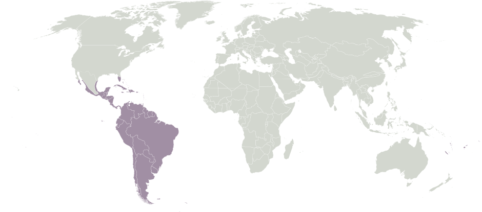 in distribution, ranging from central Mexico south to Argentina (Michener 2007Michener 2007:
Michener, C.D. 2007. The Bees of the World (2nd ed.). Johns Hopkins University Press, Baltimore and London, 953 pp.).
Distribution map generated by Discover Life -- click on map for details, credits, and terms of use.
Cilla, G. and G. Rolón. 2012. Macroscopic and microscopic studies of the nests and the stages involved in the nesting process of Centris muralis Burmeister (Hymenoptera: Apidae: Centridini) bee in the abode walls, in La Rioja, Argentina. Biologia 67: 573-583.
Michener, C.D. 2007. The Bees of the World (2nd ed.). Johns Hopkins University Press, Baltimore and London, 953 pp.
Parizotto, D.R. and G.A.R. Melo. 2015. Nests of the bees of the Anthidiine genus Ananthidium Urban (Hymenoptera, Apidae, Megachilinae). Journal of Hymenoptera Research 47:115-122.
Stange, L.A. 1983. Synopsis of the genus Epanthidium Moure with the description of a new species from northeastern Mexico (Hymenoptera: Megachilidae). Pan-Pacific Entomologist 59:281-297.
Authors: S. Burrows, C. Ritner, M. Christman, L. Spears, A. Smith-Pardo, S. Price, R. Ramirez, T. Griswold, A. Redford
Edition 3 - last updated November 2021
tool images at ITP Node
idtools.org


.jpg)