
Head voluminous.
Frons convex with genagena:
the part of the cranium on each side below the eye fully inflated; vertexvertex:
the top of the head between the eyes, frons and occiput, anterior to the occipital suture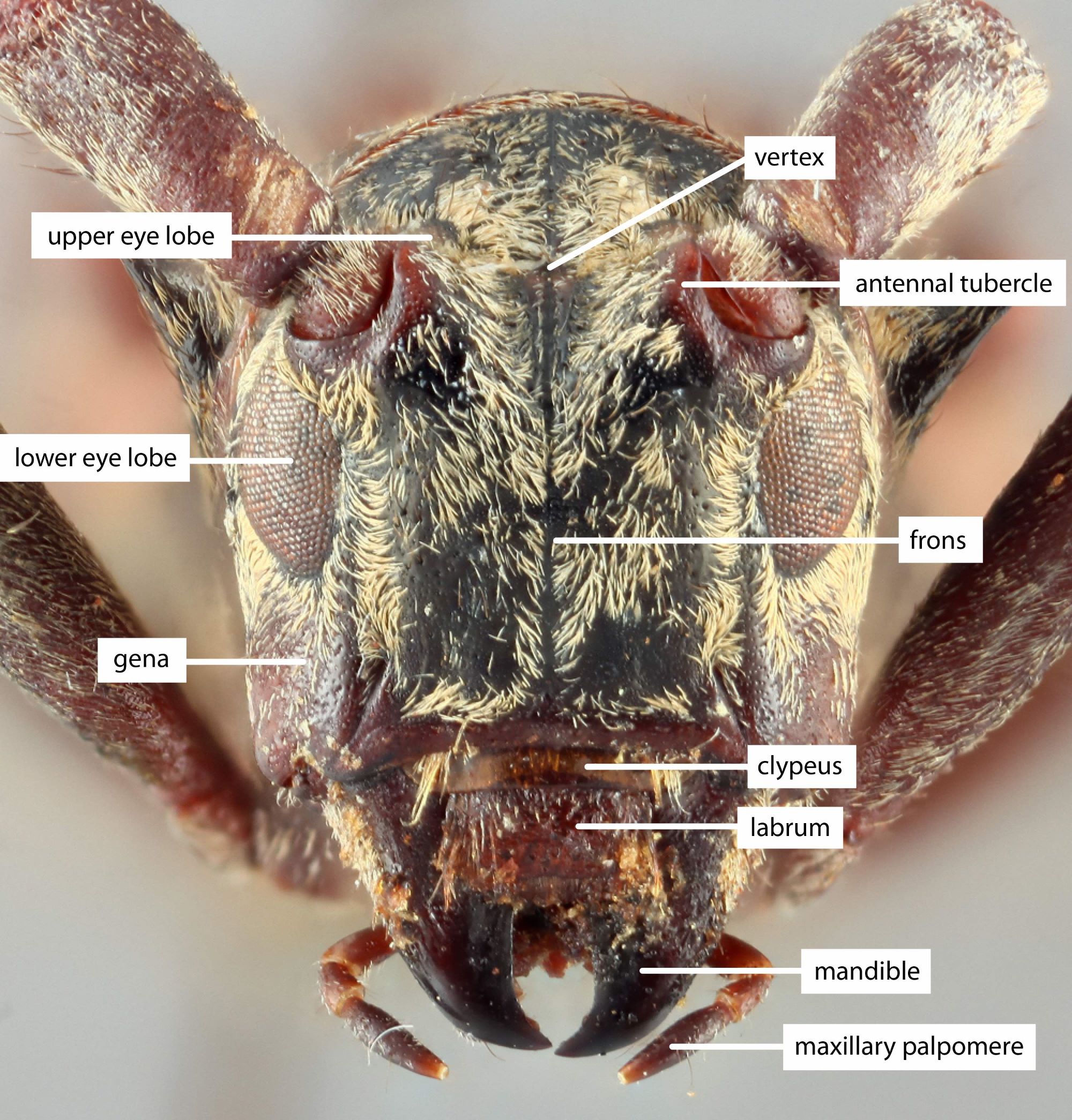 broad with inter-antennal area flattened; occiputocciput:
dorsal part of the head between the occipital sulcus and the postoccipital sulcus
slightly convex.
Eyes large, deeply emarginateemarginate:
notched at the margin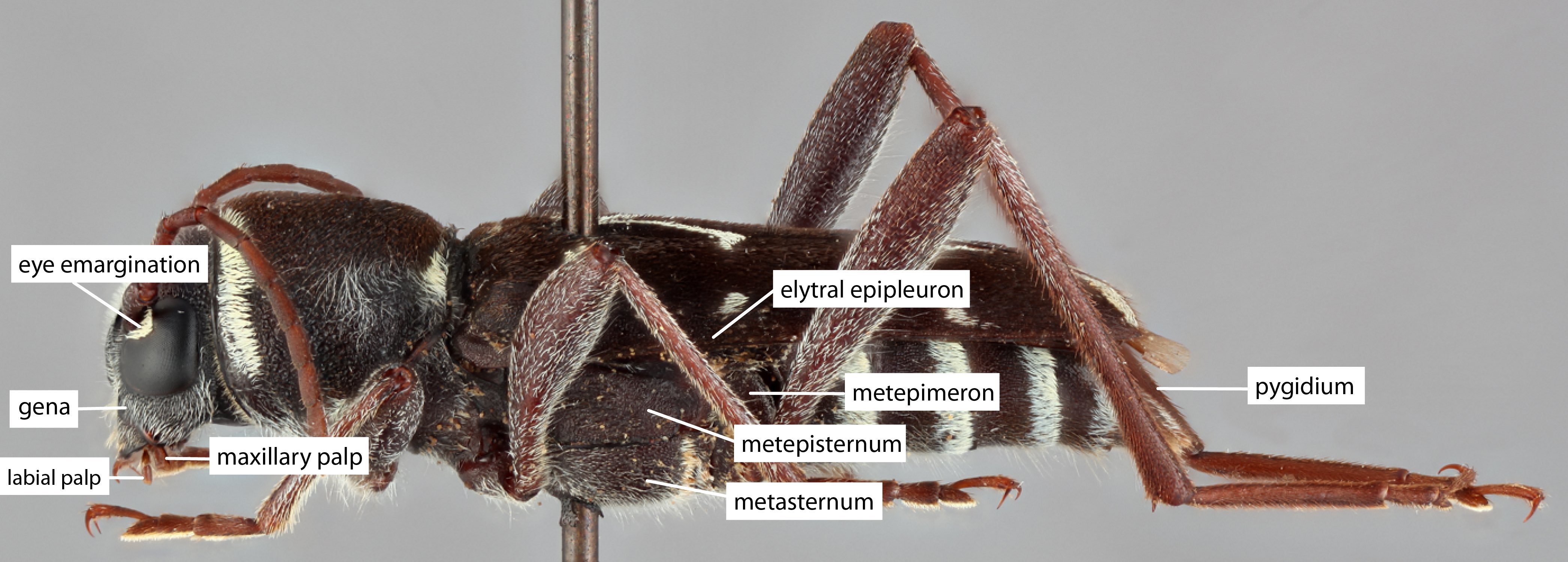 .
.
Antennae stout, the last segment reaching the elytral apexapex:
end of any structure distad to the base
; scapescape:
the first proximal segment of the antenna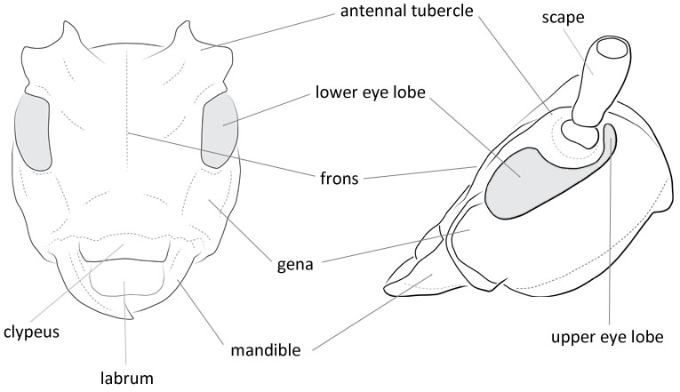 strongly clavateclavate:
thickening gradually toward the tip
with long erect hairs, shorter than third; third same length as fourth; underside of 2nd to last segment provided with long erect hairs.
Pronotum longer than wide; each lateral side provided with a spinous tubercletubercle:
a small knoblike or rounded protuberance
; disc gibbous with a large swelling; the area between swelling and lateral tubercletubercle:
a small knoblike or rounded protuberance
smooth.
Prosternum with intercoxal process narrow. about 1/4 as wide as coxal cavities.
Mesosternum with intercoxal process almost plane. about 1/3 as wide as coxal cavities.
Elytraelytron:
the leathery forewing of beetles, serving as a covering for the hind wings, commonly meeting opposite elytron in a straight line down the middle of the dorsum in repose
2.4–2.6 times as long as humeral width; sides strongly dilated on apical half; disk provided with a pair of large spinous callosities at basal area, but without small tubercles throughout; area just behind the callosities broadly and obliquely impressed; each elytronelytron:
the leathery forewing of beetles, serving as a covering for the hind wings, commonly meeting opposite elytron in a straight line down the middle of the dorsum in repose
with seven rows of punctures.
Legs short; femora clavateclavate:
thickening gradually toward the tip
; middle and hind tibiaetibia:
the leg segment distal to the femur, proximal to the tarsus
with a distinct external sinus at apical one-fourth; first segment of each tarsustarsus:
the leg segment distal to the apex of the tibia, bearing the pretarsus; consists of one to five tarsomeres (including pretarsus)
almost same as long as the following two segments combined; claws provided with distinct appendage (Hasegawa and Ohbayashi 2001Hasegawa and Ohbayashi 2001:
Hasegawa M and Ohbayashi N. 2001. A revisional study on the genus Miccolamia of Japan (Coleoptera, Cerambycidae, Lamiinae). The Japanese Journal of Systematic Entomology 7(1): 1–28, 84 figs.).
The tarsal clawtarsal claw:
usually paired claws of the pretarsus, at the distal end of the leg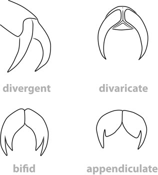 appendage distinguishes this subgenus from Isomiccolamia. This is unique among conifer feeding genera. The tarsal clawtarsal claw:
usually paired claws of the pretarsus, at the distal end of the leg appendage, inflated clavateclavate:
thickening gradually toward the tip
scapescape:
the first proximal segment of the antenna, very small size, and rounded elytral apicesapex:
end of any structure distad to the base
will distinguish from Pogonocherus.
Asia, Indomalaya
broadleaf; Picea
17 spp. Conifers: M. (M.) cleroides.
Miccolamia Bates, 1884
Edition 1
Authors: P.S. Gorring, S.M. Smith, A.I. Cognato, A.J. Redford
Content last updated August 2024
idtools.org
tool images at ITP Node
