
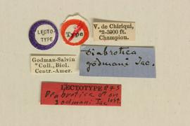
Diabrotica godmani Jacoby 1887: 510
Volcan de Chiriqui, Panama
BMNH, lectotype, male, verified
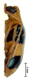
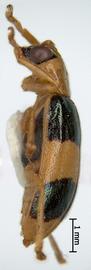
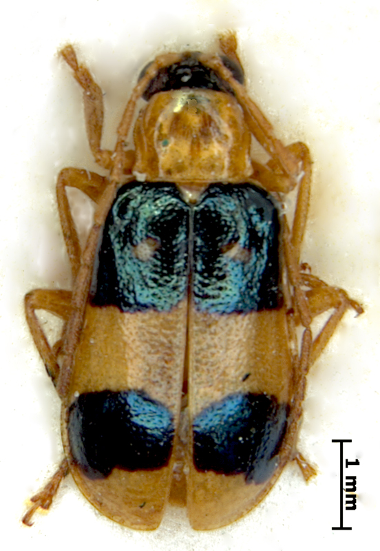
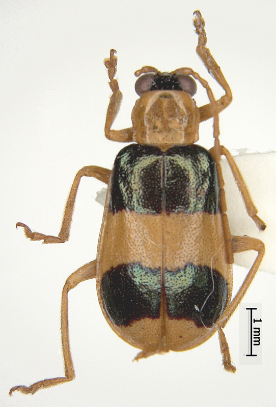
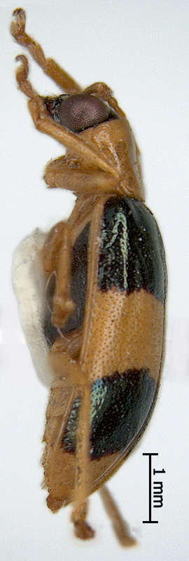
Body length 5.3-6.5 mm. Body width 2.6-3.3 mm. Head basic color black. Antennae shape filiformfiliform:
slender antennae with antennomeres of similar shape
, uniformly yellow ocher. Maxillary palpi yellow or yellow ocher, labrumlabrum:
the "upper lip" of beetles, a movable sclerite joined under clypeus
black or piceous. Pronotumpronotum:
the notum of the prothorax with highly sclerotized pronotal disc
yellow or yellow ocher, subquadrate, bifoveate, with small round foveae, not shagreened. Scutellumscutellum:
small, usually triangular shield between the bases of elytra
yellow. Elytra yellow or rufous, with two metallic black green bandsbands:
(here) transverse maculae on the beetle elytra
, one on the base, another behind the middle. Elytral epipleura completely yellow, sutural anglesutural angle:
the posterior angle or apex of the elytron near the suture
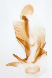
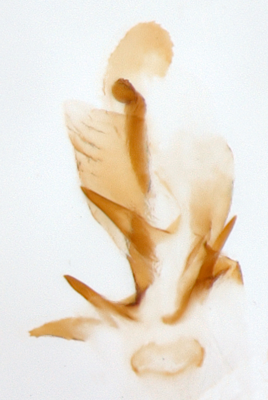
of elytra round, punctation scattered, fine. Abdomen yellow or yellow ocher. Legs yellow or yellow ocher. Aedeagusaedeagus:
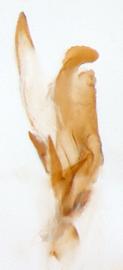
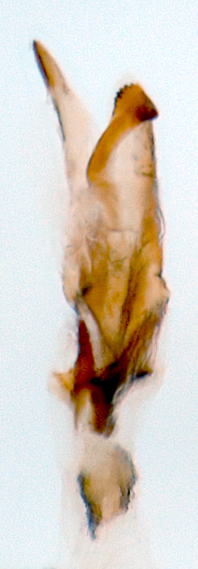
the main sclerotized part of the male genitalia; "aedeagus" is used here instead of "median lobe of aedeagus"
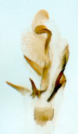
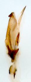
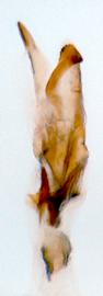
symmetric, with five internal sac scleritessclerites:
(here) the sclerotized hooks, spines or plates in the internal sac
.
Honduras, Guatemala, Panama, Costa Rica, Suriname, Peru
Unknown
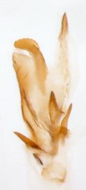
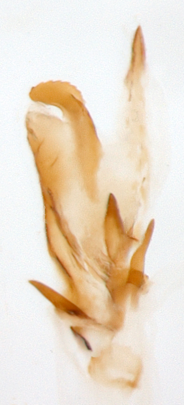
Diabrotica godmani is very similar to D. pulchella (Jacquelin-du-Val). We could not recognize any substantial morphological differences in two species except for the smaller size of D. godmani, with thinner and longer than in D. pulchella, slightly moniliformmoniliform:
(here) antennae with bead-like, slightly elongate and swollen antennomeres
antennae, in males usually bearing longitudinal costae on the inner margin of the antennomeres. The shape and arrangement of the black maculae on the elytra is very variable in both species. We have seen specimens of both species with about the same coloration patterns. The most reliable feature allowing to distinguish these two species is the shape of the scleritessclerites:
(here) the sclerotized hooks, spines or plates in the internal sac
5C-5D in internal sac of the aedeagusaedeagus:
the main sclerotized part of the male genitalia; "aedeagus" is used here instead of "median lobe of aedeagus"
. Sclerite 5C in D. godmani is a short thick hook with "collar" of well-sclerotized leaf, but it looks like a simple long, pointed hook in D. pulchella. Sclerite 5D is a flat plate pointed on the apexapex:
<em>(pl. apices)</em> the far distal end of a structure; opposite of base
in D. godmani, while it is a long pointed hook of round section in D. pulchella. Shape and size of the internal sac plate vary in D. godmani, but in all cases it is a flat plate, sometimes slightly elongate from basis to apexapex:
<em>(pl. apices)</em> the far distal end of a structure; opposite of base
. We suspect that D. godmani and D. pulchella are recently diverged species and it is possible to find more intermediate specimens. Diabrotica pulchella is more common in northern part of Central America and in Mexico, D. godmani is more common in southern part, for example, in Panama.
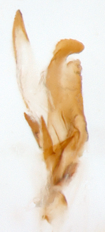
Diabrotica godmani Jacoby is very similar to D. pulchella (Jacquelin-du-Val), D. g1n and D. g2n. They can be separated by the following features: penultimate joint of maxillary palpi in D. godmani is slightly incrassate, but not incrassate in D. g2n; scutellumscutellum:
small, usually triangular shield between the bases of elytra
is yellow in D. godmani, but black in D. g2n; internal sac scleritessclerites:
(here) the sclerotized hooks, spines or plates in the internal sac
5A and 5B are clearly different in D. g1n and D. g2n.
Authors: A. Derunkov, A. Konstantinov, A. Tishechkin, L. Hartje, and A.J. Redford
Last updated Feb. 12, 2015
idtools.org | tool images at ITP Node


