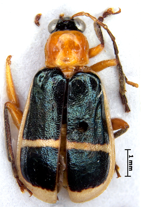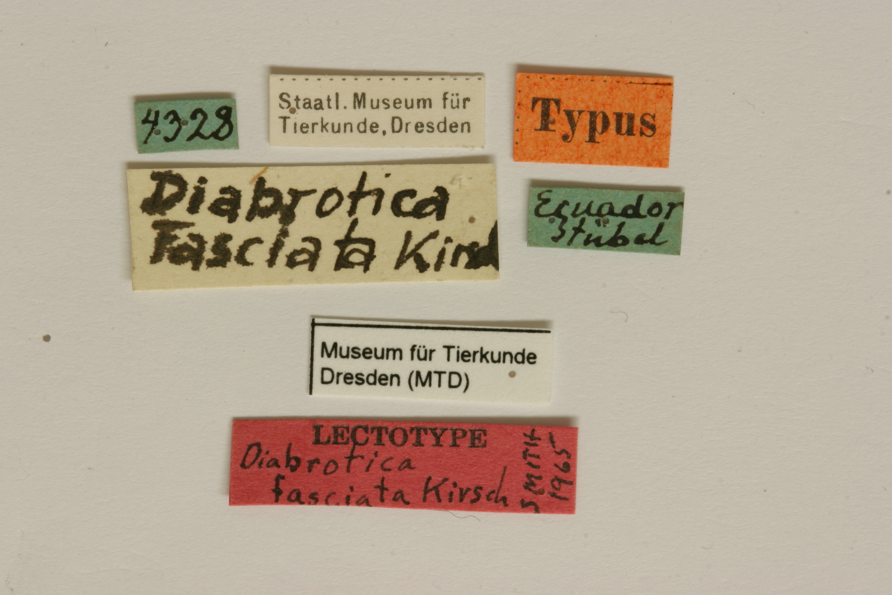
Diabrotica fasciata Kirsch 1883: 200
Ecuador
MTD, lectotype, male, verified
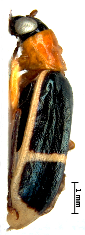
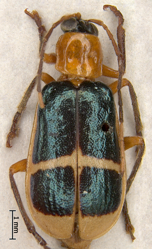
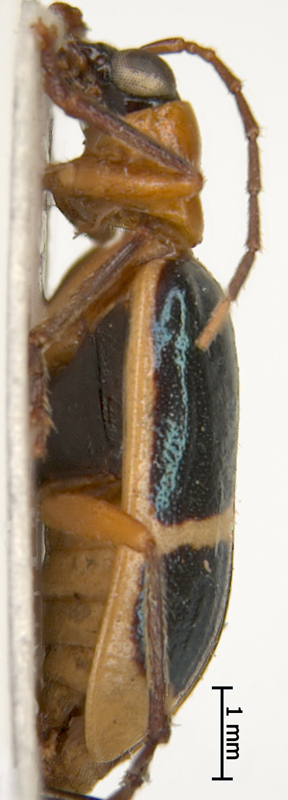
Body length 7.1-7.2 mm. Body width 3.4-3.6 mm. Head basic color black. Antennae filiformfiliform:
slender antennae with antennomeres of similar shape
, bi- or tricolored, antennomeres 1-3 yellow, upper sides darkened, antennomeres 4-8 cinnamon brown, antennomeres 9-10 light cadmium, antennomereantennomere:
"segment" of antenna, more or less clearly separated
11 dark apically. Maxillary palpi and labrumlabrum:
the "upper lip" of beetles, a movable sclerite joined under clypeus
black or chestnut. Pronotumpronotum:
the notum of the prothorax with highly sclerotized pronotal disc
yellow or ochraceous-orange, quadrate, weakly bifoveate, with wide shallow foveae, shagreened with minute wrinkles. Scutellumscutellum:
small, usually triangular shield between the bases of elytra
yellow. Elytra yellow or rufous, with 2 metallic black green bandsbands:
(here) transverse maculae on the beetle elytra
, one on basis of elytronelytron:
<em>(pl. elytra)</em> the fore highly sclerotized wing of beetle
, another - behind middle. Elytral epipleura completely yellow, sutural anglesutural angle:
the posterior angle or apex of the elytron near the suture
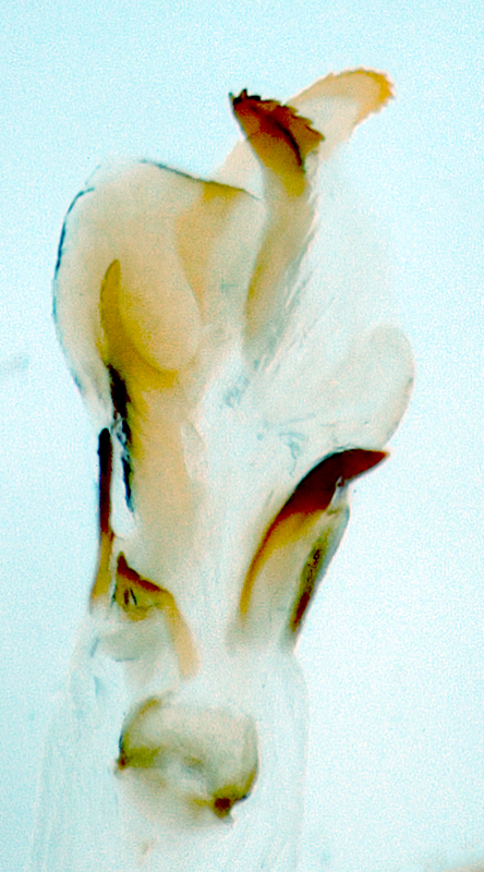
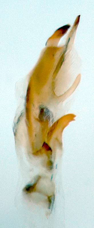
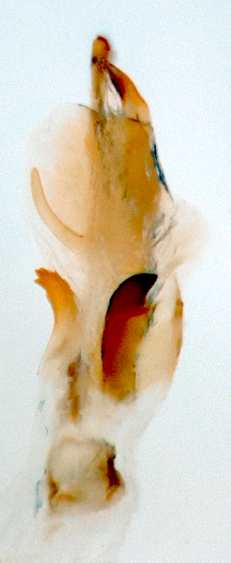
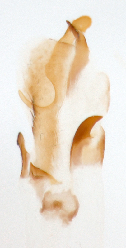
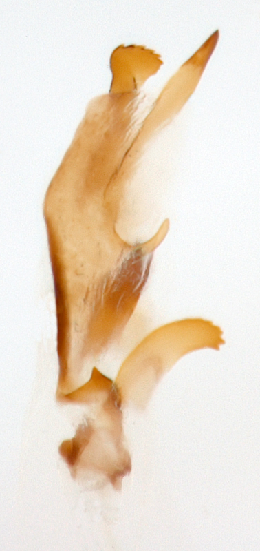
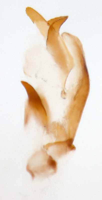
of elytra round, punctation scattered, fine. Abdomen yellow. Tarsi and tibiae black or chestnut, femora yellow or sulphur yellow. Aedeagusaedeagus:
the main sclerotized part of the male genitalia; "aedeagus" is used here instead of "median lobe of aedeagus"
symmetric, with five internal sac scleritessclerites:
(here) the sclerotized hooks, spines or plates in the internal sac
.
Costa Rica, Panama, Colombia, Ecuador
Unknown
Smith and Lawrence (1967) transfered D. fasciata to the genus Paranapiacaba Bechyné. They noted that the lectotype is a female. We studied the lectotype in MTD and found that it is a male. The internal sac armament of Di. fasciata is typical for Diabrotica species with 5 internal sac scleritessclerites:
(here) the sclerotized hooks, spines or plates in the internal sac
. The most important external morphological feature allowing to distinguish Diabrotica and Paranapiacaba is the length of the third antennomereantennomere:
"segment" of antenna, more or less clearly separated
. This antennomereantennomere:
"segment" of antenna, more or less clearly separated
is around 2 times as long as the second in Paranapiacaba and subequal (notmore than 1.5 times as long as second) in Diabrotica (Smith & Lawrence, 1967). The third antennomereantennomere:
"segment" of antenna, more or less clearly separated
in the lectotype and in all other studied specimens is 1.5 time as long as second. Based on the internal sac armament and morphological features we restore the previous combination and treat Paranapiacaba fasciata in the genus Diabrotica .
Diabrotica fasciata Kirsch is very similar to D. militaris Jacoby. They can be separated by the following features: the body is more slender with narrow shoulders in D. fasciata, while it is wider, of round-oval shape in D. militaris; pronotumpronotum:
the notum of the prothorax with highly sclerotized pronotal disc
is narrower in D. fasciata, but wider, slightly transverse in D. militaris; the size and the shape of elytral bandsbands:
(here) transverse maculae on the beetle elytra
is different in both species. The shape of the internal sac scleritessclerites:
(here) the sclerotized hooks, spines or plates in the internal sac
allows distinguishing Diabrotica fasciata from D. militaris.
Authors: A. Derunkov, A. Konstantinov, A. Tishechkin, L. Hartje, and A.J. Redford
Last updated Feb. 12, 2015
idtools.org | tool images at ITP Node