
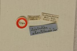
Diabrotica chontalensis Jacoby 1887: 515
Nicaragua, Chontales
BMNH, holotype, male, verified
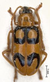
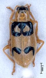
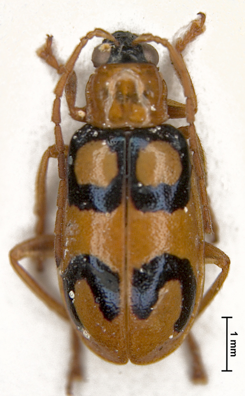
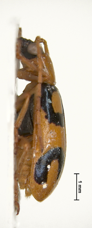
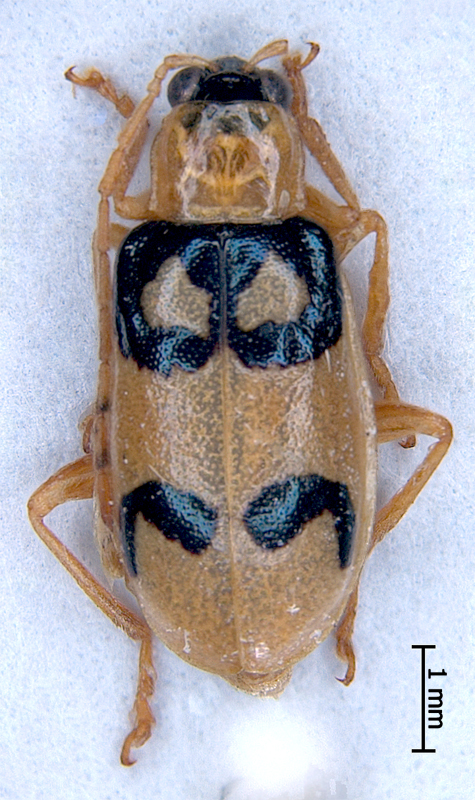
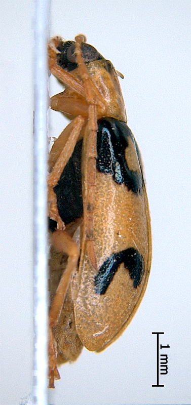
Body length 5.5-6.3 mm, width 2.7-3.0 mm. Head basic color black. Antennae filiformfiliform:
slender antennae with antennomeres of similar shape
, uniformly yellow or bi- or tricolored, antennomeres 1-3 yellow, antennomeres 4-8 uniformly yellow or gradually infuscated, antennomeres 9-10 light cadmium, antennomereantennomere:
"segment" of antenna, more or less clearly separated
11 completely light (light cadmium) or dark apically. Antennomereantennomere:
"segment" of antenna, more or less clearly separated
3 length subequal to length of antennomereantennomere:
"segment" of antenna, more or less clearly separated
2, 2nd and 3d together equal to half or to less than a half length of 4th antennomereantennomere:
"segment" of antenna, more or less clearly separated
. Male antennal length exceed two thirds of elytronelytron:
<em>(pl. elytra)</em> the fore highly sclerotized wing of beetle
length. Maxillary palpi black, piceous or chestnut, labrumlabrum:
the "upper lip" of beetles, a movable sclerite joined under clypeus
black. Pronotumpronotum:
the notum of the prothorax with highly sclerotized pronotal disc
yellow or mustard yellow, subquadrate or quadrate, nonfoveate or bifoveate with small round foveae, not shagreened. Scutellumscutellum:
small, usually triangular shield between the bases of elytra
black. Elytra yellow or rufous, with three black or metallic black blue bandsbands:
(here) transverse maculae on the beetle elytra
, basalbasal:
of or pertaining to the base, as in the first, or basal segment of an appendage; opposite of apical
and middle bandsbands:
(here) transverse maculae on the beetle elytra
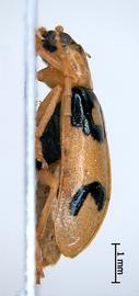
connected together, so two yellow maculae are bounded on elytra basis. Posteriorposterior:
the region of the body parts of the beetle furthest from the head
band on each elytronelytron:
<em>(pl. elytra)</em> the fore highly sclerotized wing of beetle
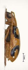
ring-shaped (sometimes opened). Epipleura completely yellow, sutural anglesutural angle:
the posterior angle or apex of the elytron near the suture
of elytronelytron:
<em>(pl. elytra)</em> the fore highly sclerotized wing of beetle
round, punctation scattered, coarse. Abdomen yellow. Legs yellow or yellow ocher, male protibiaeprotibiae:
tibiae of fore legs
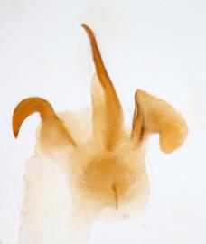
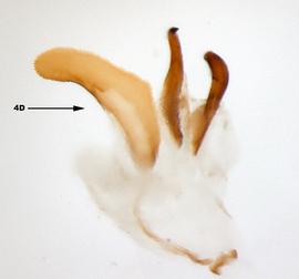
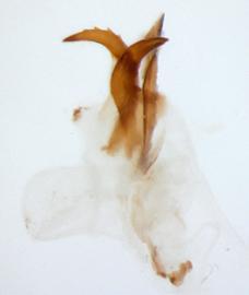
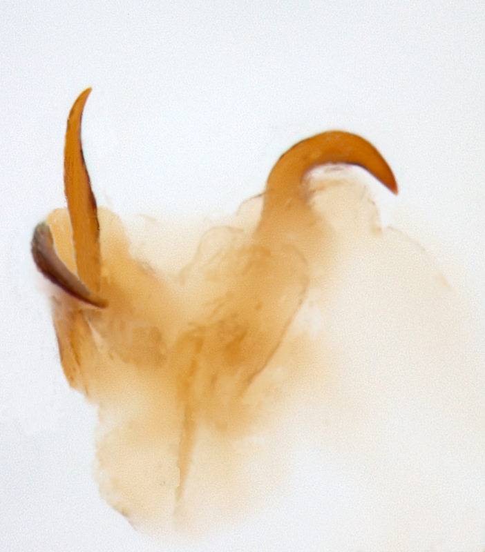
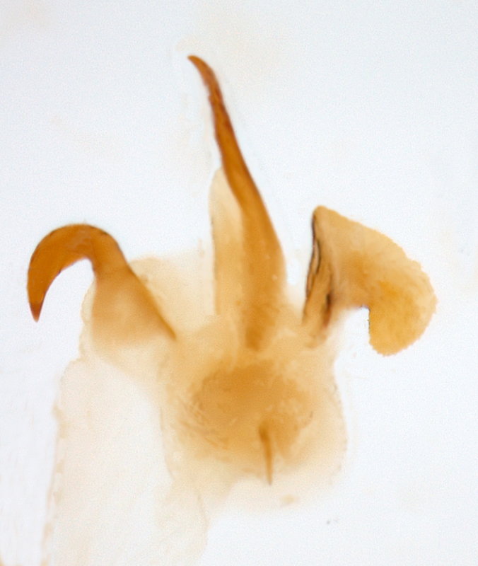
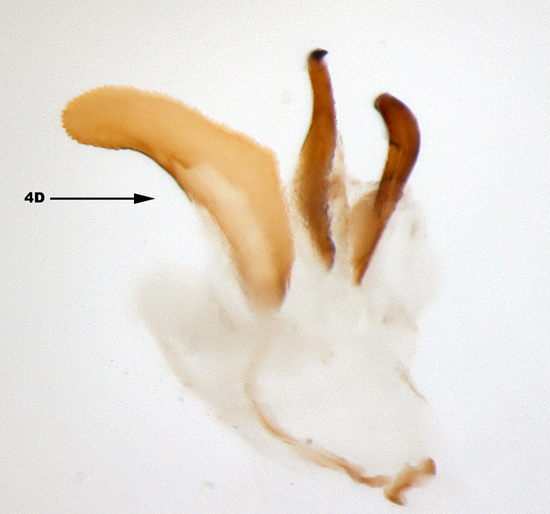
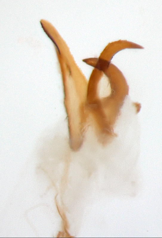
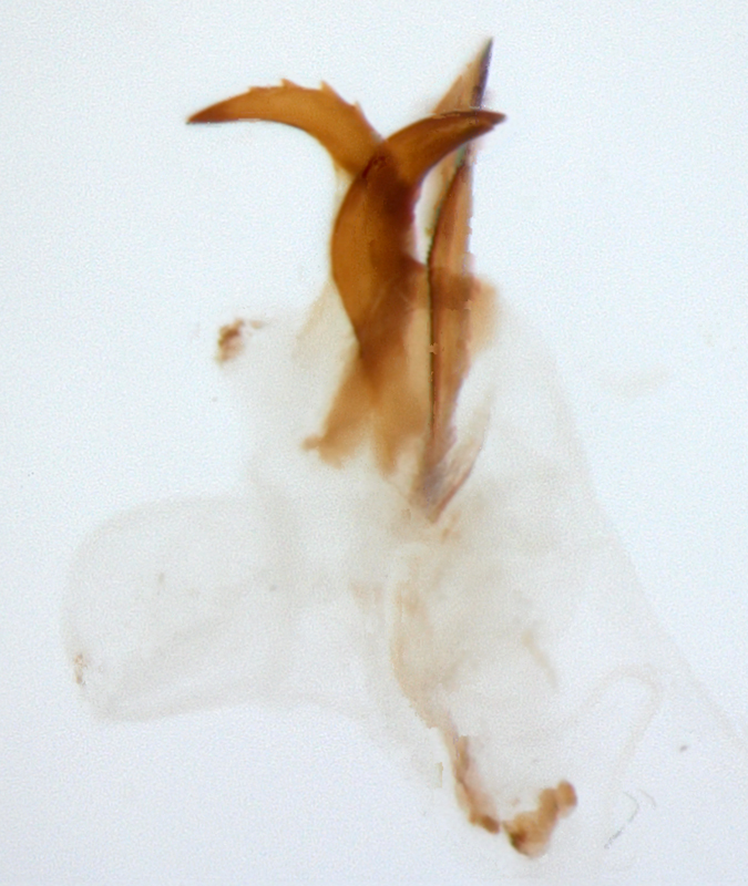
thickened. Aedeagusaedeagus:
the main sclerotized part of the male genitalia; "aedeagus" is used here instead of "median lobe of aedeagus"
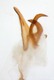
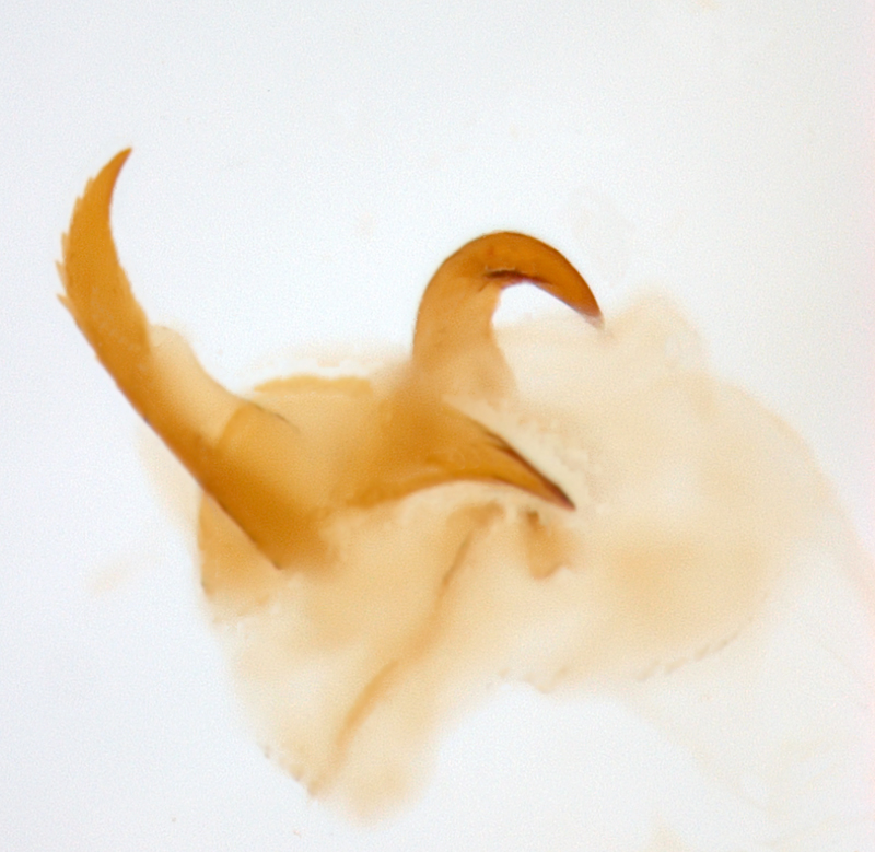
symmetric,with four internal sac scleritessclerites:
(here) the sclerotized hooks, spines or plates in the internal sac
.
Honduras, Nicaragua, Costa Rica
Unknown
Diabrotica chontalensis is very similar to many of the "oculate" species such as D. gratiosa Baly, D. adelpha Harold, D. bioculata Bowditch, D. pulchella (Jacquelin-du-Val) and D. godmani Jacoby. They can be separated by the following features: scutellumscutellum:
small, usually triangular shield between the bases of elytra
of D. chontalensis is black, but yellow in D. bioculata, D. pulchella and D. godmani. The shape of the internal sac scleritessclerites:
(here) the sclerotized hooks, spines or plates in the internal sac
allows to distinguish D. chontalensis from all other species too. Sclerite 4A is an elongate spine. Sclerite 4C is quite remarkable; it is a thick, bent, and pointed hook bearing 4-6 large teeth apically.
Diabrotica chontalensis is particularly similar to D. biannularis Harold and D. spilota Baly. The only feature allowing to distinguish D. chontalensis and D. biannularis is female antennae length. Female antennae are very short in D. biannularis, do not exceed a half of elytronelytron:
<em>(pl. elytra)</em> the fore highly sclerotized wing of beetle
length, in D. chontalensis female antennae exceed two thirds of elytronelytron:
<em>(pl. elytra)</em> the fore highly sclerotized wing of beetle
length. Antennae in D. biannularis are thinner than in D. chontalensis. There is no difference in the internal sac armament of the aedeagusaedeagus:
the main sclerotized part of the male genitalia; "aedeagus" is used here instead of "median lobe of aedeagus"
in both species. According to Jacoby (1887) the only feature allowing to distinguish D. chontalensis is "very short second and third joints of the antennae, these joints being of exactly the same length". The antennomeres 2 and 3 are about the same lenght in D. biannularis and D. chontalensis. The antennomereantennomere:
"segment" of antenna, more or less clearly separated
3 is only slightly longer than 2 in D. spilota, however length may vary between specimens. Sclerite 4C in the internal sac of aedeagusaedeagus:
the main sclerotized part of the male genitalia; "aedeagus" is used here instead of "median lobe of aedeagus"
is slightly longer and slender in D. spilota than in D. biannularis and D. chontalensis. Diabrotica spilota is slightly larger than D. biannularis and D. chontalensis. It is possible that all three names belong to the same species. Jacoby (1887) noted about D. spilota: "there are, however, varieties before me in which the spots of the elytra are either transversely or longitudinally connected". Thus D. spilota is most likely an example of an extreme pattern reduction in D. biannularis. Finally, Baly (1886) gave Mexico as a locality for D. spilota. Smith and Lawrence (1967) wrote that "It is possible that the Mexico record given by Baly is a misinterpretation of a [Magd] handwritten label". It seems that Mexico record was true and D. spilota range may be from Mexico to South America.
Authors: A. Derunkov, A. Konstantinov, A. Tishechkin, L. Hartje, and A.J. Redford
Last updated Feb. 12, 2015
idtools.org | tool images at ITP Node


