
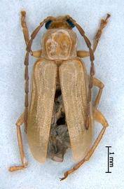
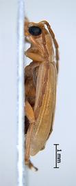
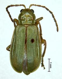
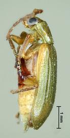
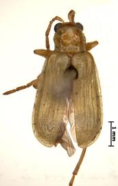
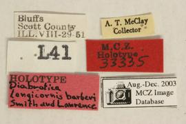
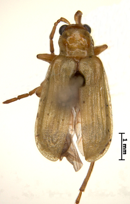
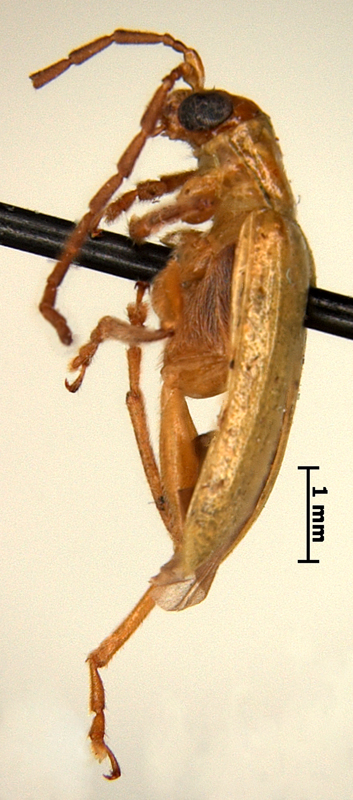
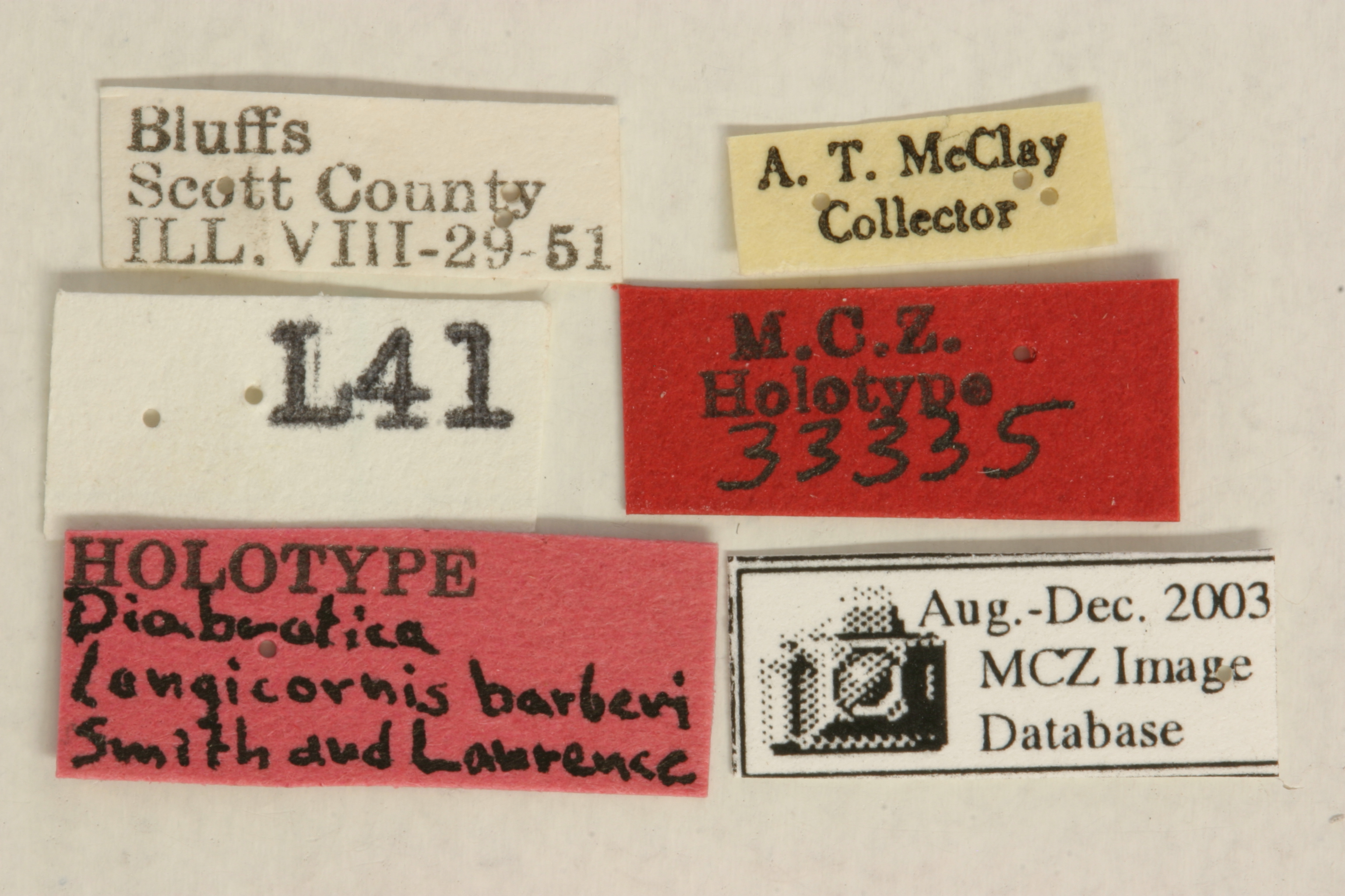
Diabrotica barberi Smith & Lawrence 1967: 87
Northern corn rootworm
U.S.A., Illinois, Scott County, Bluffs
MCZ, holotype, male, verified
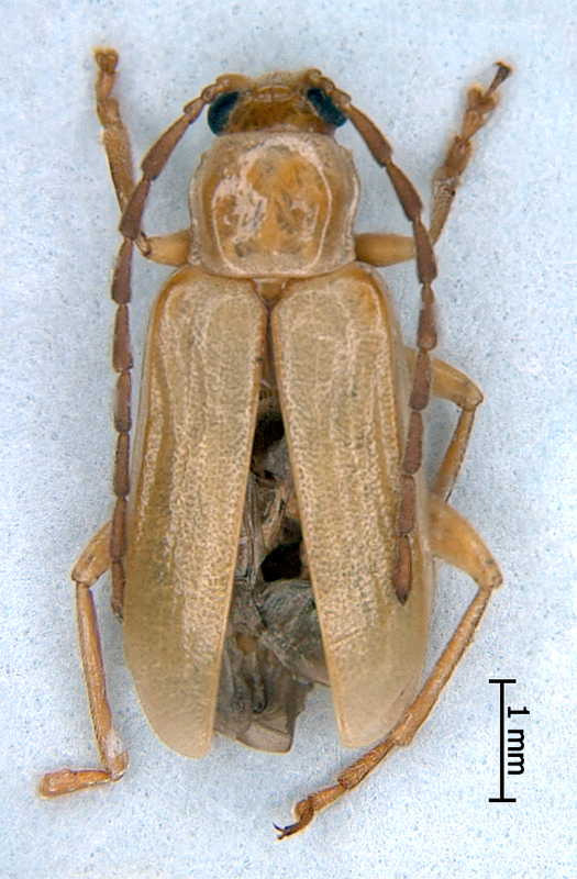
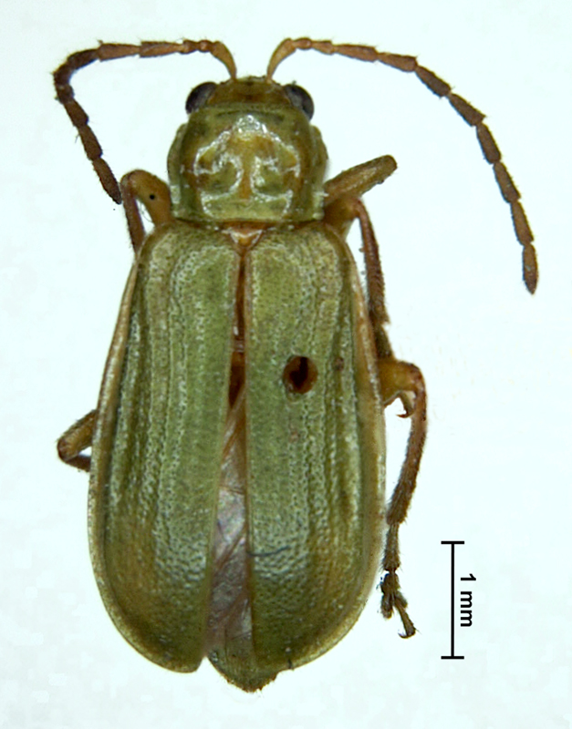
Body length 4.8-5.6 mm. Body width 2.0-2.5 mm. Head basic color yellow. Antennae filiformfiliform:
slender antennae with antennomeres of similar shape
, bi- or tricolored, antennomereantennomere:
"segment" of antenna, more or less clearly separated
1 yellow, testaceous or greenish brown, antennomeres 2-11 brussels brown. Maxillary palpi and labrumlabrum:
the "upper lip" of beetles, a movable sclerite joined under clypeus
black or piceous. Pronotumpronotum:
the notum of the prothorax with highly sclerotized pronotal disc
paris green, green or yellow, quadrate, deeply bifoveate, not shagreened. Scutellumscutellum:
small, usually triangular shield between the bases of elytra
yellow or amber yellow. Elytra green, sometimes humeri and basalbasal:
of or pertaining to the base, as in the first, or basal segment of an appendage; opposite of apical
third of suture tinged with amber, with five distinct sinuatesinuate:
curved in some way
sulci, strongest behind the humeral callushumeral callus:
<em>(pl. calli)</em> more or less marked tubercle or knob on the anterobasal angle of elytron
and extending beyond the middle. Elytral epipleura green, sutural anglesutural angle:
the posterior angle or apex of the elytron near the suture
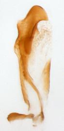
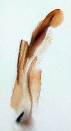
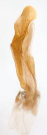
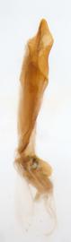
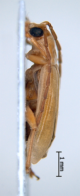
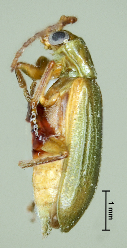
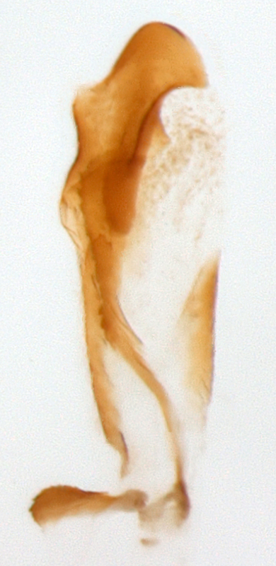
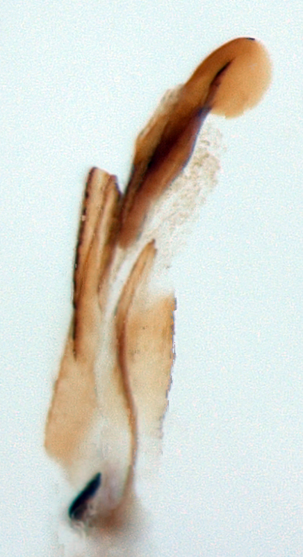
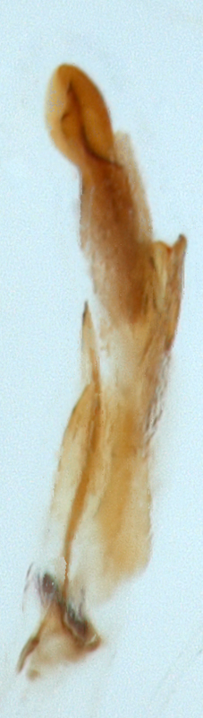
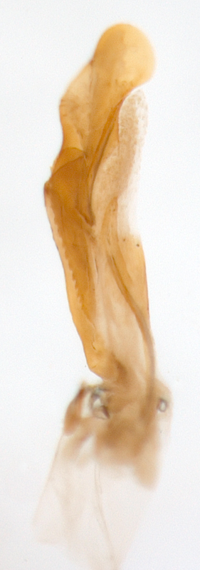
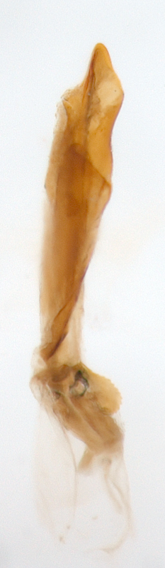
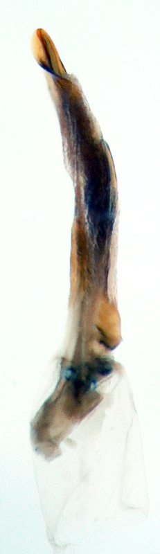
of elytra round, punctation scattered, fine. Abdomen yellow, pale olivine or green. Tarsi black, amber brown or chestnut. Tibiae bicolored yellow, outer edge with piceous or testaceous line, or extensively darkened. Femora uniform yellow or olive ocher. Aedeagusaedeagus:
the main sclerotized part of the male genitalia; "aedeagus" is used here instead of "median lobe of aedeagus"
symmetric, with four internal sac scleritessclerites:
(here) the sclerotized hooks, spines or plates in the internal sac
.
CANADA: Manitoba, New Brunswick, Ontario, Quebec; USA: AL, AR, CO, CT, DE, GA, IL, IN, IA, KS, KY, MD, MA, MI, MN, MO, NE, NH, NJ, NY, NC, ND, OH, OK, PA, RI, SC, SD, TN, VT, VA, WV, WI. (Distribution Maps of Plant Pests, http://www.cabi.org/dmpp)
Maize (Zea mays L.), Cucurbitaceae, Poaceae, Asteraceae and Fabaceae (Clark et al., 2004)
Diabrotica barberi Smith & Lawrence is similar to D. longicornis (Say) and D. virgifera LeConte. They can be separated by the following features: in D. barberi the head, tibiatibia:
<em>(pl. tibiae)</em> the forth part of the beetle leg articulated with femur on the one side and with tarsus on the other side
and tarsi are paler than in D. longicornis; femora unicolorous green or flavous in D. barberi, while femora of D. virgifera as a rule bicolored, with outer edges dark, chestnut or piceous; distance from apexapex:
<em>(pl. apices)</em> the far distal end of a structure; opposite of base
to ventral flange of aedeagusaedeagus:
the main sclerotized part of the male genitalia; "aedeagus" is used here instead of "median lobe of aedeagus"
in D. v. virgifera is 1.5 - 2.0 times that of D. barberi. The shapes of the internal sac scleritessclerites:
(here) the sclerotized hooks, spines or plates in the internal sac
(especially sclerite 4B) differentiates all three species very well.
Authors: A. Derunkov, A. Konstantinov, A. Tishechkin, L. Hartje, and A.J. Redford
Last updated Feb. 12, 2015
idtools.org | tool images at ITP Node


