
Gallery
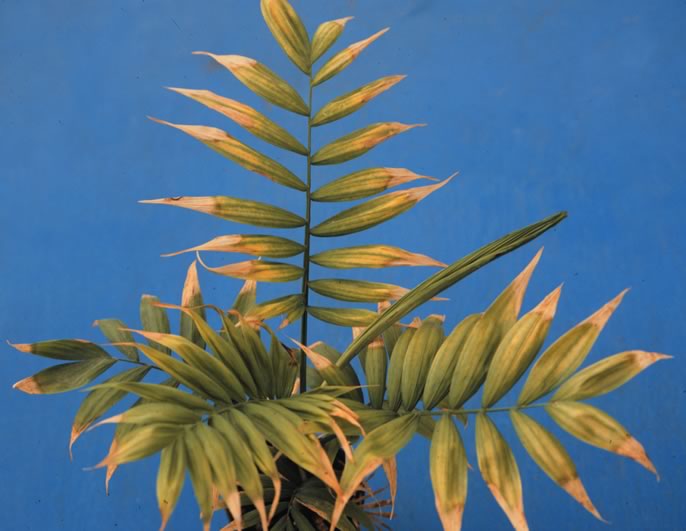
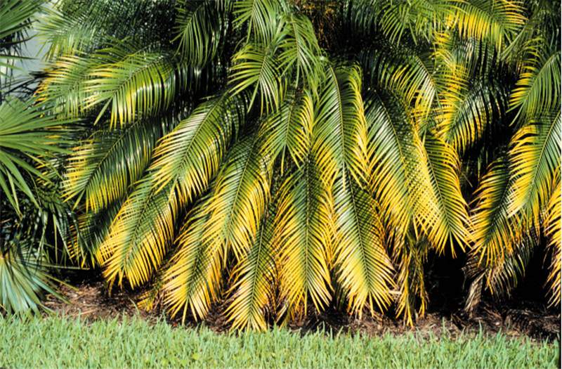 Figure 1. Magnesium deficiency symptoms on Phoenix roebelenii . Note the broad yellow bands along the outer margins of the leaves, but distinctly green central portions of the leaves. Photo by T.K. Broschat.
Magnesium Deficiency
Figure 1. Magnesium deficiency symptoms on Phoenix roebelenii. Note the broad yellow bands along the outer margins of the leaves, but distinctly green central portions of the leaves. Photo by T.K. Broschat.
Figure 1. Magnesium deficiency symptoms on Phoenix roebelenii. Note the broad yellow bands along the outer margins of the leaves, but distinctly green central portions of the leaves. Photo by T.K. Broschat.
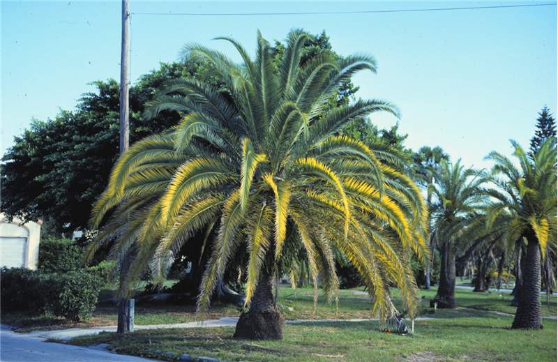 Figure 2. Magnesium and potassium deficiency symptoms on Phoenix canariensis . The broad yellow-banded mid canopy leaves show Mg deficiency, while the oldest leaves show leaflet tip necrosis (K deficiency). Photo by T.K. Broschat.
Magnesium Deficiency
Figure 2. Magnesium and potassium deficiency symptoms on Phoenix canariensis. The broad yellow-banded mid canopy leaves show Mg deficiency, while the oldest leaves show leaflet tip necrosis (K deficiency). Photo by T.K. Broschat.
Figure 2. Magnesium and potassium deficiency symptoms on Phoenix canariensis. The broad yellow-banded mid canopy leaves show Mg deficiency, while the oldest leaves show leaflet tip necrosis (K deficiency). Photo by T.K. Broschat.
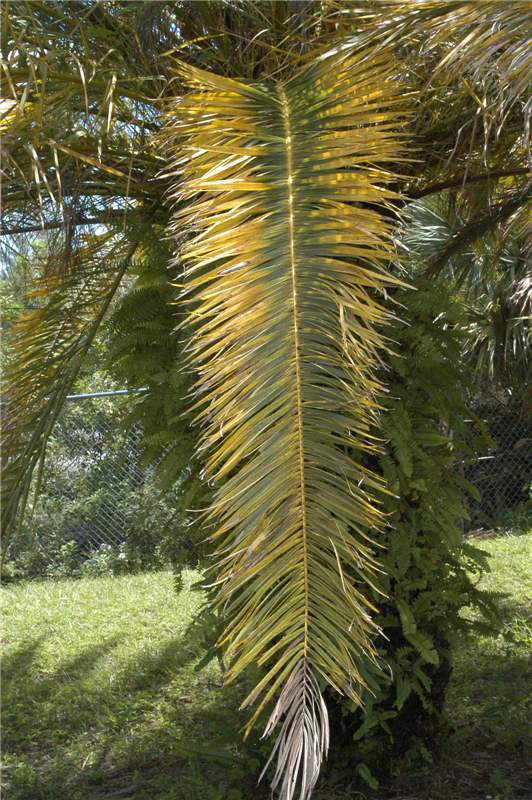 Figure 3. Older leaf of Phoenix canariensis showing Mg deficiency symptoms towards the leaf base and K deficiency symptoms towards the leaf tip. Photo by T.K. Broschat.
Magnesium Deficiency
Figure 3. Older leaf of Phoenix canariensis showing Mg deficiency symptoms towards the leaf base and K deficiency symptoms towards the leaf tip. Photo by T.K. Broschat.
Figure 3. Older leaf of Phoenix canariensis showing Mg deficiency symptoms towards the leaf base and K deficiency symptoms towards the leaf tip. Photo by T.K. Broschat.
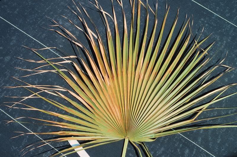
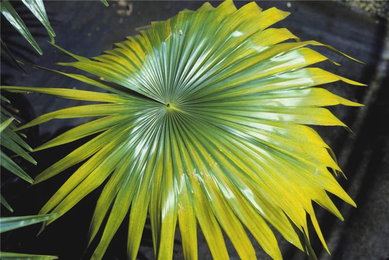 Figure 4. Magnesium-deficient older leaf of Livistona rotundifolia showing broad yellow band around entire leaf as well as a yellow band along margins of single leaf segments in upper left. Photo by T.K. Broschat.
Magnesium Deficiency
Figure 4. Magnesium-deficient older leaf of Livistona rotundifolia showing broad yellow band around entire leaf as well as a yellow band along margins of single leaf segments in upper left. Photo by T.K. Broschat.
Figure 4. Magnesium-deficient older leaf of Livistona rotundifolia showing broad yellow band around entire leaf as well as a yellow band along margins of single leaf segments in upper left. Photo by T.K. Broschat.
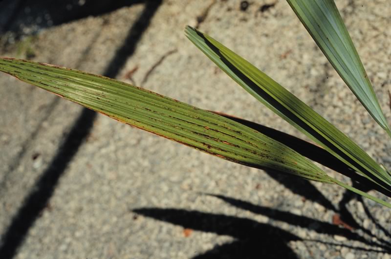
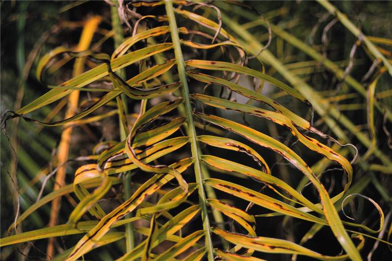 Figure 2. Close-up of Mn-deficient leaf of Phoenix roebelenii showing longitudinal necrotic streaking. Photo by T.K. Broschat.
Manganese Deficiency
Figure 2. Close-up of Mn-deficient leaf of Phoenix roebelenii showing longitudinal necrotic streaking. Photo by T.K. Broschat.
Figure 2. Close-up of Mn-deficient leaf of Phoenix roebelenii showing longitudinal necrotic streaking. Photo by T.K. Broschat.
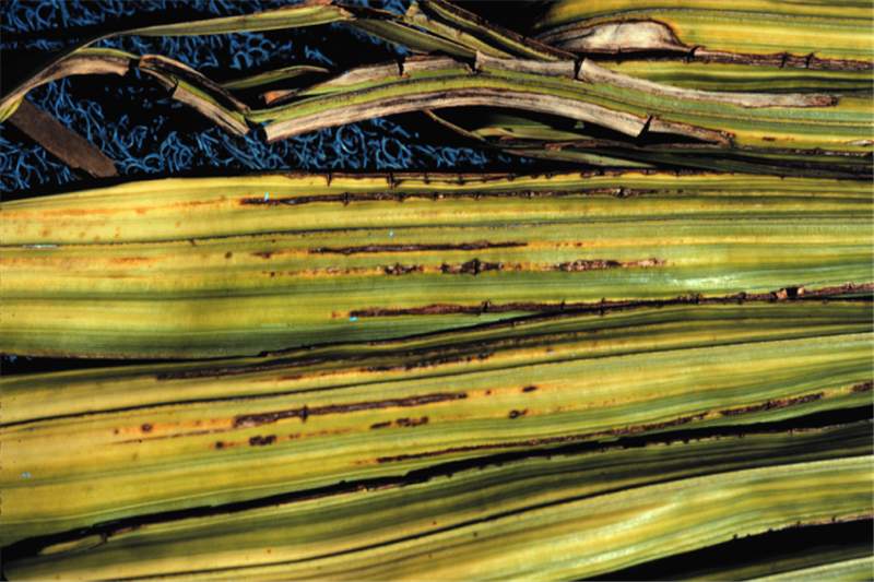 Figure 1. Close-up of leaflets of Mn-deficient Archontophoenix alexandrae leaf. Note the longitudinal necrotic streaking. Photo by T.K. Broschat.
Manganese Deficiency
Figure 1. Close-up of leaflets of Mn-deficient Archontophoenix alexandrae leaf. Note the longitudinal necrotic streaking. Photo by T.K. Broschat.
Figure 1. Close-up of leaflets of Mn-deficient Archontophoenix alexandrae leaf. Note the longitudinal necrotic streaking. Photo by T.K. Broschat.
Manganese Deficiency
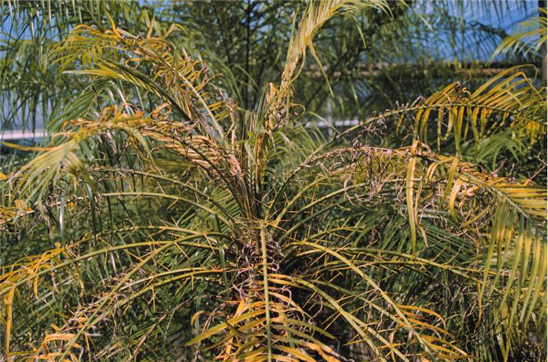
Figure 4. Manganese deficiency symptoms on Phoenix roebelenii. Photo by T.K. Broschat.
Figure 4. Manganese deficiency symptoms on Phoenix roebelenii. Photo by T.K. Broschat.
Manganese Deficiency
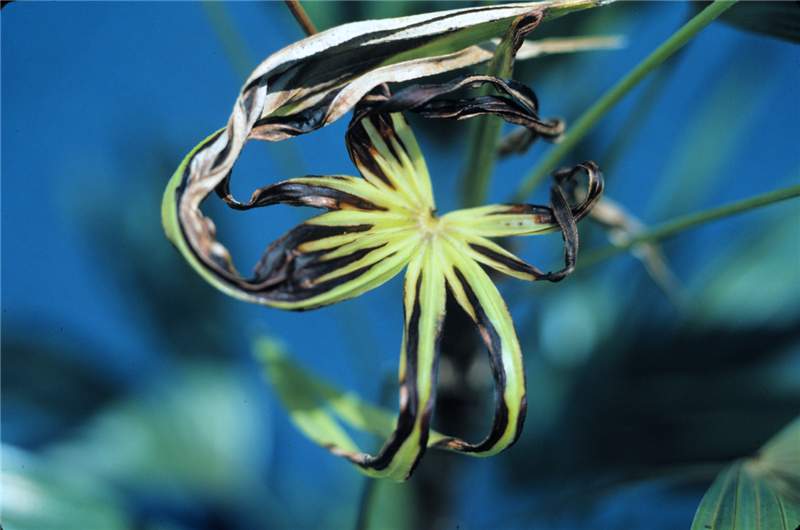
Figure 3. Mangenese-deficient new leaf of Rhapis excelsa. Photo by T.K. Broschat.
Figure 3. Mangenese-deficient new leaf of Rhapis excelsa. Photo by T.K. Broschat.
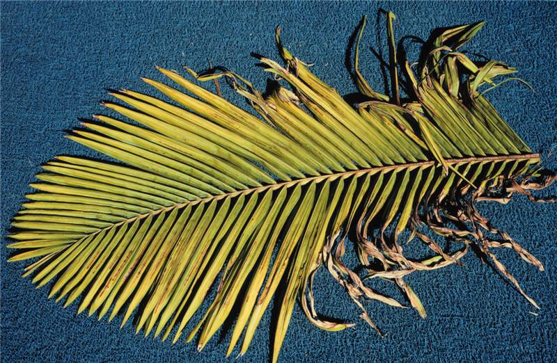 Figure 6. Manganese-deficient new leaf of Archontophoenix alexandrae . Note that symptoms are most severe towards the leaf base and least so towards the tip. Photo by T.K. Broschat.
Manganese Deficiency
Figure 6. Manganese-deficient new leaf of Archontophoenix alexandrae. Note that symptoms are most severe towards the leaf base and least so towards the tip. Photo by T.K. Broschat.
Figure 6. Manganese-deficient new leaf of Archontophoenix alexandrae. Note that symptoms are most severe towards the leaf base and least so towards the tip. Photo by T.K. Broschat.
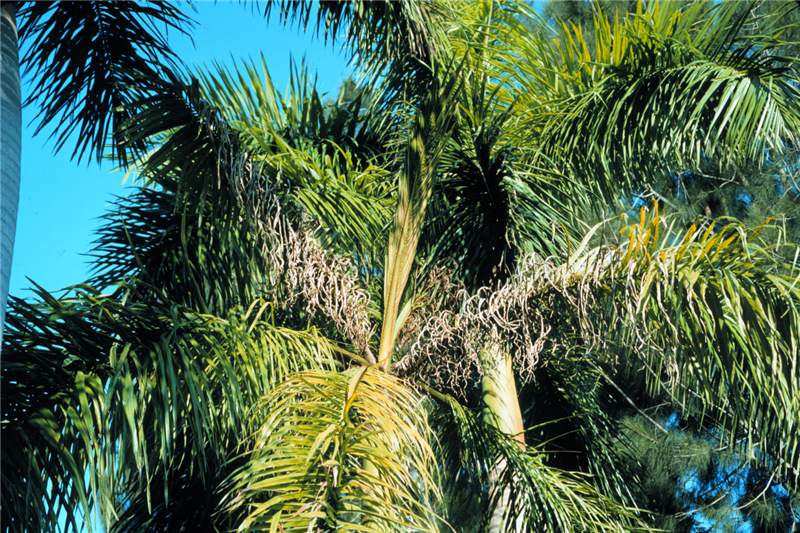 Figure 5. Manganese deficiency on Roystonea regia . Note that frizzling is most severe towards the base of the leaves. Photo by T.K. Broschat.
Manganese Deficiency
Figure 5. Manganese deficiency on Roystonea regia. Note that frizzling is most severe towards the base of the leaves. Photo by T.K. Broschat.
Figure 5. Manganese deficiency on Roystonea regia. Note that frizzling is most severe towards the base of the leaves. Photo by T.K. Broschat.
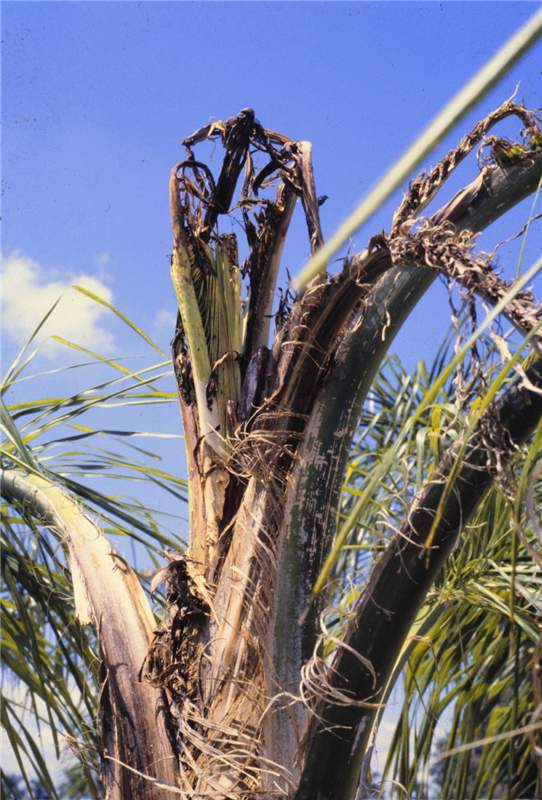 Figure 8. Severe Mn deficiency symptoms on Syagrus romanzoffiana with only necrotic petiole stubs emerging. Photo by T.K. Broschat.
Manganese Deficiency
Figure 8. Severe Mn deficiency symptoms on Syagrus romanzoffiana with only necrotic petiole stubs emerging. Photo by T.K. Broschat.
Figure 8. Severe Mn deficiency symptoms on Syagrus romanzoffiana with only necrotic petiole stubs emerging. Photo by T.K. Broschat.
Manganese Deficiency
Figure 7. Severe Mn deficiency symptoms on Cocos nucifera. Photo by T.K. Broschat.
Figure 7. Severe Mn deficiency symptoms on Cocos nucifera. Photo by T.K. Broschat.
Micronutrient or Chemical Toxicity
Figure 1. Boron toxicity on Chamaedorea leaves. Photo by T.K. Broschat.
Figure 1. Boron toxicity on Chamaedorea leaves. Photo by T.K. Broschat.
Micronutrient or Chemical Toxicity
Figure 2. Copper toxicity on Acoelorrhaphe wrightii leaves. Photo by T.K. Broschat.
Figure 2. Copper toxicity on Acoelorrhaphe wrightii leaves. Photo by T.K. Broschat.
Micronutrient or Chemical Toxicity
Figure 3. Close-up of copper toxicity on Acoelorrhaphe wrightii leaves. Photo by T.K. Broschat.
Figure 3. Close-up of copper toxicity on Acoelorrhaphe wrightii leaves. Photo by T.K. Broschat.
Mite Damage
Figure 1. Mite damage on Veitchia sp. seedling. Photo by T.K. Broschat.
Figure 1. Mite damage on Veitchia sp. seedling. Photo by T.K. Broschat.
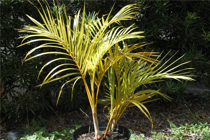 Figure 1. N deficient Dypsis lutescens . Note the golden yellow petioles and rachides and light green colored leaflets. Photo by T.K. Broschat
Nitrogen Deficiency
Figure 1. N deficient Dypsis lutescens. Note the golden yellow petioles and rachides and light green colored leaflets. Photo by T.K. Broschat
Figure 1. N deficient Dypsis lutescens. Note the golden yellow petioles and rachides and light green colored leaflets. Photo by T.K. Broschat
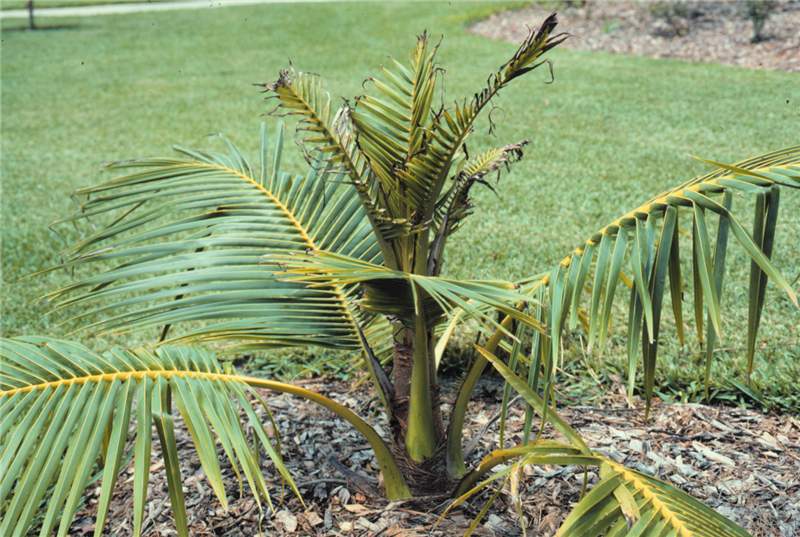
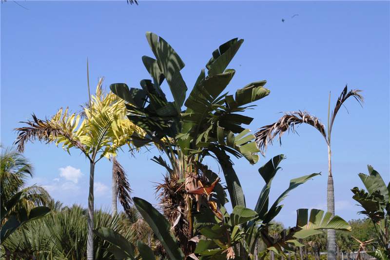 Figure 2. Severe nitrogen deficiency in Veitchia sp. The palm on the right has died from chronic N deficiency. Photo by T.K. Broschat
Nitrogen Deficiency
Figure 2. Severe nitrogen deficiency in Veitchia sp. The palm on the right has died from chronic N deficiency. Photo by T.K. Broschat
Figure 2. Severe nitrogen deficiency in Veitchia sp. The palm on the right has died from chronic N deficiency. Photo by T.K. Broschat
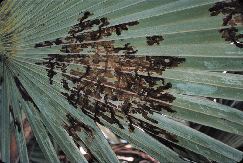 Figure 1. Damage caused by leaf skeletonizer ( Homaledra sabalella ) larvae on Latania lontaroides . Photo by T.K. Broschat
Palm Leaf Skeletonizer Damage
Figure 1. Damage caused by leaf skeletonizer (Homaledra sabalella) larvae on Latania lontaroides. Photo by T.K. Broschat
Figure 1. Damage caused by leaf skeletonizer (Homaledra sabalella) larvae on Latania lontaroides. Photo by T.K. Broschat
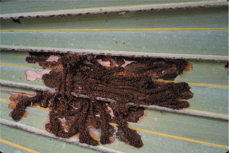 Figure 2. Frass tubes of palm leaf skeletonizer ( Homaledra sabalella ) on Latania lontaroides . Photo by T.K. Broschat
Palm Leaf Skeletonizer Damage
Figure 2. Frass tubes of palm leaf skeletonizer (Homaledra sabalella) on Latania lontaroides. Photo by T.K. Broschat
Figure 2. Frass tubes of palm leaf skeletonizer (Homaledra sabalella) on Latania lontaroides. Photo by T.K. Broschat
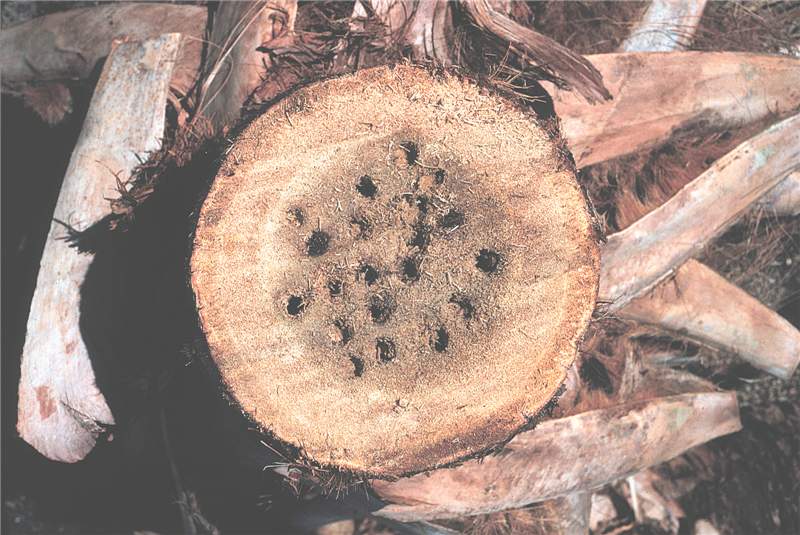 Figure 1. Cross section through Sabal palmetto infested with palmetto weevil ( Rhynchophorus cruentatus ). Photo by R. Giblin-Davis.
Palmetto Weevil Damage
Figure 1. Cross section through Sabal palmetto infested with palmetto weevil (Rhynchophorus cruentatus). Photo by R. Giblin-Davis.
Figure 1. Cross section through Sabal palmetto infested with palmetto weevil (Rhynchophorus cruentatus). Photo by R. Giblin-Davis.
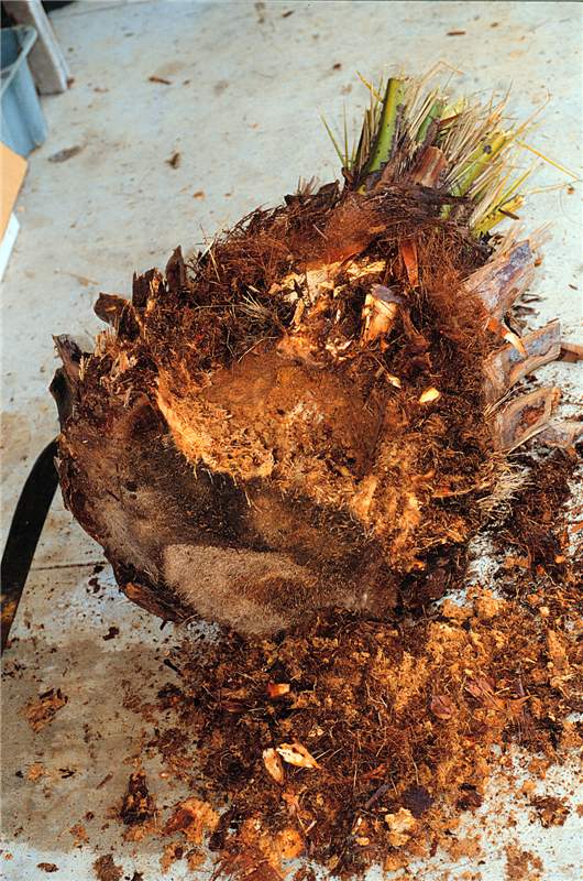 Figure 2. Inside of Phoenix canariensis infested with palmetto weevil ( Rhyncophorus cruentatus ). Photo by R. Giblin-Davis.
Palmetto Weevil Damage
Figure 2. Inside of Phoenix canariensis infested with palmetto weevil (Rhyncophorus cruentatus). Photo by R. Giblin-Davis.
Figure 2. Inside of Phoenix canariensis infested with palmetto weevil (Rhyncophorus cruentatus). Photo by R. Giblin-Davis.
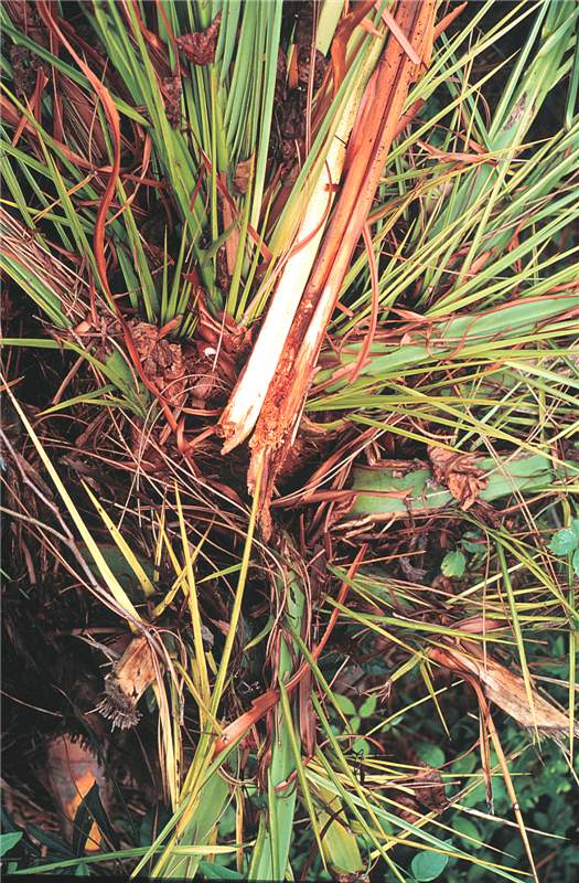 Figure 3. Damage caused by palmetto weevil ( Rhynchophorus cruentatus ) to new growth of Phoenix canariensis . Photo by R.Giblin-Davis.
Palmetto Weevil Damage
Figure 3. Damage caused by palmetto weevil (Rhynchophorus cruentatus) to new growth of Phoenix canariensis. Photo by R.Giblin-Davis.
Figure 3. Damage caused by palmetto weevil (Rhynchophorus cruentatus) to new growth of Phoenix canariensis. Photo by R.Giblin-Davis.
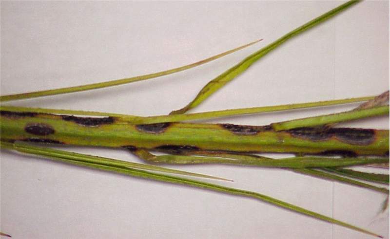 Figure 1. The rachis blight lesions appear black and sunken on this palm rachis. As the lesions expand together, larger areas of blighted rachis tissue develop. Photo by M. L. Elliott.
Pestalotiopsis Rachis Blight and Crown Rot
Figure 1. The rachis blight lesions appear black and sunken on this palm rachis. As the lesions expand together, larger areas of blighted rachis tissue develop. Photo by M. L. Elliott.
Figure 1. The rachis blight lesions appear black and sunken on this palm rachis. As the lesions expand together, larger areas of blighted rachis tissue develop. Photo by M. L. Elliott.
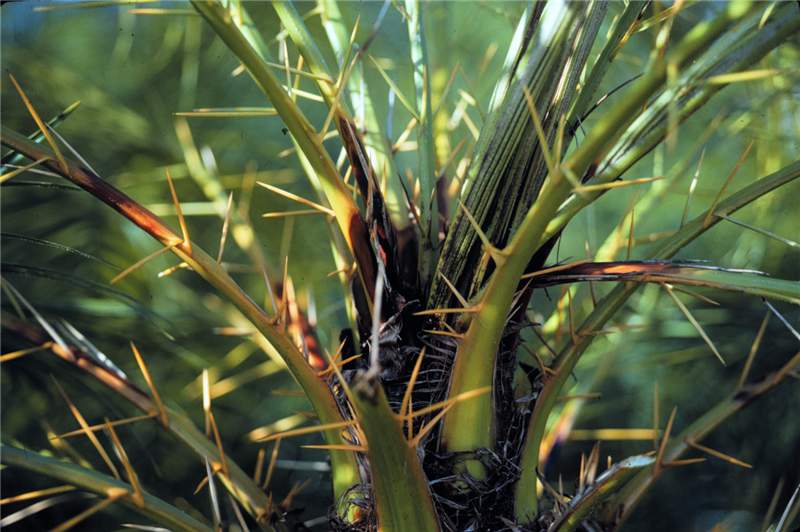 Figure 2. Large, dark lesions have formed near or on the leaf base of all the rachides of this Phoenix roebelenii canopy. These lesions often continue to expand, and the fungus will infect the spear leaf and the apical meristem causing a bud and crown rot. Photo by T. K. Broschat.
Pestalotiopsis Rachis Blight and Crown Rot
Figure 2. Large, dark lesions have formed near or on the leaf base of all the rachides of this Phoenix roebelenii canopy. These lesions often continue to expand, and the fungus will infect the spear leaf and the apical meristem causing a bud and crown rot. Photo by T. K. Broschat.
Petiole (Rachis) Blight
Figure 2. Internal discoloration due to a petiole (rachis) blight pathogen. Photo by A. J. Downer
Figure 2. Internal discoloration due to a petiole (rachis) blight pathogen. Photo by A. J. Downer
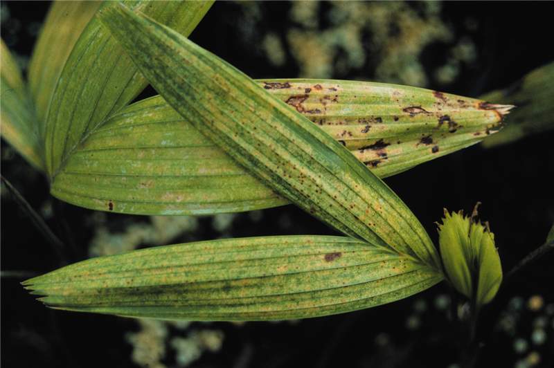
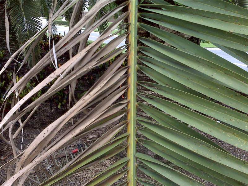 Figure 1. This Phoenix canariensis leaf has green leaflets on one side of the rachis and necrotic leaflets on the opposite side. There is a reddish-brown stripe on the rachis that corresponds to the necrotic leaflets. Photo by M. L. Elliott.
Petiole (Rachis) Blight
Figure 1. This Phoenix canariensis leaf has green leaflets on one side of the rachis and necrotic leaflets on the opposite side. There is a reddish-brown stripe on the rachis that corresponds to the necrotic leaflets. Photo by M. L. Elliott.
Figure 1. This Phoenix canariensis leaf has green leaflets on one side of the rachis and necrotic leaflets on the opposite side. There is a reddish-brown stripe on the rachis that corresponds to the necrotic leaflets. Photo by M. L. Elliott.
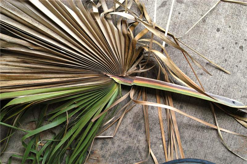 Figure 4. A single Washingtonia robusta leaf where a quarter of the leaf blade is still green, but the remaining leaf segments are necrotic. The petiole has a reddish-brown stripe on the same side where the necrotic leaf segments are located. Photo by M. L. Elliott.
Petiole (Rachis) Blight
Figure 4. A single Washingtonia robusta leaf where a quarter of the leaf blade is still green, but the remaining leaf segments are necrotic. The petiole has a reddish-brown stripe on the same side where the necrotic leaf segments are located. Photo by M. L. Elliott.
Figure 4. A single Washingtonia robusta leaf where a quarter of the leaf blade is still green, but the remaining leaf segments are necrotic. The petiole has a reddish-brown stripe on the same side where the necrotic leaf segments are located. Photo by M. L. Elliott.
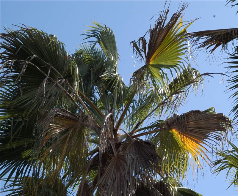 Figure 3. Washingtonia robusta canopy with older leaves exhibiting leaf blades with mixture of healthy, chlorotic and necrotic leaflet segments. Photo by M. L. Elliott.
Petiole (Rachis) Blight
Figure 3. Washingtonia robusta canopy with older leaves exhibiting leaf blades with mixture of healthy, chlorotic and necrotic leaflet segments. Photo by M. L. Elliott.
Figure 3. Washingtonia robusta canopy with older leaves exhibiting leaf blades with mixture of healthy, chlorotic and necrotic leaflet segments. Photo by M. L. Elliott.
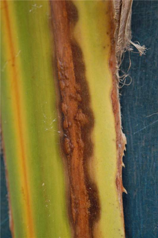 Figure 6. This petiole has a reddish-brown stripe that is accompanied by "blisters". The fungal pathogen is forming spore structures that will erupt through the epidermis. Photo by M. L. Elliott.
Petiole (Rachis) Blight
Figure 6. This petiole has a reddish-brown stripe that is accompanied by "blisters". The fungal pathogen is forming spore structures that will erupt through the epidermis. Photo by M. L. Elliott.
Figure 6. This petiole has a reddish-brown stripe that is accompanied by "blisters". The fungal pathogen is forming spore structures that will erupt through the epidermis. Photo by M. L. Elliott.
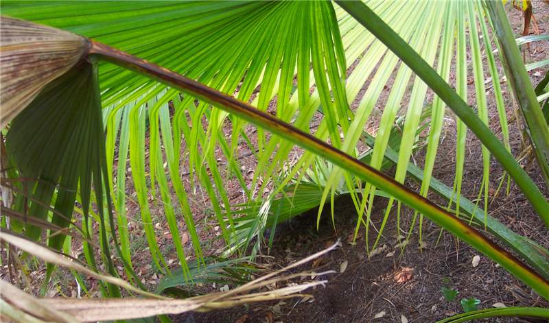 Figure 5. This Livistona chinensis leaf has a reddish-brown stripe on only one side of the petiole due to petiole blight. Part of the leaf blade is necrotic and the other portion is still green. Photo by M. L. Elliott.
Petiole (Rachis) Blight
Figure 5. This Livistona chinensis leaf has a reddish-brown stripe on only one side of the petiole due to petiole blight. Part of the leaf blade is necrotic and the other portion is still green. Photo by M. L. Elliott.
Figure 5. This Livistona chinensis leaf has a reddish-brown stripe on only one side of the petiole due to petiole blight. Part of the leaf blade is necrotic and the other portion is still green. Photo by M. L. Elliott.
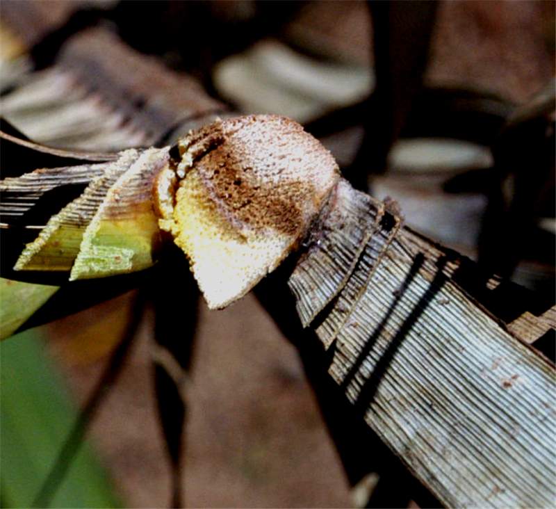
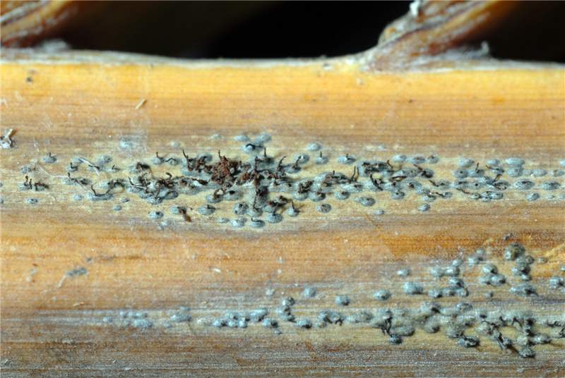 Figure 7. The "bumps" are spore structures (pycnidia) of Dothiorella. In the center of the photo, you can observe spore masses emerging from the pycnidia. Photo by A. J. Downer.
Petiole (Rachis) Blight
Figure 7. The "bumps" are spore structures (pycnidia) of Dothiorella. In the center of the photo, you can observe spore masses emerging from the pycnidia. Photo by A. J. Downer.
Figure 7. The "bumps" are spore structures (pycnidia) of Dothiorella. In the center of the photo, you can observe spore masses emerging from the pycnidia. Photo by A. J. Downer.
