
Gallery
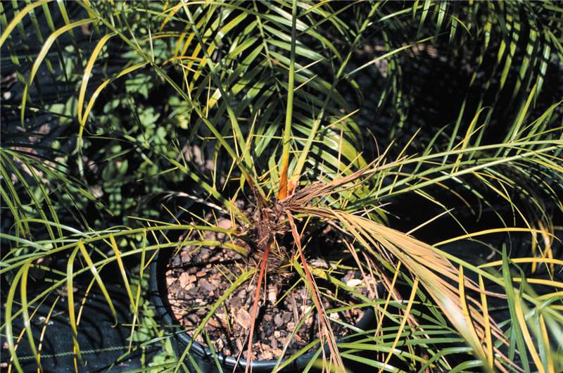
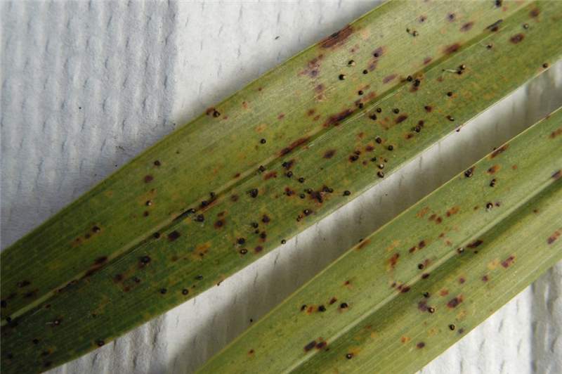 Figure 4. The brown spots are not early symptoms of Graphiola leaf spot, but are symptoms of potassium deficiency. Photo by M. L. Elliott.
Graphiola Leaf Spot
Figure 4. The brown spots are not early symptoms of Graphiola leaf spot, but are symptoms of potassium deficiency. Photo by M. L. Elliott.
Figure 4. The brown spots are not early symptoms of Graphiola leaf spot, but are symptoms of potassium deficiency. Photo by M. L. Elliott.
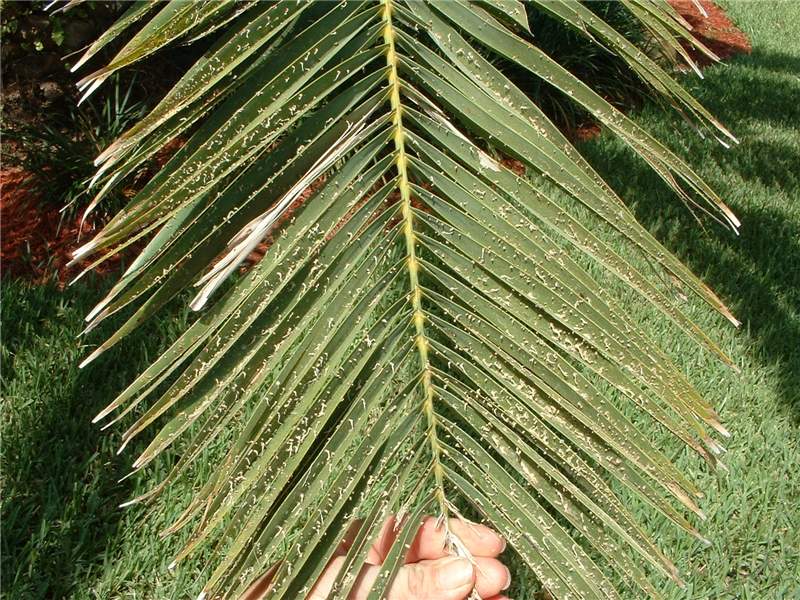 Figure 3. Palm leaflets with numerous sori and emerging filaments (look like tiny strings) with spores. There are no leaf spot symptoms, only the sori of the fungus. Photo by M. L. Elliott.
Graphiola Leaf Spot
Figure 3. Palm leaflets with numerous sori and emerging filaments (look like tiny strings) with spores. There are no leaf spot symptoms, only the sori of the fungus. Photo by M. L. Elliott.
Figure 3. Palm leaflets with numerous sori and emerging filaments (look like tiny strings) with spores. There are no leaf spot symptoms, only the sori of the fungus. Photo by M. L. Elliott.
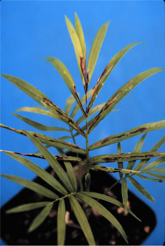
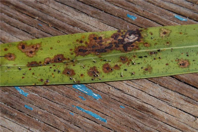 Figure 5. A leaf affected by both Graphiola leaf spot and Stigmina leaf spot. Signs of Graphiola phoenicis are the small back bodies (sori), many with filaments emerging from the sori. Stigmina palmivora symptoms are the large brown spots with dark edges and darker but flat centers. In some cases, a G. phoenicis sorus is superimposed upon the S. palmivora leaf spot. Photo by M. L. Elliott.
Graphiola Leaf Spot
Figure 5. A leaf affected by both Graphiola leaf spot and Stigmina leaf spot. Signs of Graphiola phoenicis are the small back bodies (sori), many with filaments emerging from the sori. Stigmina palmivora symptoms are the large brown spots with dark edges and darker but flat centers. In some cases, a G. phoenicis sorus is superimposed upon the S. palmivora leaf spot. Photo by M. L. Elliott.
Figure 5. A leaf affected by both Graphiola leaf spot and Stigmina leaf spot. Signs of Graphiola phoenicis are the small back bodies (sori), many with filaments emerging from the sori. Stigmina palmivora symptoms are the large brown spots with dark edges and darker but flat centers. In some cases, a G. phoenicis sorus is superimposed upon the S. palmivora leaf spot. Photo by M. L. Elliott.
 Figure 1. Chlorosis and necrosis of leaf tip of older leaf of Elaeis guineensis affected by Marchitez Sorpresiva/sudden wilt/lethal wilt. Photo by M. L. Elliott.
Hartrot and Marchitez Sorpresiva
Figure 1. Chlorosis and necrosis of leaf tip of older leaf of Elaeis guineensis affected by Marchitez Sorpresiva/sudden wilt/lethal wilt. Photo by M. L. Elliott.
Figure 1. Chlorosis and necrosis of leaf tip of older leaf of Elaeis guineensis affected by Marchitez Sorpresiva/sudden wilt/lethal wilt. Photo by M. L. Elliott.
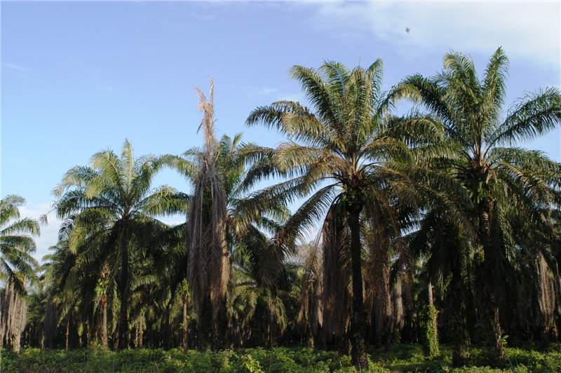 Figure 2. Elaeis guineensis affected by Marchitez Sorpresiva/sudden wilt/lethal wilt in late stages of disease surrounded by healthy palms. Dead leaves have formed skirt around trunk. Photo by M. L. Elliott.
Hartrot and Marchitez Sorpresiva
Figure 2. Elaeis guineensis affected by Marchitez Sorpresiva/sudden wilt/lethal wilt in late stages of disease surrounded by healthy palms. Dead leaves have formed skirt around trunk. Photo by M. L. Elliott.
Figure 2. Elaeis guineensis affected by Marchitez Sorpresiva/sudden wilt/lethal wilt in late stages of disease surrounded by healthy palms. Dead leaves have formed skirt around trunk. Photo by M. L. Elliott.
Herbicide Toxicity
Figure 2. Glyphosate (Roundup) injury on Phoenix sylvestris. Photo by T.K. Broschat.
Figure 2. Glyphosate (Roundup) injury on Phoenix sylvestris. Photo by T.K. Broschat.
 Figure 1. Glyphosate (Roundup) injury on Phoenix roebelenii . Note that the palm has since grown out of the injury. Photo by T.K. Broschat
Herbicide Toxicity
Figure 1. Glyphosate (Roundup) injury on Phoenix roebelenii. Note that the palm has since grown out of the injury. Photo by T.K. Broschat
Figure 1. Glyphosate (Roundup) injury on Phoenix roebelenii. Note that the palm has since grown out of the injury. Photo by T.K. Broschat
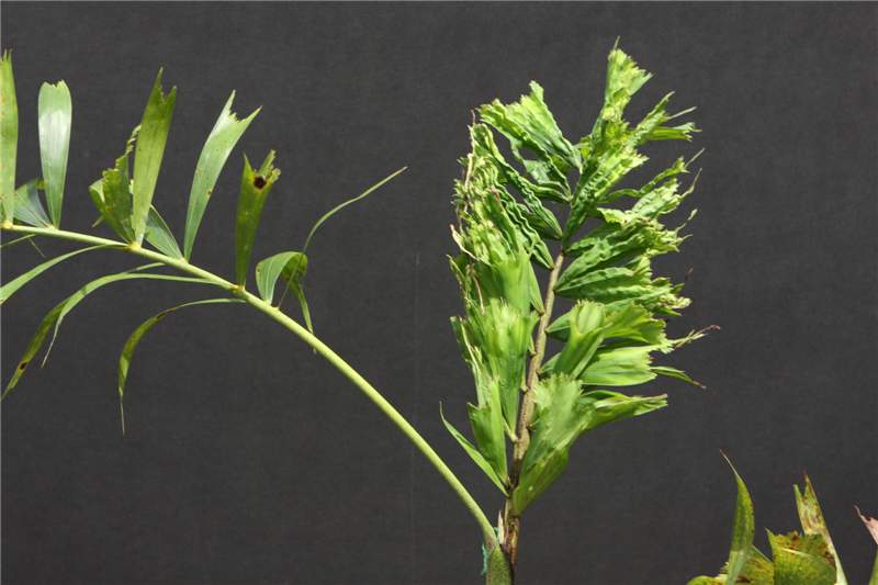 Figure 4. Metsulfuron (Manor or Blade) injury on Wodyetia bifurcata . Damage became visible about 7 months following application to the soil. Photo by T.K. Broschat.
Herbicide Toxicity
Figure 4. Metsulfuron (Manor or Blade) injury on Wodyetia bifurcata. Damage became visible about 7 months following application to the soil. Photo by T.K. Broschat.
Figure 4. Metsulfuron (Manor or Blade) injury on Wodyetia bifurcata. Damage became visible about 7 months following application to the soil. Photo by T.K. Broschat.
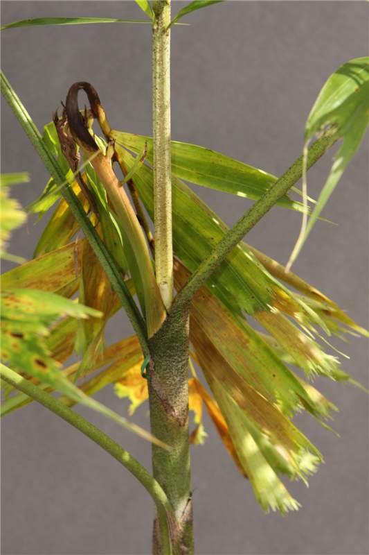 Figure 3. Metsulfuron (Manor or Blade) injury on Wodyetia bifurcata . Damage became visible about 7 months following application to the soil. Photo by T.K. Broschat.
Herbicide Toxicity
Figure 3. Metsulfuron (Manor or Blade) injury on Wodyetia bifurcata. Damage became visible about 7 months following application to the soil. Photo by T.K. Broschat.
Figure 3. Metsulfuron (Manor or Blade) injury on Wodyetia bifurcata. Damage became visible about 7 months following application to the soil. Photo by T.K. Broschat.
Herbicide Toxicity
Figure 6. Isoxaben plus oryzalin (Snapshot) injury on Phoenix roebelenii. Photo by T.K. Broschat.
Figure 6. Isoxaben plus oryzalin (Snapshot) injury on Phoenix roebelenii. Photo by T.K. Broschat.
Herbicide Toxicity
Figure 5. Oryzalin plus oxyfluorfen (Rout) injury on Chamaedorea elegans. Photo by T.K. Broschat.
Figure 5. Oryzalin plus oxyfluorfen (Rout) injury on Chamaedorea elegans. Photo by T.K. Broschat.
 Figure 7. Metolachlor (Pennant) injury on Coccothrinax sp. Note branching in this normally single-stemmed species. Photo by T.K. Broschat.
Herbicide Toxicity
Figure 7. Metolachlor (Pennant) injury on Coccothrinax sp. Note branching in this normally single-stemmed species. Photo by T.K. Broschat.
Figure 7. Metolachlor (Pennant) injury on Coccothrinax sp. Note branching in this normally single-stemmed species. Photo by T.K. Broschat.
Iron Deficiency
Figure 1. Iron-deficient Rhapis excelsa on left with healthy palms on right. Photo by T.K. Broschat
Figure 1. Iron-deficient Rhapis excelsa on left with healthy palms on right. Photo by T.K. Broschat
 Figure 2. Severe Fe deficiency on Syagrus romanzoffiana seedling (left). Normal palm on right for comparision. Photo by T.K. Broschat
Iron Deficiency
Figure 2. Severe Fe deficiency on Syagrus romanzoffiana seedling (left). Normal palm on right for comparision. Photo by T.K. Broschat
Figure 2. Severe Fe deficiency on Syagrus romanzoffiana seedling (left). Normal palm on right for comparision. Photo by T.K. Broschat
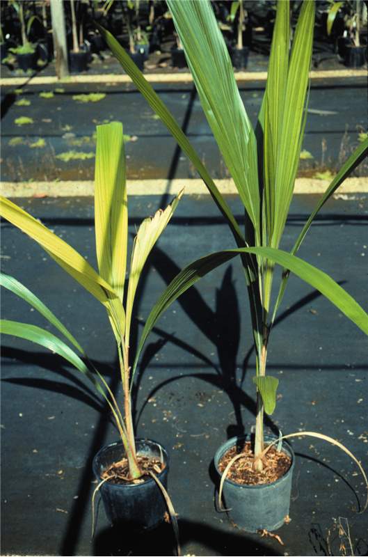 Figure 3. Severe Fe deficiency on Syagrus romanzoffiana seedling (left). Normal palm on right for comparision. Photo by T.K. Broschat
Iron Deficiency
Figure 3. Severe Fe deficiency on Syagrus romanzoffiana seedling (left). Normal palm on right for comparision. Photo by T.K. Broschat
Figure 3. Severe Fe deficiency on Syagrus romanzoffiana seedling (left). Normal palm on right for comparision. Photo by T.K. Broschat
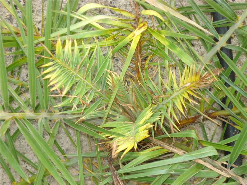
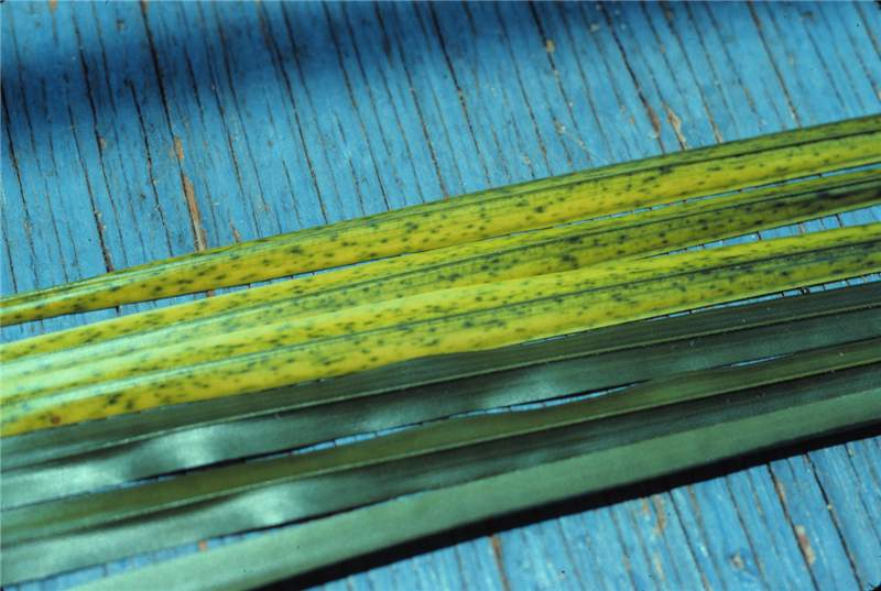 Figure 4. Mild Fe deficiency symptoms on leaflets of new leaves (two upper leaflets) and symptom-free older leaves (two lower leaflets). Photo by T.K. Broschat
Iron Deficiency
Figure 4. Mild Fe deficiency symptoms on leaflets of new leaves (two upper leaflets) and symptom-free older leaves (two lower leaflets). Photo by T.K. Broschat
Figure 4. Mild Fe deficiency symptoms on leaflets of new leaves (two upper leaflets) and symptom-free older leaves (two lower leaflets). Photo by T.K. Broschat
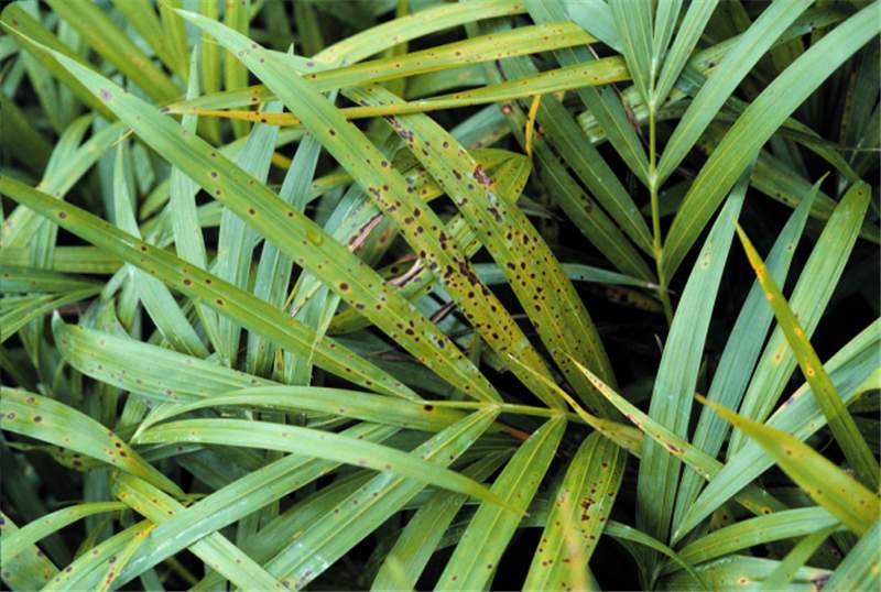 Figure 2. Leaf spot exhibitng brown or black centers with yellow halos. Spots are randomly scattered on the leaf tissue. Photo by T. K. Broschat.
Leaf Spots and Leaf Blights
Figure 2. Leaf spot exhibitng brown or black centers with yellow halos. Spots are randomly scattered on the leaf tissue. Photo by T. K. Broschat.
Figure 2. Leaf spot exhibitng brown or black centers with yellow halos. Spots are randomly scattered on the leaf tissue. Photo by T. K. Broschat.
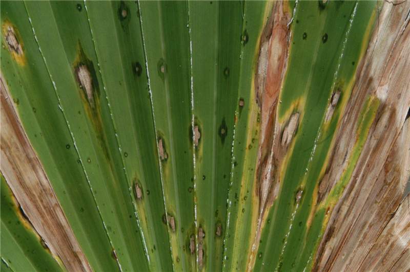 Figure 1. The initial lesions of this leaf spot disease are pin point water-soaked appearing spots. As the spot expands, the center becomes gray with water-soaked edges As the lesions continue to expand, a yellow halo may be observed. Coalescing of expanding lesions are observed until large areas of blighted tissue result. Photo by M. L. Elliott.
Leaf Spots and Leaf Blights
Figure 1. The initial lesions of this leaf spot disease are pin point water-soaked appearing spots. As the spot expands, the center becomes gray with water-soaked edges As the lesions continue to expand, a yellow halo may be observed. Coalescing of expanding lesions are observed until large areas of blighted tissue result. Photo by M. L. Elliott.
Figure 1. The initial lesions of this leaf spot disease are pin point water-soaked appearing spots. As the spot expands, the center becomes gray with water-soaked edges As the lesions continue to expand, a yellow halo may be observed. Coalescing of expanding lesions are observed until large areas of blighted tissue result. Photo by M. L. Elliott.
Figure 4. A leaf affected by both Graphiola leaf spot and Stigmina leaf spot. Signs of Graphiola phoenicis are the small back bodies (sori), many with filaments emerging from the sori. Stigmina palmivora symptoms are the large brown spots with dark edges and darker but flat centers. In some cases, a G. phoenicis sorus is superimposed upon the S. palmivora leaf spot. Photo by M. L. Elliott.
Leaf Spots and Leaf Blights
Figure 4. A leaf affected by both Graphiola leaf spot and Stigmina leaf spot. Signs of Graphiola phoenicis are the small back bodies (sori), many with filaments emerging from the sori. Stigmina palmivora symptoms are the large brown spots with dark edges and darker but flat centers. In some cases, a G. phoenicis sorus is superimposed upon the S. palmivora leaf spot. Photo by M. L. Elliott.
Figure 4. A leaf affected by both Graphiola leaf spot and Stigmina leaf spot. Signs of Graphiola phoenicis are the small back bodies (sori), many with filaments emerging from the sori. Stigmina palmivora symptoms are the large brown spots with dark edges and darker but flat centers. In some cases, a G. phoenicis sorus is superimposed upon the S. palmivora leaf spot. Photo by M. L. Elliott.
 Figure 3. Leaf spot that begins as a black spot with distinct yellow halo. As the lesion expands or lesions coalesce, the affected area becomes a leaf blight and the center becomes a gray color. Photo by T. K. Broschat.
Leaf Spots and Leaf Blights
Figure 3. Leaf spot that begins as a black spot with distinct yellow halo. As the lesion expands or lesions coalesce, the affected area becomes a leaf blight and the center becomes a gray color. Photo by T. K. Broschat.
Figure 3. Leaf spot that begins as a black spot with distinct yellow halo. As the lesion expands or lesions coalesce, the affected area becomes a leaf blight and the center becomes a gray color. Photo by T. K. Broschat.
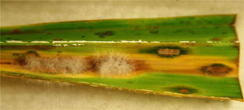 Figure 6. A leaf spot symptomatic leaf segment of the leaf in Figure 1 was placed under high humidity. The result was growth of the fungus from some of the spots. Note fungal growth is only associated with the leaf spot symptom and not with the healthy green leaf tissue. Photo by M. L. Elliott.
Leaf Spots and Leaf Blights
Figure 6. A leaf spot symptomatic leaf segment of the leaf in Figure 1 was placed under high humidity. The result was growth of the fungus from some of the spots. Note fungal growth is only associated with the leaf spot symptom and not with the healthy green leaf tissue. Photo by M. L. Elliott.
Figure 6. A leaf spot symptomatic leaf segment of the leaf in Figure 1 was placed under high humidity. The result was growth of the fungus from some of the spots. Note fungal growth is only associated with the leaf spot symptom and not with the healthy green leaf tissue. Photo by M. L. Elliott.
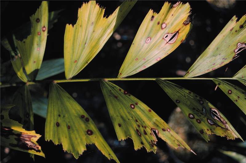 Figure 5. Spots are initially black with no halos. As the spot expands, the center becomes tan and is outlined by a brown to brownish-black edge, but no yellow halo. The leaflet in the lower-left corner has large areas of necrotic tissue. The leaf spot is superimposed on iron deficiency symptoms. Photo by M. L. Elliott.
Leaf Spots and Leaf Blights
Figure 5. Spots are initially black with no halos. As the spot expands, the center becomes tan and is outlined by a brown to brownish-black edge, but no yellow halo. The leaflet in the lower-left corner has large areas of necrotic tissue. The leaf spot is superimposed on iron deficiency symptoms. Photo by M. L. Elliott.
Figure 5. Spots are initially black with no halos. As the spot expands, the center becomes tan and is outlined by a brown to brownish-black edge, but no yellow halo. The leaflet in the lower-left corner has large areas of necrotic tissue. The leaf spot is superimposed on iron deficiency symptoms. Photo by M. L. Elliott.
 Figure 2. Leaf yellowing symptoms on Cocos nucifera. Photo by N. A. Harrison, University of Florida.
Lethal Yellowing
Figure 2. Leaf yellowing symptoms on Cocos nucifera. Photo by N. A. Harrison, University of Florida.
Figure 2. Leaf yellowing symptoms on Cocos nucifera. Photo by N. A. Harrison, University of Florida.
 Figure 1. Cocos nucifera in various stages of lethal yellowing disease development. Healthy palms are in the background. Palm in foreground is in early to mid-stages of the disease. Palm on the left has died but dead leaves are still attached to the trunk. Trunks without canopies died previously from LY. Photo by N. A. Harrison, University of Florida.
Lethal Yellowing
Figure 1. Cocos nucifera in various stages of lethal yellowing disease development. Healthy palms are in the background. Palm in foreground is in early to mid-stages of the disease. Palm on the left has died but dead leaves are still attached to the trunk. Trunks without canopies died previously from LY. Photo by N. A. Harrison, University of Florida.
Figure 1. Cocos nucifera in various stages of lethal yellowing disease development. Healthy palms are in the background. Palm in foreground is in early to mid-stages of the disease. Palm on the left has died but dead leaves are still attached to the trunk. Trunks without canopies died previously from LY. Photo by N. A. Harrison, University of Florida.
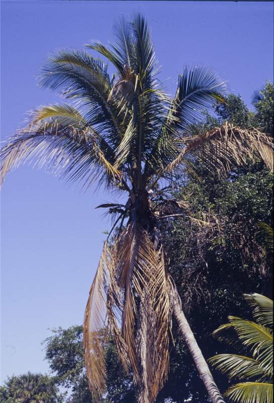 Figure 4. The leaves of dwarf cultivars of Cocos nucifera affected by lethal yellowing usually do not turn yellow. Photo by N. A. Harrison, University of Florida.
Lethal Yellowing
Figure 4. The leaves of dwarf cultivars of Cocos nucifera affected by lethal yellowing usually do not turn yellow. Photo by N. A. Harrison, University of Florida.
Figure 4. The leaves of dwarf cultivars of Cocos nucifera affected by lethal yellowing usually do not turn yellow. Photo by N. A. Harrison, University of Florida.
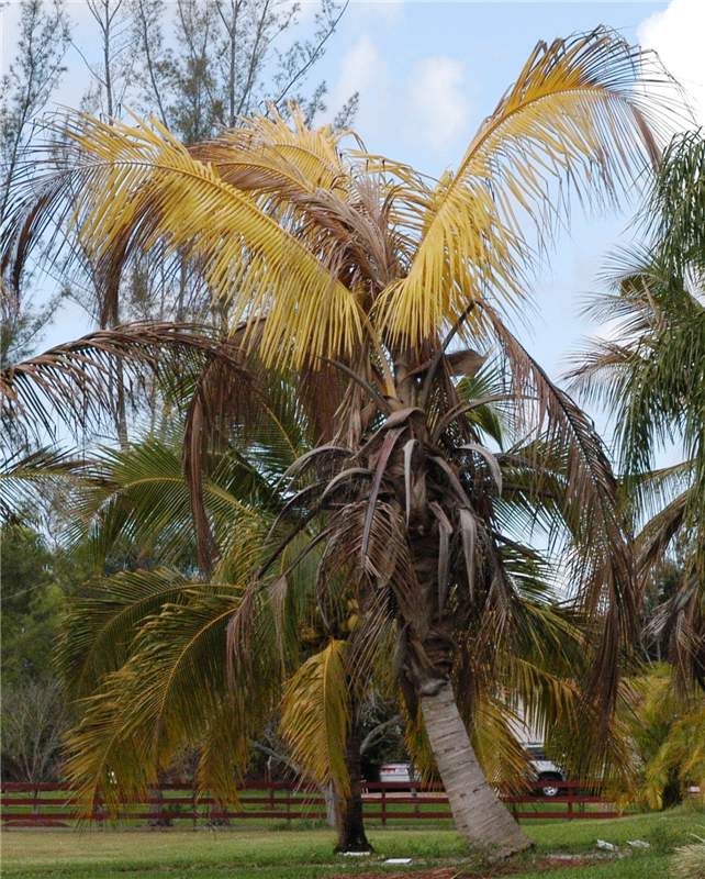 Figure 3. Cocos nucifera spear leaf is dying just as the last leaves are discoloring. Photo by M. L. Elliott.
Lethal Yellowing
Figure 3. Cocos nucifera spear leaf is dying just as the last leaves are discoloring. Photo by M. L. Elliott.
Figure 3. Cocos nucifera spear leaf is dying just as the last leaves are discoloring. Photo by M. L. Elliott.
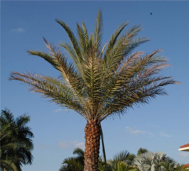 Figure 6. Spear leaf of this Phoenix sylvestris has collapsed and is hanging down out of the canopy on the right side of the trunk. Very few of the oldest leaves have discolored. Photo by M. L. Elliott.
Lethal Yellowing
Figure 6. Spear leaf of this Phoenix sylvestris has collapsed and is hanging down out of the canopy on the right side of the trunk. Very few of the oldest leaves have discolored. Photo by M. L. Elliott.
Figure 6. Spear leaf of this Phoenix sylvestris has collapsed and is hanging down out of the canopy on the right side of the trunk. Very few of the oldest leaves have discolored. Photo by M. L. Elliott.
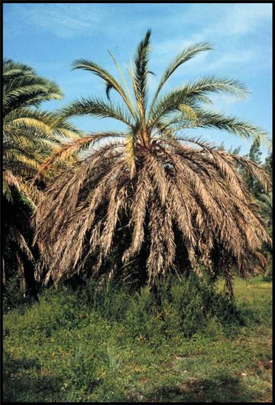 Figure 5. Leaves of a Phoenix canariensis with lethal yellowing do not turn yellow, but are various shades of reddish-brown to dark brown or gray. The spear leaf had died on this palm weeks prior to when this photo was taken. Photo by N. A. Harrison., University of Florida
Lethal Yellowing
Figure 5. Leaves of a Phoenix canariensis with lethal yellowing do not turn yellow, but are various shades of reddish-brown to dark brown or gray. The spear leaf had died on this palm weeks prior to when this photo was taken. Photo by N. A. Harrison., University of Florida
Figure 5. Leaves of a Phoenix canariensis with lethal yellowing do not turn yellow, but are various shades of reddish-brown to dark brown or gray. The spear leaf had died on this palm weeks prior to when this photo was taken. Photo by N. A. Harrison., University of Florida
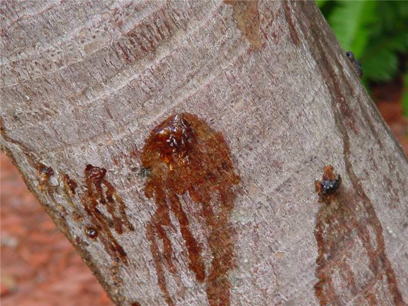
Lightning Injury
Figure 1. Trunk bleeding caused by lightning. Photo by T.K. Broschat.
Figure 1. Trunk bleeding caused by lightning. Photo by T.K. Broschat.
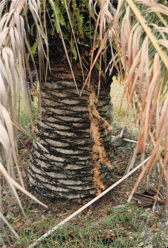
Lightning Injury
Figure 2. Trunk splitting in Phoenix canariensis caused by lightning. Photo by T.K. Broschat.
Figure 2. Trunk splitting in Phoenix canariensis caused by lightning. Photo by T.K. Broschat.
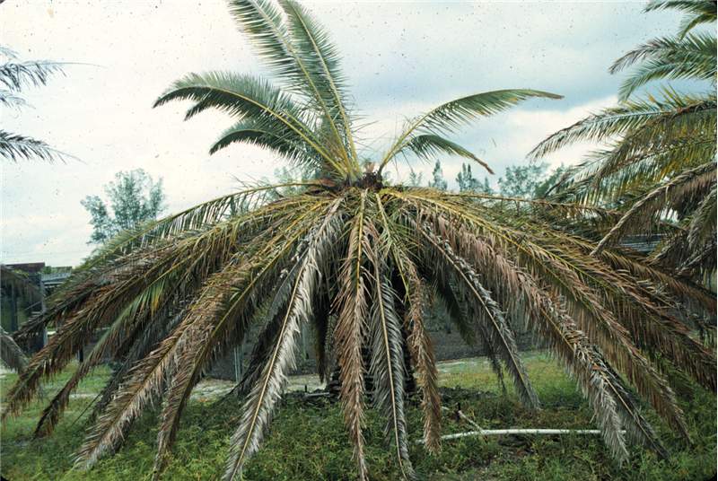
Lightning Injury
Figure 3. Sudden crown collapse in Phoenix canariensis caused by lightning. Photo by T.K. Broschat.
Figure 3. Sudden crown collapse in Phoenix canariensis caused by lightning. Photo by T.K. Broschat.
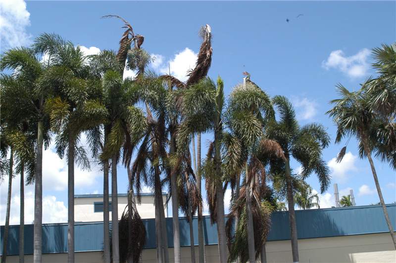 Figure 4. Collapse of twelve Veitchia mcdanielsii and Wodyetia bifurcata due to single lightning strike. Photo by T.K. Broschat.
Lightning Injury
Figure 4. Collapse of twelve Veitchia mcdanielsii and Wodyetia bifurcata due to single lightning strike. Photo by T.K. Broschat.
Figure 4. Collapse of twelve Veitchia mcdanielsii and Wodyetia bifurcata due to single lightning strike. Photo by T.K. Broschat.
