
Gallery
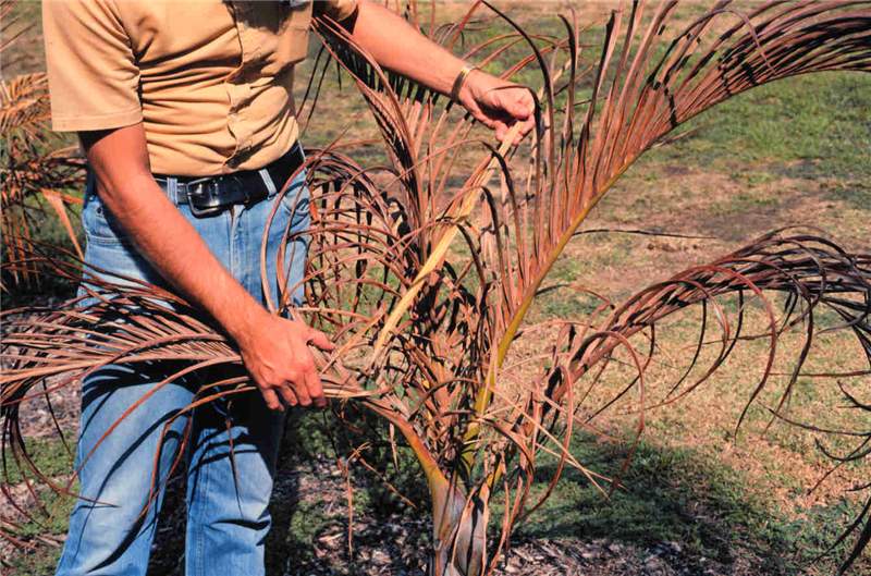 Figure 4. Basal spear leaf rot on Hyophorbe verschafeltii caused by secondary bacteria and fungi following freeze damage to the base of the spear leaf. Photo by T.K. Broschat.
Cold Damage
Figure 4. Basal spear leaf rot on Hyophorbe verschafeltii caused by secondary bacteria and fungi following freeze damage to the base of the spear leaf. Photo by T.K. Broschat.
Figure 4. Basal spear leaf rot on Hyophorbe verschafeltii caused by secondary bacteria and fungi following freeze damage to the base of the spear leaf. Photo by T.K. Broschat.
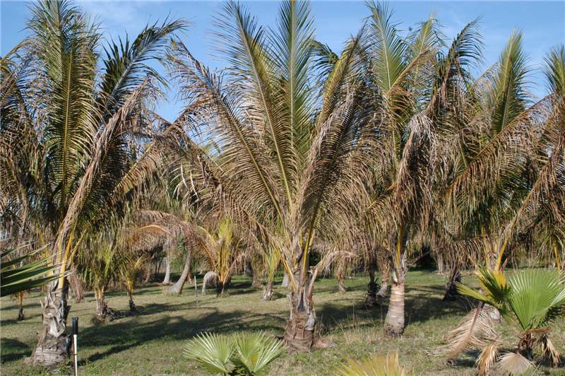 Figure 3. Chilling injury on Cocos nucifera showing the distribution of necrosis within and among leaves. Photo by T.K. Broschat.
Cold Damage
Figure 3. Chilling injury on Cocos nucifera showing the distribution of necrosis within and among leaves. Photo by T.K. Broschat.
Figure 3. Chilling injury on Cocos nucifera showing the distribution of necrosis within and among leaves. Photo by T.K. Broschat.
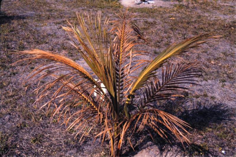 Figure 5. Wilting of living leaf tissue of Cocos nucifera following a freeze. Photo by T.K. Broschat.
Cold Damage
Figure 5. Wilting of living leaf tissue of Cocos nucifera following a freeze. Photo by T.K. Broschat.
Figure 5. Wilting of living leaf tissue of Cocos nucifera following a freeze. Photo by T.K. Broschat.
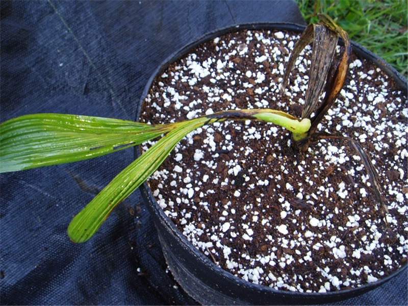 Figure 1. This is an example of post-emergence damping-off. Photo courtesy of University of Florida/IFAS.
Damping-off
Figure 1. This is an example of post-emergence damping-off. Photo courtesy of University of Florida/IFAS.
Figure 1. This is an example of post-emergence damping-off. Photo courtesy of University of Florida/IFAS.
 Figure 1. The raised black structures on this leaf are stromata, a mixture of host tissue and mycelia of Phaeochoropsis neowashingtoniae . Photo by A. J. Downer, University of California.
Diamond Scale
Figure 1. The raised black structures on this leaf are stromata, a mixture of host tissue and mycelia of Phaeochoropsis neowashingtoniae. Photo by A. J. Downer, University of California.
Figure 1. The raised black structures on this leaf are stromata, a mixture of host tissue and mycelia of Phaeochoropsis neowashingtoniae. Photo by A. J. Downer, University of California.
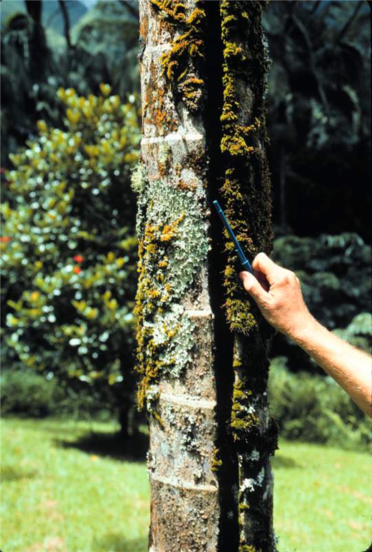 Figure 1. Trunk splitting in Archontophoenix alexandrae caused by excessive water uptake. Photo by T.K. Broschat.
Excessive Water Uptake
Figure 1. Trunk splitting in Archontophoenix alexandrae caused by excessive water uptake. Photo by T.K. Broschat.
Figure 1. Trunk splitting in Archontophoenix alexandrae caused by excessive water uptake. Photo by T.K. Broschat.
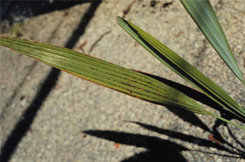 Figure 1. Foliar injury caused by a copper sulfate foliar spray on Acoelorrhaphe wrightii seedling. Photo by T.K. Broschat
Foliar Spray Toxicity
Figure 1. Foliar injury caused by a copper sulfate foliar spray on Acoelorrhaphe wrightii seedling. Photo by T.K. Broschat
Figure 1. Foliar injury caused by a copper sulfate foliar spray on Acoelorrhaphe wrightii seedling. Photo by T.K. Broschat
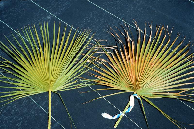 Figure 2. Foliar injury on Acoelorrhaphe wrightii caused by a copper sulfate spray (right). Untreated leaf on the left. Photo by T.K. Broschat
Foliar Spray Toxicity
Figure 2. Foliar injury on Acoelorrhaphe wrightii caused by a copper sulfate spray (right). Untreated leaf on the left. Photo by T.K. Broschat
Figure 2. Foliar injury on Acoelorrhaphe wrightii caused by a copper sulfate spray (right). Untreated leaf on the left. Photo by T.K. Broschat
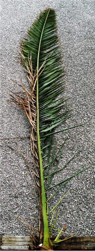 Figure 1. A Phoenix canariensis leaf with one side exhibiting necrotic leaflets and a brown stripe on the petiole and rachis due to Fusarium wilt. Photo by M. L. Elliott.
Fusarium Wilt of Canary Island Date Palm
Figure 1. A Phoenix canariensis leaf with one side exhibiting necrotic leaflets and a brown stripe on the petiole and rachis due to Fusarium wilt. Photo by M. L. Elliott.
Figure 1. A Phoenix canariensis leaf with one side exhibiting necrotic leaflets and a brown stripe on the petiole and rachis due to Fusarium wilt. Photo by M. L. Elliott.
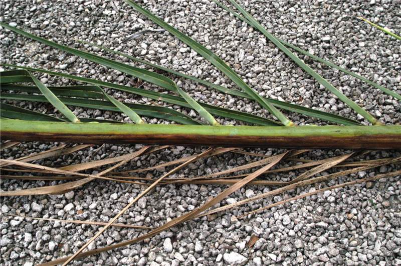 Figure 2. Close-up of Phoenix canariensis leaf with one side exhibiting necrotic leaflets and a brown stripe on the petiole and rachis due to Fusarium wilt. Photo by M. L. Elliott.
Fusarium Wilt of Canary Island Date Palm
Figure 2. Close-up of Phoenix canariensis leaf with one side exhibiting necrotic leaflets and a brown stripe on the petiole and rachis due to Fusarium wilt. Photo by M. L. Elliott.
Figure 2. Close-up of Phoenix canariensis leaf with one side exhibiting necrotic leaflets and a brown stripe on the petiole and rachis due to Fusarium wilt. Photo by M. L. Elliott.
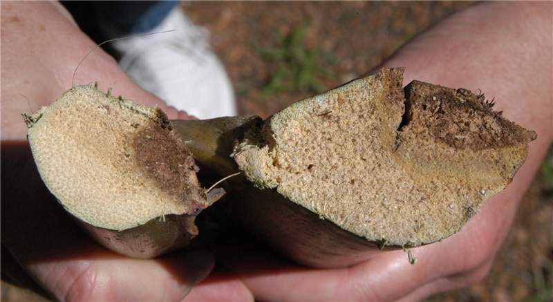 Figure 3. Cross-section of a Phoenix canariensis rachis illustrating the internal discoloration due to Fusarium wilt. Photo by T. K. Broschat.
Fusarium Wilt of Canary Island Date Palm
Figure 3. Cross-section of a Phoenix canariensis rachis illustrating the internal discoloration due to Fusarium wilt. Photo by T. K. Broschat.
Figure 3. Cross-section of a Phoenix canariensis rachis illustrating the internal discoloration due to Fusarium wilt. Photo by T. K. Broschat.
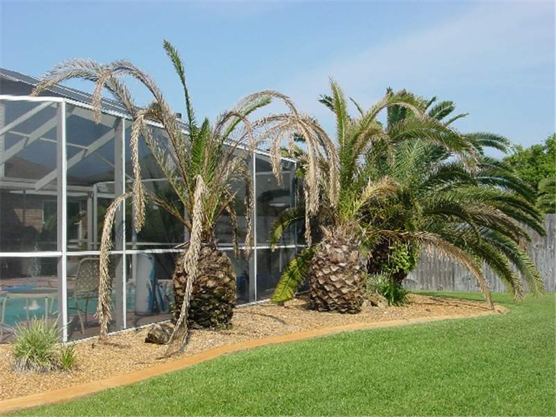 Figure 4. Phoenix canariensis in various stages of decline due to Fusarium wilt. Photo courtesy of University of Florida/IFAS.
Fusarium Wilt of Canary Island Date Palm
Figure 4. Phoenix canariensis in various stages of decline due to Fusarium wilt. Photo courtesy of University of Florida/IFAS.
Figure 4. Phoenix canariensis in various stages of decline due to Fusarium wilt. Photo courtesy of University of Florida/IFAS.
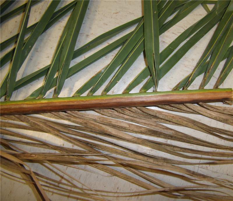 Figure 2. Leaf of Syagrus romanzoffiana with necrotic leaflets on one side and healthy leaflets on the other side of the rachis due to Fusarium wilt. Photo by M. L. Elliott
Fusarium Wilt of Queen and Mexican Fan Palms
Figure 2. Leaf of Syagrus romanzoffiana with necrotic leaflets on one side and healthy leaflets on the other side of the rachis due to Fusarium wilt. Photo by M. L. Elliott
Figure 2. Leaf of Syagrus romanzoffiana with necrotic leaflets on one side and healthy leaflets on the other side of the rachis due to Fusarium wilt. Photo by M. L. Elliott
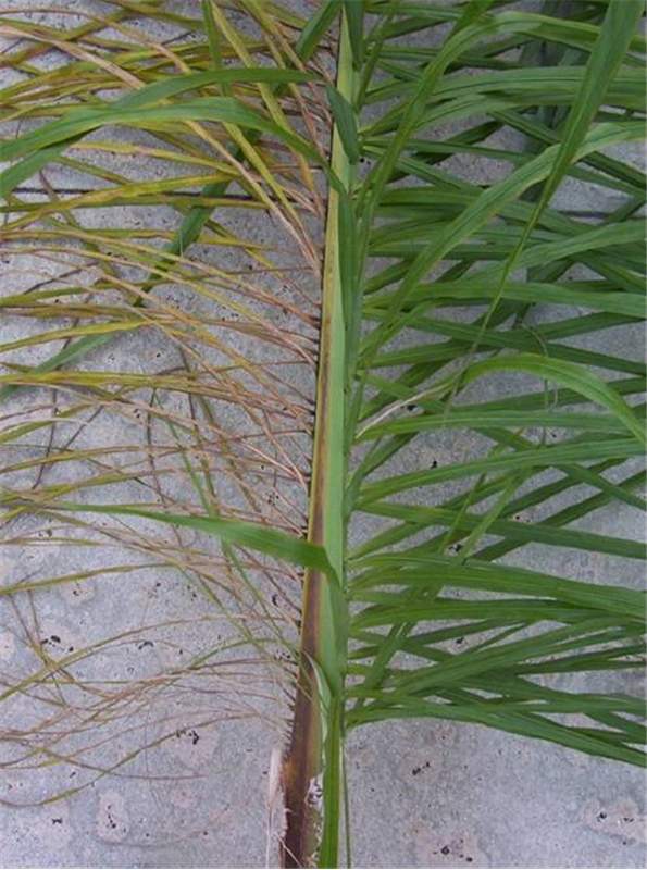 Figure 1. Initial symptoms on Syagrus romanzoffiana . Only a few leaflets on one side of the rachis have died. Note the reddish-brown stripe on the rachis. Photo by M. L. Elliott.
Fusarium Wilt of Queen and Mexican Fan Palms
Figure 1. Initial symptoms on Syagrus romanzoffiana. Only a few leaflets on one side of the rachis have died. Note the reddish-brown stripe on the rachis. Photo by M. L. Elliott.
Figure 1. Initial symptoms on Syagrus romanzoffiana. Only a few leaflets on one side of the rachis have died. Note the reddish-brown stripe on the rachis. Photo by M. L. Elliott.
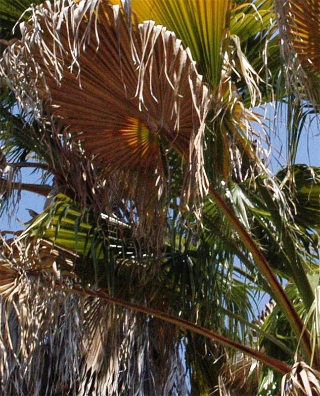 Figure 4. These dying Washingtonia robusta leaves have a brown stripe on the petiole due to Fusarium wilt. Photo by M. L. Elliott.
Fusarium Wilt of Queen and Mexican Fan Palms
Figure 4. These dying Washingtonia robusta leaves have a brown stripe on the petiole due to Fusarium wilt. Photo by M. L. Elliott.
Figure 4. These dying Washingtonia robusta leaves have a brown stripe on the petiole due to Fusarium wilt. Photo by M. L. Elliott.
 Figure 3. A cross-section of a Syagrus romanzoffiana rachis illustrating the internal discoloration due to Fusarium Wilt. Photo by T. K. Broschat.
Fusarium Wilt of Queen and Mexican Fan Palms
Figure 3. A cross-section of a Syagrus romanzoffiana rachis illustrating the internal discoloration due to Fusarium Wilt. Photo by T. K. Broschat.
Figure 3. A cross-section of a Syagrus romanzoffiana rachis illustrating the internal discoloration due to Fusarium Wilt. Photo by T. K. Broschat.
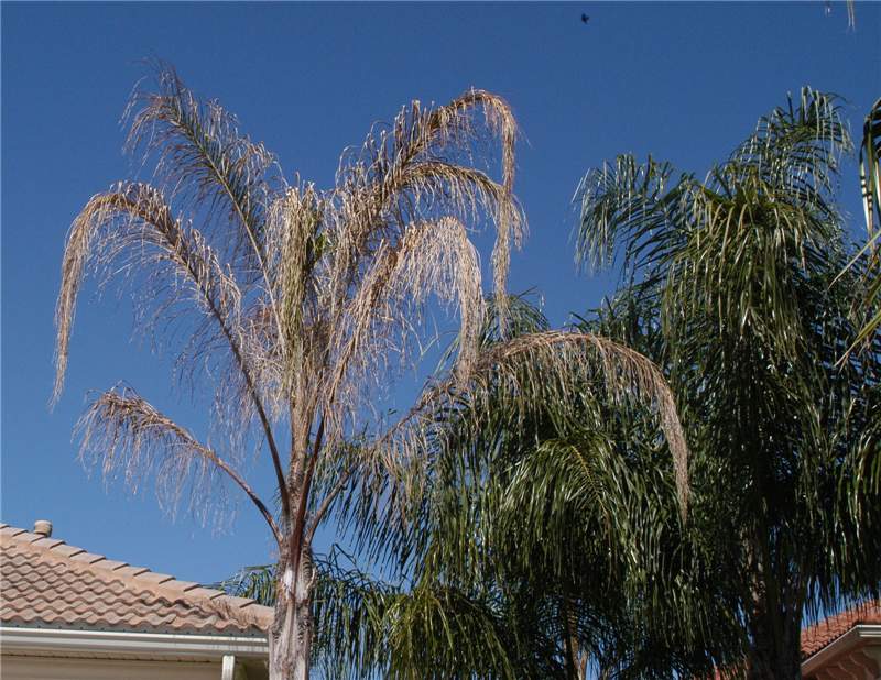 Figure 6. A nearly dead Syagrus romanzoffiana due to Fusarium wilt next to healthy palms. The palm leaves turn necrotic but do not break or collapse around the trunk. Photo by M. L. Elliott.
Fusarium Wilt of Queen and Mexican Fan Palms
Figure 6. A nearly dead Syagrus romanzoffiana due to Fusarium wilt next to healthy palms. The palm leaves turn necrotic but do not break or collapse around the trunk. Photo by M. L. Elliott.
Figure 6. A nearly dead Syagrus romanzoffiana due to Fusarium wilt next to healthy palms. The palm leaves turn necrotic but do not break or collapse around the trunk. Photo by M. L. Elliott.
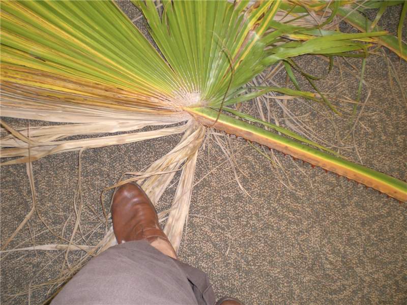 Figure 5. Washingtonia robusta leaf with one-sided discoloration and death of leaf segments due to Fusarium wilt. Note the brown stripe on the petiole is on the same side as the necrotic leaf segments. Photo courtesy of University of Florida/IFAS.
Fusarium Wilt of Queen and Mexican Fan Palms
Figure 5. Washingtonia robusta leaf with one-sided discoloration and death of leaf segments due to Fusarium wilt. Note the brown stripe on the petiole is on the same side as the necrotic leaf segments. Photo courtesy of University of Florida/IFAS.
Figure 5. Washingtonia robusta leaf with one-sided discoloration and death of leaf segments due to Fusarium wilt. Note the brown stripe on the petiole is on the same side as the necrotic leaf segments. Photo courtesy of University of Florida/IFAS.
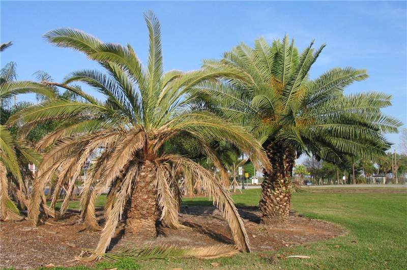 Figure 2. The Phoenix canariensis on the left is exhibiting premature death of the oldest leaves due to Ganoderma butt rot. This is in contrast to the healthy palm on the right. Photo by B. Dick, City of Lakeland, Florida.
Ganoderma Butt Rot
Figure 2. The Phoenix canariensis on the left is exhibiting premature death of the oldest leaves due to Ganoderma butt rot. This is in contrast to the healthy palm on the right. Photo by B. Dick, City of Lakeland, Florida.
Figure 2. The Phoenix canariensis on the left is exhibiting premature death of the oldest leaves due to Ganoderma butt rot. This is in contrast to the healthy palm on the right. Photo by B. Dick, City of Lakeland, Florida.
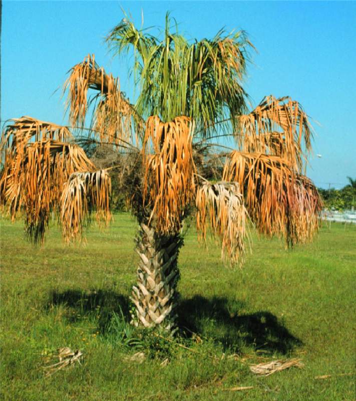 Figure 1. Sabal palmetto exhibiting an overall wilted canopy and premature death of the oldest leaves due to Ganoderma butt rot. Photo by M. L. Elliott.
Ganoderma Butt Rot
Figure 1. Sabal palmetto exhibiting an overall wilted canopy and premature death of the oldest leaves due to Ganoderma butt rot. Photo by M. L. Elliott.
Figure 1. Sabal palmetto exhibiting an overall wilted canopy and premature death of the oldest leaves due to Ganoderma butt rot. Photo by M. L. Elliott.
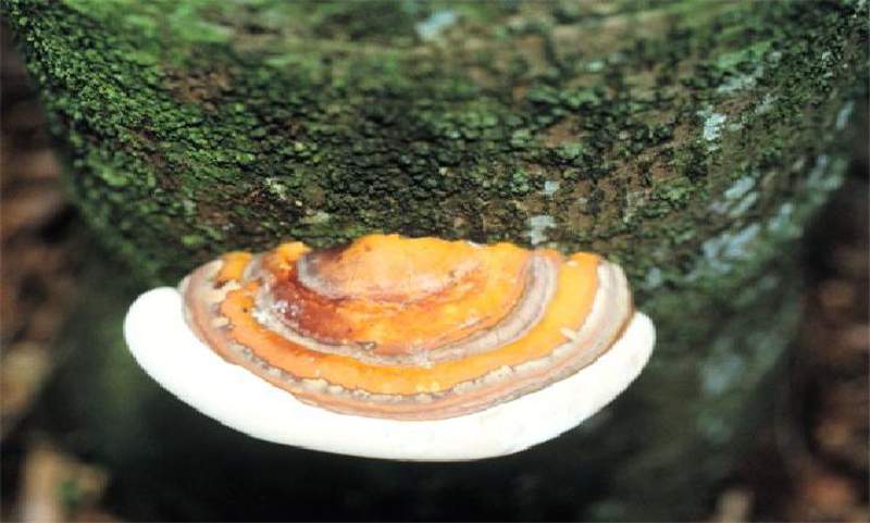 Figure 4. A developing basidiocarp of Ganoderma zonatum . Note the "zones" on the top portion of the basidiocarp, and that the fungus is directly attached to the palm trunk. Photo by M. L. Elliott.
Ganoderma Butt Rot
Figure 4. A developing basidiocarp of Ganoderma zonatum. Note the "zones" on the top portion of the basidiocarp, and that the fungus is directly attached to the palm trunk. Photo by M. L. Elliott.
Figure 4. A developing basidiocarp of Ganoderma zonatum. Note the "zones" on the top portion of the basidiocarp, and that the fungus is directly attached to the palm trunk. Photo by M. L. Elliott.
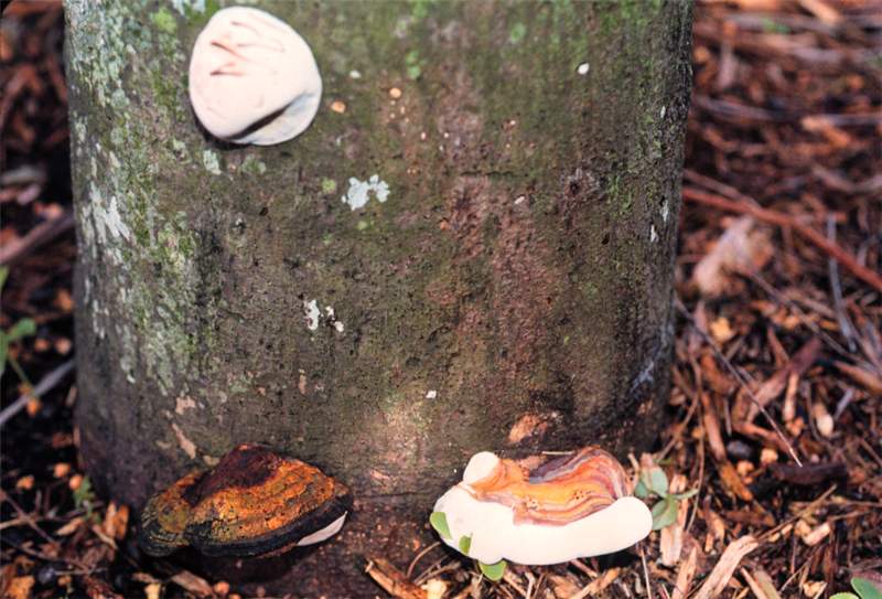 Figure 3. The white mass in the upper-left corner is an early stage of basidiocarp formation of Ganoderma zonatum . The lower-right corner illustrates the shelf or bracket structure of a developing basidiocarp. Because it is still white at the edges and underneath, it has not yet matured and has not released the basidiospores. The lower-left corner illustrates an old, decaying basidiocarp. Photo by M. L. Elliott.
Ganoderma Butt Rot
Figure 3. The white mass in the upper-left corner is an early stage of basidiocarp formation of Ganoderma zonatum. The lower-right corner illustrates the shelf or bracket structure of a developing basidiocarp. Because it is still white at the edges and underneath, it has not yet matured and has not released the basidiospores. The lower-left corner illustrates an old, decaying basidiocarp. Photo by M. L. Elliott.
Figure 3. The white mass in the upper-left corner is an early stage of basidiocarp formation of Ganoderma zonatum. The lower-right corner illustrates the shelf or bracket structure of a developing basidiocarp. Because it is still white at the edges and underneath, it has not yet matured and has not released the basidiospores. The lower-left corner illustrates an old, decaying basidiocarp. Photo by M. L. Elliott.
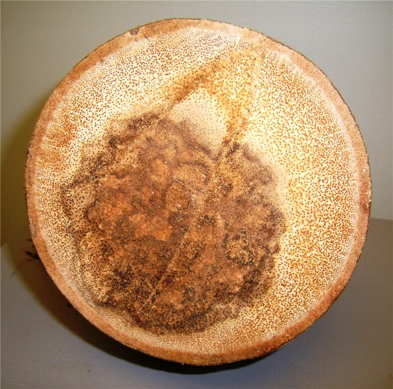 Figure 6. A cross-section through a Cocothrinax sp. trunk with Ganoderma butt rot. The darkened area in the center is a symptom of the trunk rot. Photo by M. L. Elliott.
Ganoderma Butt Rot
Figure 6. A cross-section through a Cocothrinax sp. trunk with Ganoderma butt rot. The darkened area in the center is a symptom of the trunk rot. Photo by M. L. Elliott.
Figure 6. A cross-section through a Cocothrinax sp. trunk with Ganoderma butt rot. The darkened area in the center is a symptom of the trunk rot. Photo by M. L. Elliott.
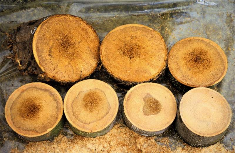 Figure 5. A series of cross-sections of a Ganoderma butt rot affected palm trunk. The section in the upper-left corner is the lowest section (at the soil line). The section in the lower-right corner is the furthest section from the soil line. The remaining sections are in the correct sequence from the soil line upwards. Note how the discoloration due to degrading tissue remains centralized within the trunk, but decreases as the cross-section ascends from the soil line. Photo by M. L. Elliott.
Ganoderma Butt Rot
Figure 5. A series of cross-sections of a Ganoderma butt rot affected palm trunk. The section in the upper-left corner is the lowest section (at the soil line). The section in the lower-right corner is the furthest section from the soil line. The remaining sections are in the correct sequence from the soil line upwards. Note how the discoloration due to degrading tissue remains centralized within the trunk, but decreases as the cross-section ascends from the soil line. Photo by M. L. Elliott.
Figure 5. A series of cross-sections of a Ganoderma butt rot affected palm trunk. The section in the upper-left corner is the lowest section (at the soil line). The section in the lower-right corner is the furthest section from the soil line. The remaining sections are in the correct sequence from the soil line upwards. Note how the discoloration due to degrading tissue remains centralized within the trunk, but decreases as the cross-section ascends from the soil line. Photo by M. L. Elliott.
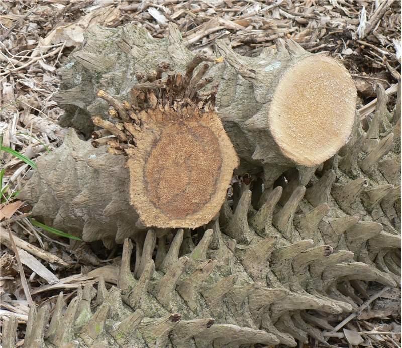 Figure 7. The section of Phoenix roebelenii on the left is the end of the trunk at the soil line and illustrates the typical trunk discoloration associated with Ganoderma butt rot. The section on the right was located further up the trunk and illustrates a trunk section unaffected by Ganoderma zonatum . Photo by M. L. Elliott.
Ganoderma Butt Rot
Figure 7. The section of Phoenix roebelenii on the left is the end of the trunk at the soil line and illustrates the typical trunk discoloration associated with Ganoderma butt rot. The section on the right was located further up the trunk and illustrates a trunk section unaffected by Ganoderma zonatum. Photo by M. L. Elliott.
Figure 7. The section of Phoenix roebelenii on the left is the end of the trunk at the soil line and illustrates the typical trunk discoloration associated with Ganoderma butt rot. The section on the right was located further up the trunk and illustrates a trunk section unaffected by Ganoderma zonatum. Photo by M. L. Elliott.
 Figure 2. Pink spore mass of Nalanthamala vermoesenii (= Gliocladium vermoesenii ) on inflorescence sheath. Photo by A. J. Downer, University of California.
Gliocladium Blight and Gliocladium Trunk Rot
Figure 2. Pink spore mass of Nalanthamala vermoesenii (=Gliocladium vermoesenii) on inflorescence sheath. Photo by A. J. Downer, University of California.
Figure 2. Pink spore mass of Nalanthamala vermoesenii (=Gliocladium vermoesenii) on inflorescence sheath. Photo by A. J. Downer, University of California.
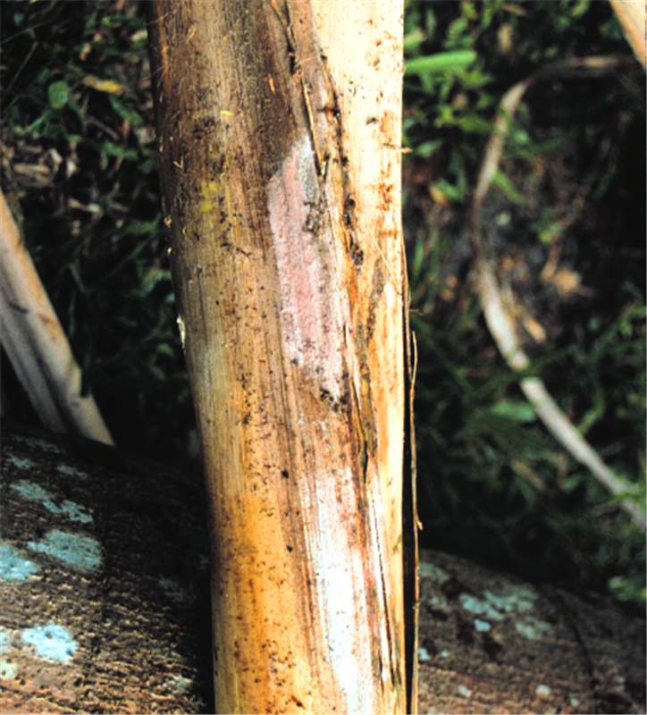 Figure 1. Pink spore mass of Nalanthamala vermoesenii (= Gliocladium vermoesenii ) on palm leaf base. Photo by T. K. Broschat.
Gliocladium Blight and Gliocladium Trunk Rot
Figure 1. Pink spore mass of Nalanthamala vermoesenii (=Gliocladium vermoesenii) on palm leaf base. Photo by T. K. Broschat.
Figure 1. Pink spore mass of Nalanthamala vermoesenii (=Gliocladium vermoesenii) on palm leaf base. Photo by T. K. Broschat.
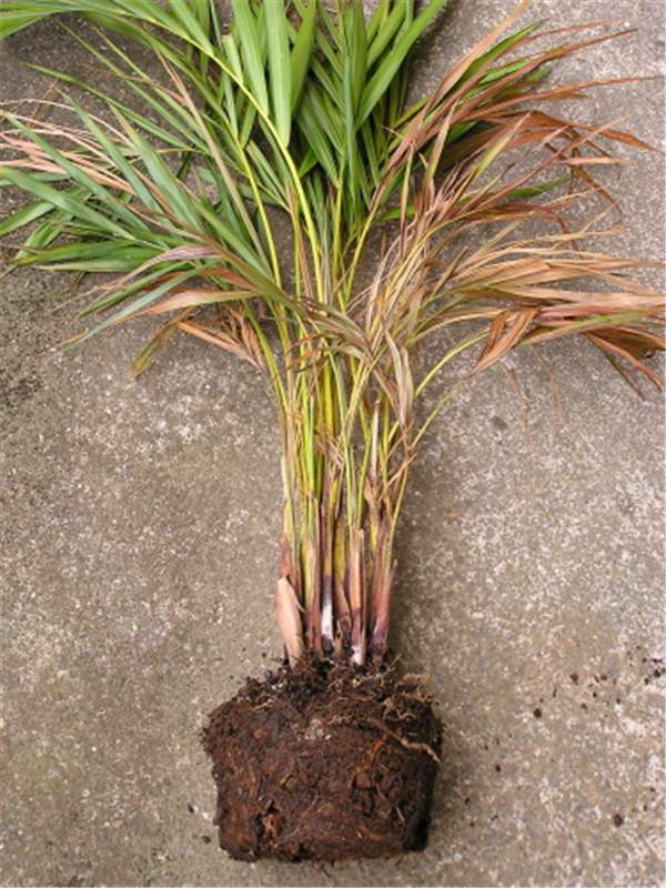 Figure 4. Leaf sheath rot of Dypsis lutescens caused by Gliocladium blight. Photo by S. Nelson, University of Hawaii at Manoa.
Gliocladium Blight and Gliocladium Trunk Rot
Figure 4. Leaf sheath rot of Dypsis lutescens caused by Gliocladium blight. Photo by S. Nelson, University of Hawaii at Manoa.
Figure 4. Leaf sheath rot of Dypsis lutescens caused by Gliocladium blight. Photo by S. Nelson, University of Hawaii at Manoa.
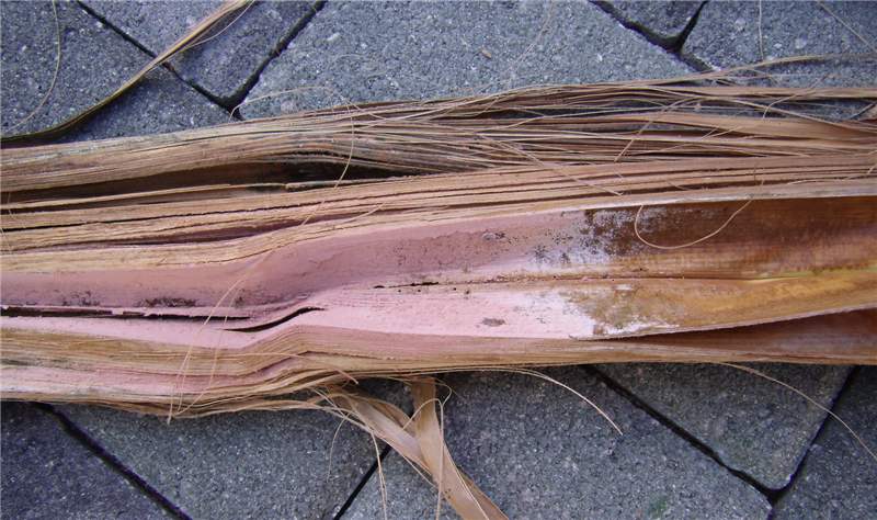 Figure 3. Pink spore mass of Nalanthamala vermoesenii (= Gliocladium vermoesenii ) on spear leaf. Photo by M. L. Elliott.
Gliocladium Blight and Gliocladium Trunk Rot
Figure 3. Pink spore mass of Nalanthamala vermoesenii (=Gliocladium vermoesenii) on spear leaf. Photo by M. L. Elliott.
Figure 3. Pink spore mass of Nalanthamala vermoesenii (=Gliocladium vermoesenii) on spear leaf. Photo by M. L. Elliott.
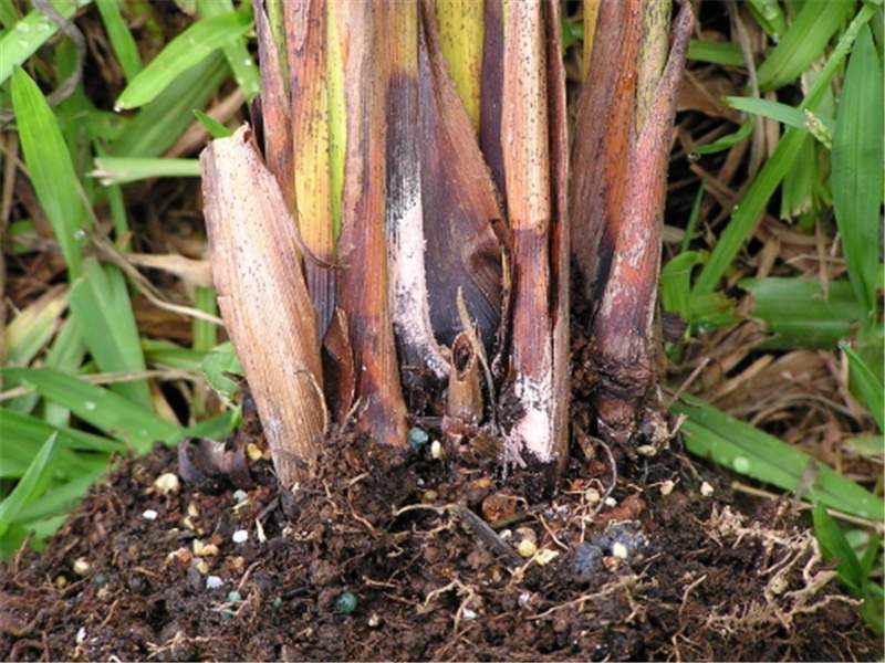 Figure 5. Close-up of leaf sheath rot from Fig. 4 illustrating pink spore mass of Nalanthamala vermoesenii (= Gliocladium vermoesenii ). Photo by S. Nelson, University of Hawaii at Manoa.
Gliocladium Blight and Gliocladium Trunk Rot
Figure 5. Close-up of leaf sheath rot from Fig. 4 illustrating pink spore mass of Nalanthamala vermoesenii (=Gliocladium vermoesenii). Photo by S. Nelson, University of Hawaii at Manoa.
Figure 5. Close-up of leaf sheath rot from Fig. 4 illustrating pink spore mass of Nalanthamala vermoesenii (=Gliocladium vermoesenii). Photo by S. Nelson, University of Hawaii at Manoa.
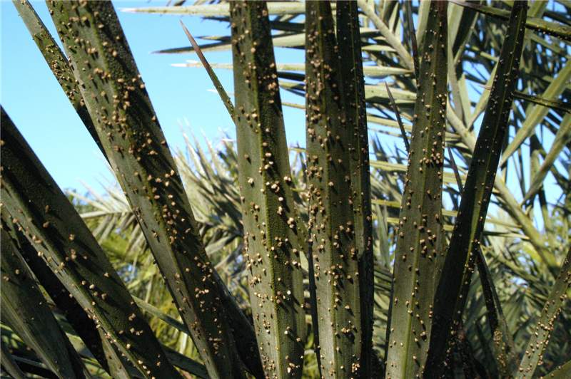 Figure 2. Palm leaflets with numerous sori and emerging spores. Note that this leaf does not have potassium deficiency. There are no leaf spot symptoms, only the sori of the fungus. Photo by M. L. Elliott.
Graphiola Leaf Spot
Figure 2. Palm leaflets with numerous sori and emerging spores. Note that this leaf does not have potassium deficiency. There are no leaf spot symptoms, only the sori of the fungus. Photo by M. L. Elliott.
Figure 2. Palm leaflets with numerous sori and emerging spores. Note that this leaf does not have potassium deficiency. There are no leaf spot symptoms, only the sori of the fungus. Photo by M. L. Elliott.
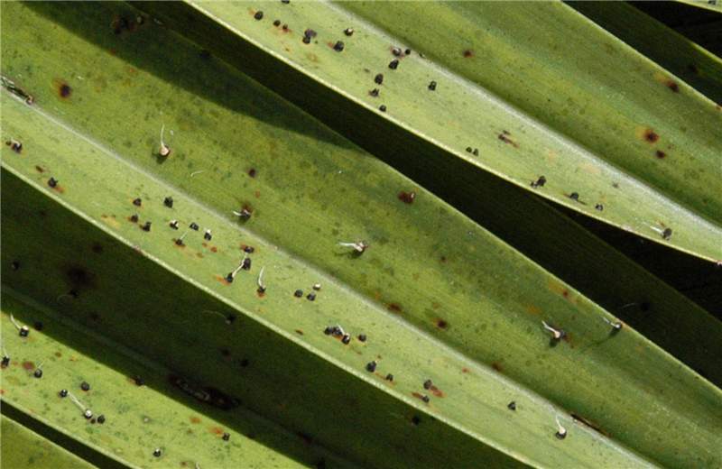 Figure 1. Sori of Graphiola phoenicis have erupted through the leaf epidermis. Some of the sori have filaments emerging from them. The mostly brown spots are not a symptom of Graphiola leaf spot, but a symptom of potassium deficiency. Photo by M. L. Elliott.
Graphiola Leaf Spot
Figure 1. Sori of Graphiola phoenicis have erupted through the leaf epidermis. Some of the sori have filaments emerging from them. The mostly brown spots are not a symptom of Graphiola leaf spot, but a symptom of potassium deficiency. Photo by M. L. Elliott.
Figure 1. Sori of Graphiola phoenicis have erupted through the leaf epidermis. Some of the sori have filaments emerging from them. The mostly brown spots are not a symptom of Graphiola leaf spot, but a symptom of potassium deficiency. Photo by M. L. Elliott.
