
Gallery
Image Type
Morphological Structure
Clades
 Phytophthora austrocedri (CPHST BL 5) colony of the ex-type grown for 7 days on (a) V8reg; Agar, (b) potato dextrose agar, and (c) malt extract agar; photo by Krysta Jennings and Leandra Knight, USDA-APHIS-PPQ
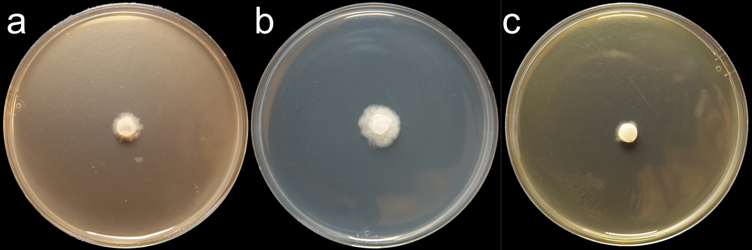
Phytophthora austrocedri (CPHST BL 5) colony of the ex-type grown for 7 days on (a) V8® Agar, (b) potato dextrose agar, and (c) malt extract agar; photo by Krysta Jennings and Leandra Knight, USDA-APHIS-PPQ
 Phytophthora austrocedri (CPHST BL 5, ex-type) asexual phase (a-d):nbsp;sporangia semipapillate (a-b), persistent (a), caducous (b); typical hyphal swellings (c, e); photos by Gloria Abad, USDA-APHIS-PPQ
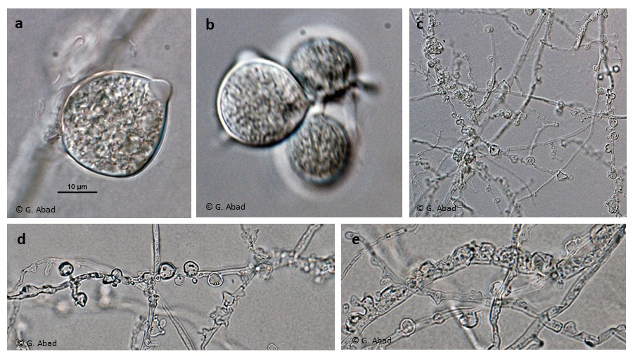
Phytophthora austrocedri (CPHST BL 5, ex-type) asexual phase (a-d): sporangia semipapillate (a-b), persistent (a), caducous (b); typical hyphal swellings (c, e); photos by Gloria Abad, USDA-APHIS-PPQ
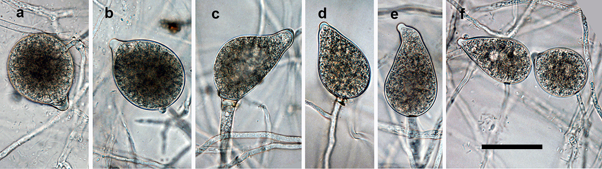
 Phytophthora austrocedri (CPHST BL 5, ex-type) sexual phase (a-d):nbsp;smooth-walled oogonia (a-c), sometimes with finger-like wall projections (d), amphigynous antheridia with one big cell (a-b amp; d), paragynous antheridium (c) also observed; photos by Gloria Abad, USDA-APHIS-PPQ
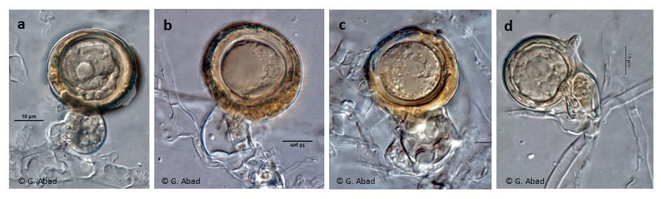
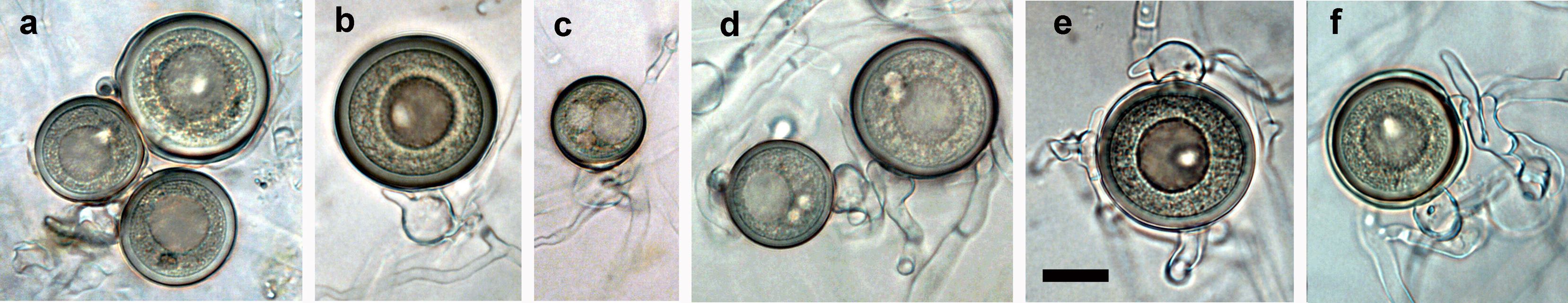
Phytophthora austrocedri (CPHST BL 5, ex-type) sexual phase (a-d): smooth-walled oogonia (a-c), sometimes with finger-like wall projections (d), amphigynous antheridia with one big cell (a-b & d), paragynous antheridium (c) also observed; photos by Gloria Abad, USDA-APHIS-PPQ
 Participants of the 7th IUFRO Phytophthora Working Group (November 10-14, 2014, Esquel, Patagonia, Argentina) attending presentations at Los Alerces National Park. Participants observed native forest sanitary problems, including the mortality of Austrocedrus chilensis affected with mal del cipres caused by Phytophthora austrocedri . Typical Argentinian asado in bottom right photo. photos by G. Abad
Participants of the 7th IUFRO Phytophthora Working Group (November 10-14, 2014, Esquel, Patagonia, Argentina) attending presentations at Los Alerces National Park. Participants observed native forest sanitary problems, including the mortality of Austrocedrus chilensis affected with mal del cipres caused by Phytophthora austrocedri. Typical Argentinian asado in bottom right photo. photos by G. Abad
 Panoramic view of Los Alerces National Park where the mortality of Austrocedrus chilensis affected with mal del cipreacute;s is observed. The disease was first detected in the area in 1935, and it was attributed to different causes. Many decades after, in 2005, the cause was attributed to Phytophthora. photo by Gloria Abad ndash; USDA-APHIS-PPQ
Panoramic view of Los Alerces National Park where the mortality of Austrocedrus chilensis affected with mal del ciprés is observed. The disease was first detected in the area in 1935, and it was attributed to different causes. Many decades after, in 2005, the cause was attributed to Phytophthora. photo by Gloria Abad – USDA-APHIS-PPQ
 Symptoms of ldquo;mal del cipreacute;srdquo; caused by Phytophthora autrocedri and taking action to prevent the spread of the disease into healthy areas. Views in Los Alercesnbsp;National Park, Patagonia Argentina during the 7th IUFRO Phytophthora; photos by Gloria Abad, USDA-APHIS-PPQnbsp;
Symptoms of “mal del ciprés” caused by Phytophthora autrocedri and taking action to prevent the spread of the disease into healthy areas. Views in Los Alerces National Park, Patagonia Argentina during the 7th IUFRO Phytophthora; photos by Gloria Abad, USDA-APHIS-PPQ
 Juniperus communis infected with Phytophthora austrocedri at Glenartney Perthshire Scotland (a, c), Whitewell Cairngorms National Park Scotland (b), Juniper Gill Yorkshire England (d), and Whitewell (e). Lesion extends up the trunk forming a cinnamon-colored tongue with the healthy, white phloem around it. Photos courtesy of April Amstrong and Sara Green at Centre for Forestry and Climate Change (CFCC), Forest Research, Northern Res. Station, Roslin, Scotland
Juniperus communis infected with Phytophthora austrocedri at Glenartney Perthshire Scotland (a, c), Whitewell Cairngorms National Park Scotland (b), Juniper Gill Yorkshire England (d), and Whitewell (e). Lesion extends up the trunk forming a cinnamon-colored tongue with the healthy, white phloem around it. Photos courtesy of April Amstrong and Sara Green at Centre for Forestry and Climate Change (CFCC), Forest Research, Northern Res. Station, Roslin, Scotland
.JPG) Phytophthora spp. in subclade 2b: portion of the seven-loci ML phylogeny featuring the type cultures of 212 described species (by T. Bourret). Notice the position o f P. aysenensis Ex-type CBS 146737 = Samp;T BL 56. Gloria Abad, USDA Samp;T.
Phytophthora spp. in subclade 2b: portion of the seven-loci ML phylogeny featuring the type cultures of 212 described species (by T. Bourret). Notice the position o f P. aysenensis Ex-type CBS 146737 = Samp;T BL 56. Gloria Abad, USDA Samp;T.
Phytophthora spp. in subclade 2b: portion of the seven-loci ML phylogeny featuring the type cultures of 212 described species (by T. Bourret). Notice the position of P. aysenensis Ex-type CBS 146737 = S&T BL 56. Gloria Abad, USDA S&T.
.JPG) Phytophthora spp. in subclade 2b: Morphological Tabular key (PDF) and Tabular key legends (PDF) in IDphy2 KEY SECTION. Notice the data of P. aysenensis Ex-type CBS 146737 = Samp;T BL 56 . Gloria Abad, USDA Samp;T.nbsp;
Phytophthora spp. in subclade 2b: Morphological Tabular key (PDF) and Tabular key legends (PDF) in IDphy2 KEY SECTION. Notice the data of P. aysenensis Ex-type CBS 146737 = Samp;T BL 56 . Gloria Abad, USDA Samp;T.nbsp;
Phytophthora spp. in subclade 2b: Morphological Tabular key (PDF) and Tabular key legends (PDF) in IDphy2 KEY SECTION. Notice the data of P. aysenensis Ex-type CBS 146737 = S&T BL 56. Gloria Abad, USDA S&T.
.JPG) Phytophthora spp. in subclade 6a: portion of the seven-loci ML phylogeny featuring the type cultures of 212 described species (by T. Bourret). Notice the position of P. balyanboodja Ex-type CBS 143058 = Samp;T BL 186 . Gloria Abad, USDA Samp;T.
Phytophthora spp. in subclade 6a: portion of the seven-loci ML phylogeny featuring the type cultures of 212 described species (by T. Bourret). Notice the position of P. balyanboodja Ex-type CBS 143058 = Samp;T BL 186 . Gloria Abad, USDA Samp;T.
Phytophthora spp. in subclade 6a: portion of the seven-loci ML phylogeny featuring the type cultures of 212 described species (by T. Bourret). Notice the position of P. balyanboodja Ex-type CBS 143058 = S&T BL 186. Gloria Abad, USDA S&T.
.JPG) Phytophthora spp. in subclade 6a: Morphological Tabular key (PDF) and Tabular key legends (PDF) in IDphy2 KEY SECTION. Notice the data of P. balyanboodja Ex-type CBS 143058 = Samp;T BL 186 . Gloria Abad, USDA Samp;T.nbsp;
Phytophthora spp. in subclade 6a: Morphological Tabular key (PDF) and Tabular key legends (PDF) in IDphy2 KEY SECTION. Notice the data of P. balyanboodja Ex-type CBS 143058 = Samp;T BL 186 . Gloria Abad, USDA Samp;T.nbsp;
Phytophthora spp. in subclade 6a: Morphological Tabular key (PDF) and Tabular key legends (PDF) in IDphy2 KEY SECTION. Notice the data of P. balyanboodja Ex-type CBS 143058 = S&T BL 186. Gloria Abad, USDA S&T.
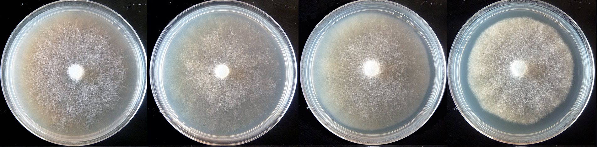
colony morphology after 5 d growth at 20ºC on CA, V8 agar, MEA, and PDA (from left to right)
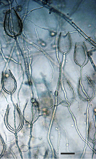
 sporangia typically ovoid and nonpapillate, although many had apical protrusions which continued to grow leading to direct germination of sporangia; scale bar = 25micro;m
sporangia typically ovoid and nonpapillate, although many had apical protrusions which continued to grow leading to direct germination of sporangia; scale bar = 25µm
.JPG) Phytophthora spp. in subclade 1c: portion of the seven-loci ML phylogeny featuring the type cultures of 212 described species (by T. Bourret). Notice the position of P. betacei Ex-type MFM-P8084 . Gloria Abad, USDA Samp;T.
Phytophthora spp. in subclade 1c: portion of the seven-loci ML phylogeny featuring the type cultures of 212 described species (by T. Bourret). Notice the position of P. betacei Ex-type MFM-P8084 . Gloria Abad, USDA Samp;T.
Phytophthora spp. in subclade 1c: portion of the seven-loci ML phylogeny featuring the type cultures of 212 described species (by T. Bourret). Notice the position of P. betacei Ex-type MFM-P8084. Gloria Abad, USDA S&T.
.JPG) Phytophthora spp. in subclade 1c: Morphological Tabular key (PDF) and Tabular key legends (PDF) in IDphy2 KEY SECTION. Notice the data of P. betacei Ex-type MFM-P8084 . Gloria Abad, USDA Samp;T. nbsp;
Phytophthora spp. in subclade 1c: Morphological Tabular key (PDF) and Tabular key legends (PDF) in IDphy2 KEY SECTION. Notice the data of P. betacei Ex-type MFM-P8084 . Gloria Abad, USDA Samp;T. nbsp;
Phytophthora spp. in subclade 1c: Morphological Tabular key (PDF) and Tabular key legends (PDF) in IDphy2 KEY SECTION. Notice the data of P. betacei Ex-type MFM-P8084. Gloria Abad, USDA S&T.
.JPG) Phytophthora spp. in subclade 6c: portion of the seven-loci ML phylogeny featuring the type cultures of 212 described species (by T. Bourret). Notice the position of P. bilorbang Ex-type CBS 131653 = Samp;T BL 101 . Gloria Abad, USDA Samp;T.
Phytophthora spp. in subclade 6c: portion of the seven-loci ML phylogeny featuring the type cultures of 212 described species (by T. Bourret). Notice the position of P. bilorbang Ex-type CBS 131653 = Samp;T BL 101 . Gloria Abad, USDA Samp;T.
Phytophthora spp. in subclade 6c: portion of the seven-loci ML phylogeny featuring the type cultures of 212 described species (by T. Bourret). Notice the position of P. bilorbang Ex-type CBS 131653 = S&T BL 101. Gloria Abad, USDA S&T.
.JPG) Phytophthora spp. in subclade 6c: Morphological Tabular key (PDF) and Tabular key legends (PDF) in IDphy2 KEY SECTION. Notice the data of P. bilorbang Ex-type CBS 131653 = Samp;T BL 101 . Gloria Abad, USDA Samp;T.
Phytophthora spp. in subclade 6c: Morphological Tabular key (PDF) and Tabular key legends (PDF) in IDphy2 KEY SECTION. Notice the data of P. bilorbang Ex-type CBS 131653 = Samp;T BL 101 . Gloria Abad, USDA Samp;T.
Phytophthora spp. in subclade 6c: Morphological Tabular key (PDF) and Tabular key legends (PDF) in IDphy2 KEY SECTION. Notice the data of P. bilorbang Ex-type CBS 131653 = S&T BL 101. Gloria Abad, USDA S&T.
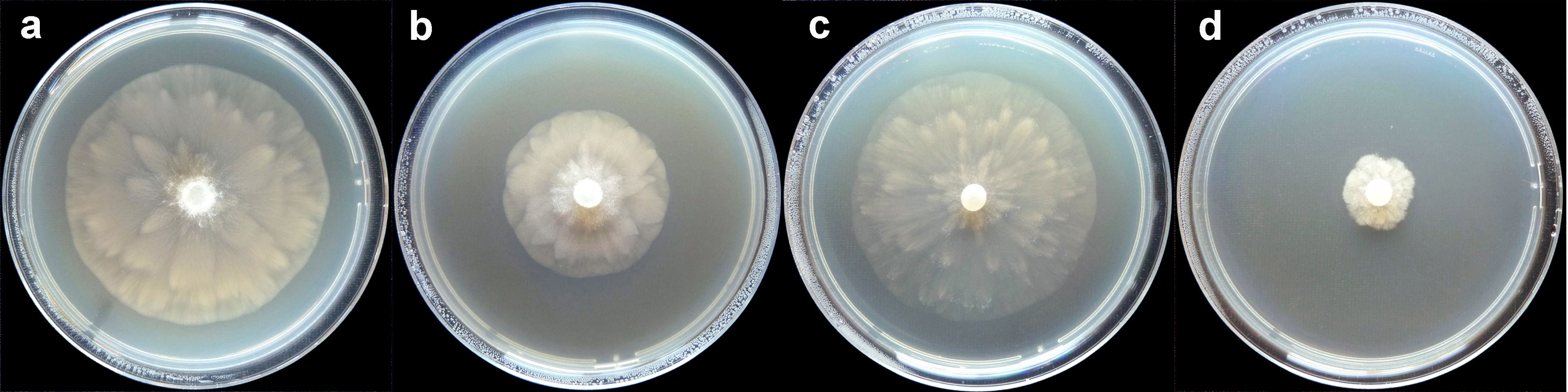 Phytophthora bilorbang nbsp;colonies of the ex-type grown for 7 days at 20deg;C on: (a) V8reg; agar (b) carrot agar (c) malt extract agar (d) potato-dextrose agar
Phytophthora bilorbang colonies of the ex-type grown for 7 days at 20°C on: (a) V8® agar (b) carrot agar (c) malt extract agar (d) potato-dextrose agar
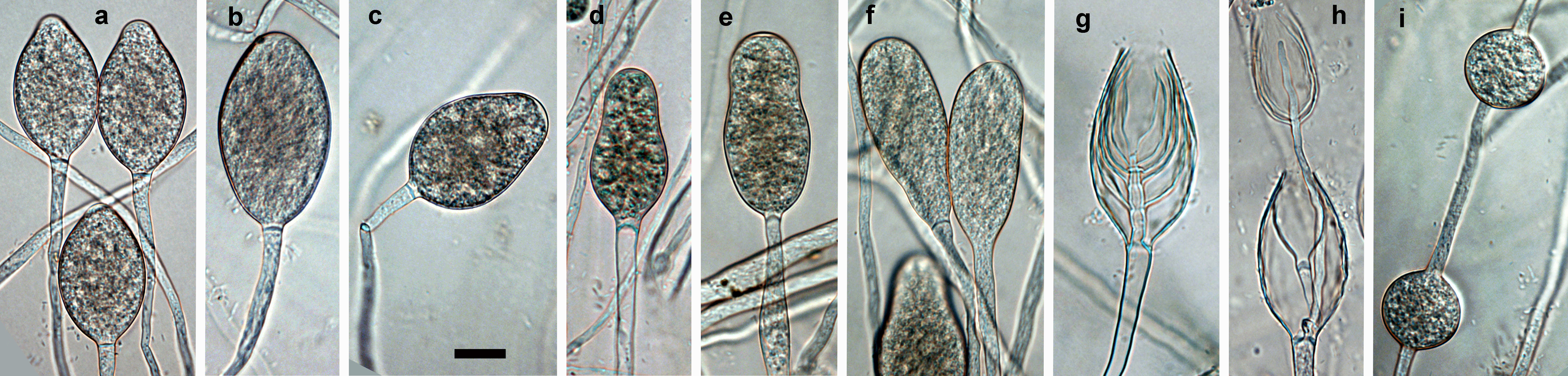 non-papillate sporangia formed on V8 agar flooded with soil extract; (a) limoniform (b) ellipsoid, (c) ovoid, (d) obpyriform (e) peanut shaped (f) club shaped (g) nested internal proliferation (h) an external proliferation arising from the internally proliferating sporangiophore, (i) catenulate, globose to subglobose hyphal swellings; scale bar = 25 micro;m
non-papillate sporangia formed on V8 agar flooded with soil extract; (a) limoniform (b) ellipsoid, (c) ovoid, (d) obpyriform (e) peanut shaped (f) club shaped (g) nested internal proliferation (h) an external proliferation arising from the internally proliferating sporangiophore, (i) catenulate, globose to subglobose hyphal swellings; scale bar = 25 µm
 (a) oospores with large ooplast bodies; (b) a pleorotic oospore and paragynous and spherical antheridium with a short projection; (c) oospores with two ooplast bodies; (d) oospores with two nuclei; (e-f) paragynous antheridia with short finger-like projections; scale bar = 25 mu;m
(a) oospores with large ooplast bodies; (b) a pleorotic oospore and paragynous and spherical antheridium with a short projection; (c) oospores with two ooplast bodies; (d) oospores with two nuclei; (e-f) paragynous antheridia with short finger-like projections; scale bar = 25 μm
.JPG) Phytophthora spp. in Clade 2e: portion of the seven-loci ML phylogeny featuring the type cultures of 212 described species (by T. Bourret). Notice the position of P. bishii Ex-type CBS 122081 = Samp;T BL 6 . Gloria Abad, USDA Samp;T.
Phytophthora spp. in Clade 2e: portion of the seven-loci ML phylogeny featuring the type cultures of 212 described species (by T. Bourret). Notice the position of P. bishii Ex-type CBS 122081 = Samp;T BL 6 . Gloria Abad, USDA Samp;T.
Phytophthora spp. in Clade 2e: portion of the seven-loci ML phylogeny featuring the type cultures of 212 described species (by T. Bourret). Notice the position of P. bishii Ex-type CBS 122081 = S&T BL 6. Gloria Abad, USDA S&T.
.JPG) Phytophthora spp. in Clade 2e: Morphological Tabular key (PDF) and Tabular key legends (PDF) in IDphy2 KEY SECTION. Notice the data of P. bishi i Ex-type CBS 122081 = Samp;T BL 6 . Gloria Abad, USDA Samp;T.
Phytophthora spp. in Clade 2e: Morphological Tabular key (PDF) and Tabular key legends (PDF) in IDphy2 KEY SECTION. Notice the data of P. bishi i Ex-type CBS 122081 = Samp;T BL 6 . Gloria Abad, USDA Samp;T.
Phytophthora spp. in Clade 2e: Morphological Tabular key (PDF) and Tabular key legends (PDF) in IDphy2 KEY SECTION. Notice the data of P. bishii Ex-type CBS 122081 = S&T BL 6. Gloria Abad, USDA S&T.
 Phytophthora bishii nbsp;(CPHST BL 6) colony of the ex-type grown for 7 days on (a) V8reg; Agar, (b) potato dextrose agar, and (c) malt extract agar; photo by Krysta Jennings and Leandra Knight, USDA-APHIS-PPQ
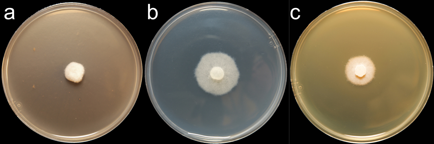
Phytophthora bishii (CPHST BL 6) colony of the ex-type grown for 7 days on (a) V8® Agar, (b) potato dextrose agar, and (c) malt extract agar; photo by Krysta Jennings and Leandra Knight, USDA-APHIS-PPQ
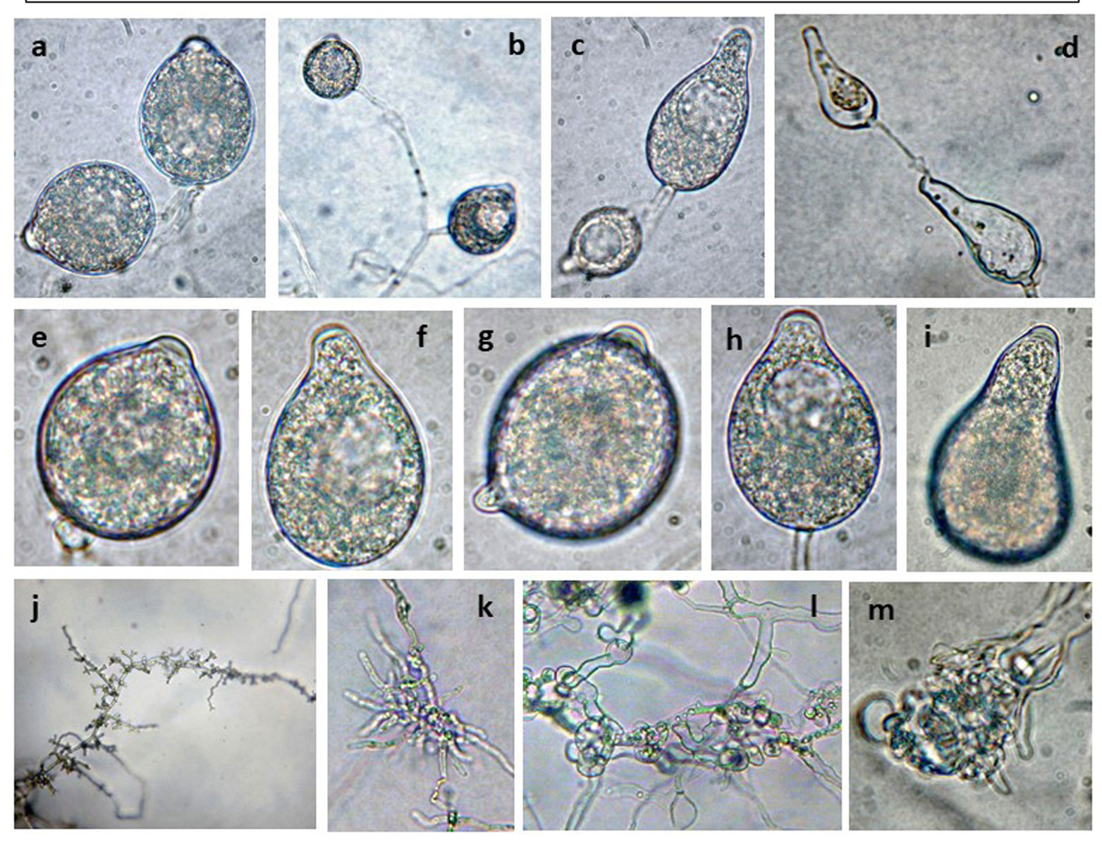 Phytophthora bishii (CPHST BL 6) asexual phase of the ex-type: (a-i) semipapillate persistent sporangia with different shapes, originated in simple sympodial sporangiophore (a-c) and (d) by external proliferation; (j-m) hyphal swellings; photos by G. Abad, USDA-APHIS-PPQ.
Phytophthora bishii (CPHST BL 6) asexual phase of the ex-type: (a-i) semipapillate persistent sporangia with different shapes, originated in simple sympodial sporangiophore (a-c) and (d) by external proliferation; (j-m) hyphal swellings; photos by G. Abad, USDA-APHIS-PPQ.
 Phytophthora bishii (CPHST BL 6) sexual phase of the ex-type (a-h): photos by G. Abad, USDA-APHIS-PPQ.
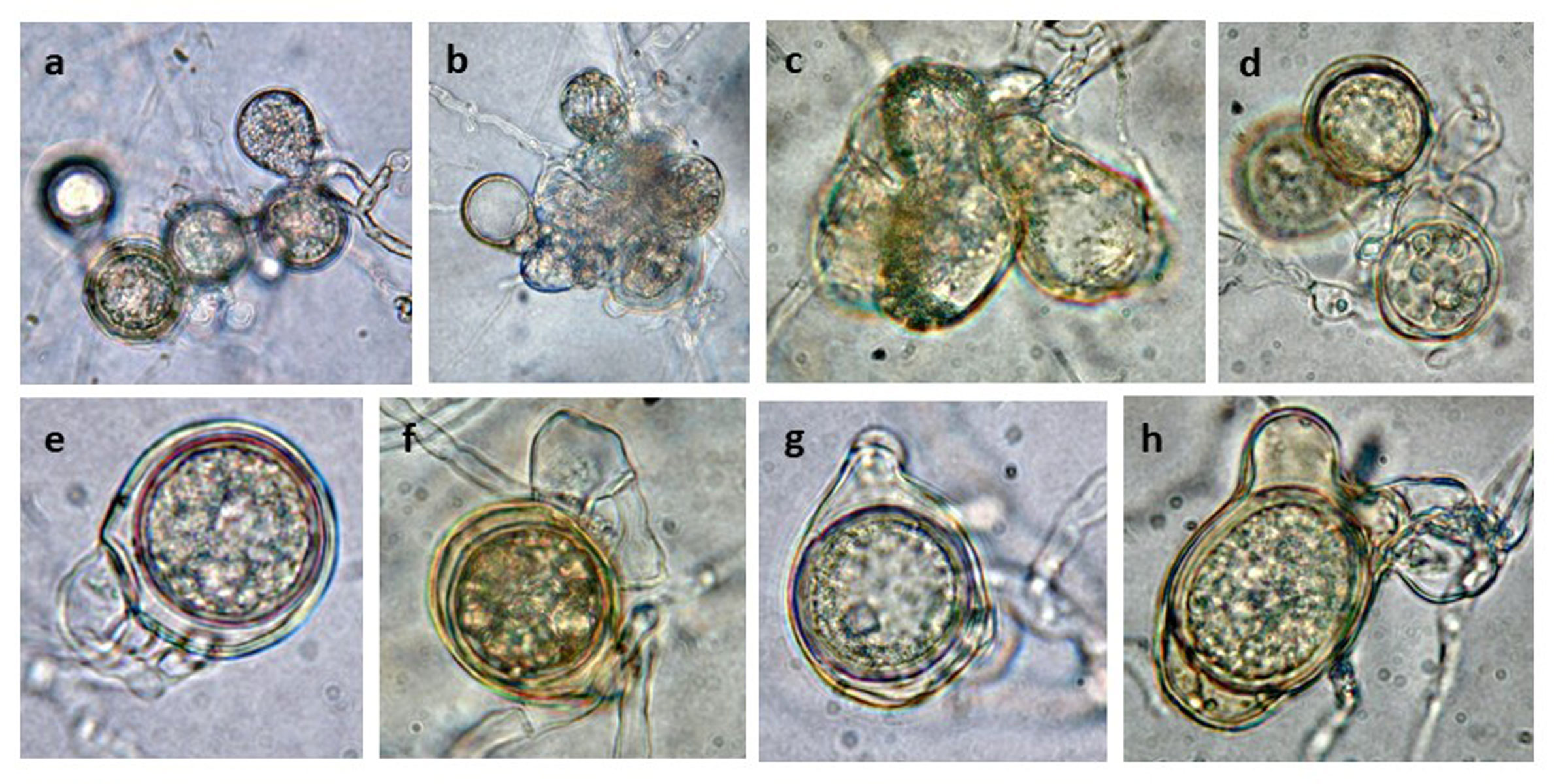
Phytophthora bishii (CPHST BL 6) sexual phase of the ex-type (a-h): photos by G. Abad, USDA-APHIS-PPQ.
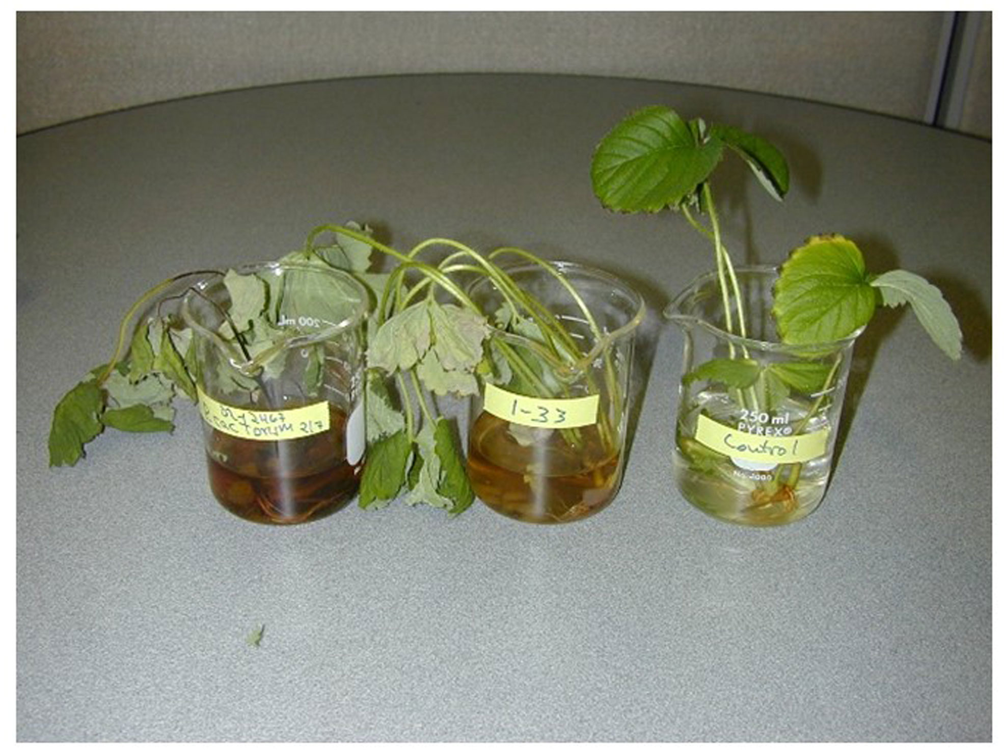 pathogenicity test of Phytophthora bishii (Abad 1-33) selected specimen (center) as compared with an isolate of P. cactorum nbsp;(Abad Ph338) (left) and healthy control (right); photo by Gloria Abad at the former Plant Pathogen Identification Laboratory (PPIL), Department of Plant Pathology, North Carolina State University,nbsp;USA (2001-2006)
pathogenicity test of Phytophthora bishii (Abad 1-33) selected specimen (center) as compared with an isolate of P. cactorum (Abad Ph338) (left) and healthy control (right); photo by Gloria Abad at the former Plant Pathogen Identification Laboratory (PPIL), Department of Plant Pathology, North Carolina State University, USA (2001-2006)
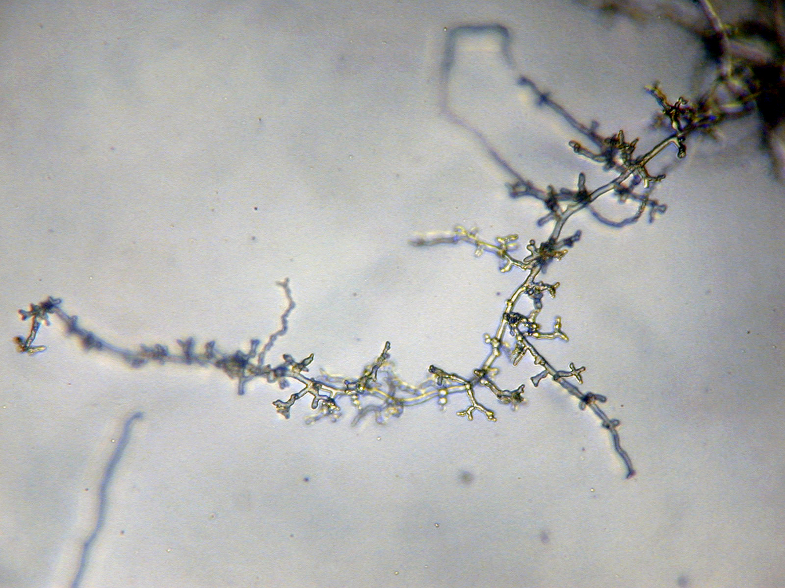 Phytophthora bishii nbsp;(CPHST BL 6) asexual phase of the ex-type:nbsp;hyphal swellings; photonbsp;by G. Abad, USDA-APHIS-PPQ.
Phytophthora bishii (CPHST BL 6) asexual phase of the ex-type: hyphal swellings; photo by G. Abad, USDA-APHIS-PPQ.
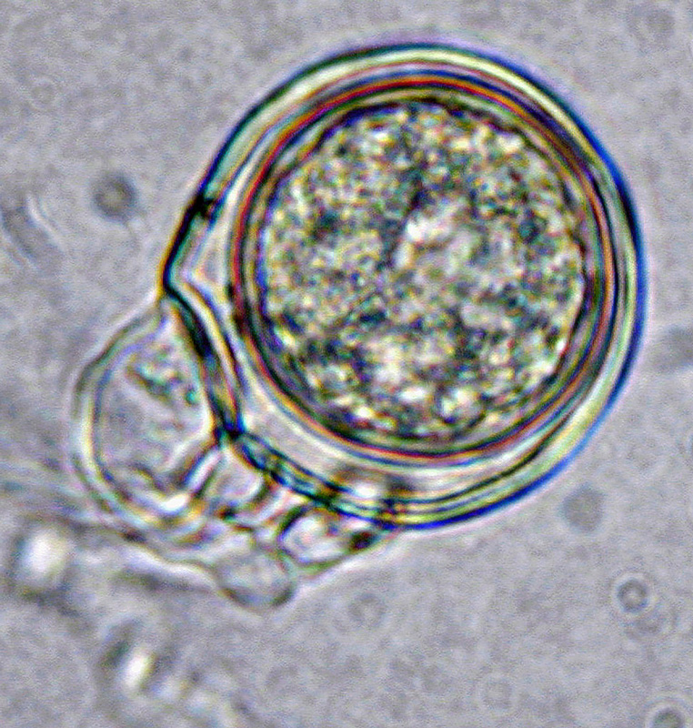 Phytophthora bishii nbsp;(CPHST BL 6) sexual phase of the ex-type: oospore with paragynousnbsp;antheridium;nbsp;photonbsp;by G. Abad, USDA-APHIS-PPQ.
Phytophthora bishii (CPHST BL 6) sexual phase of the ex-type: oospore with paragynous antheridium; photo by G. Abad, USDA-APHIS-PPQ.
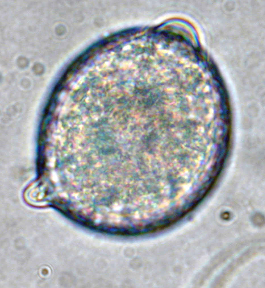
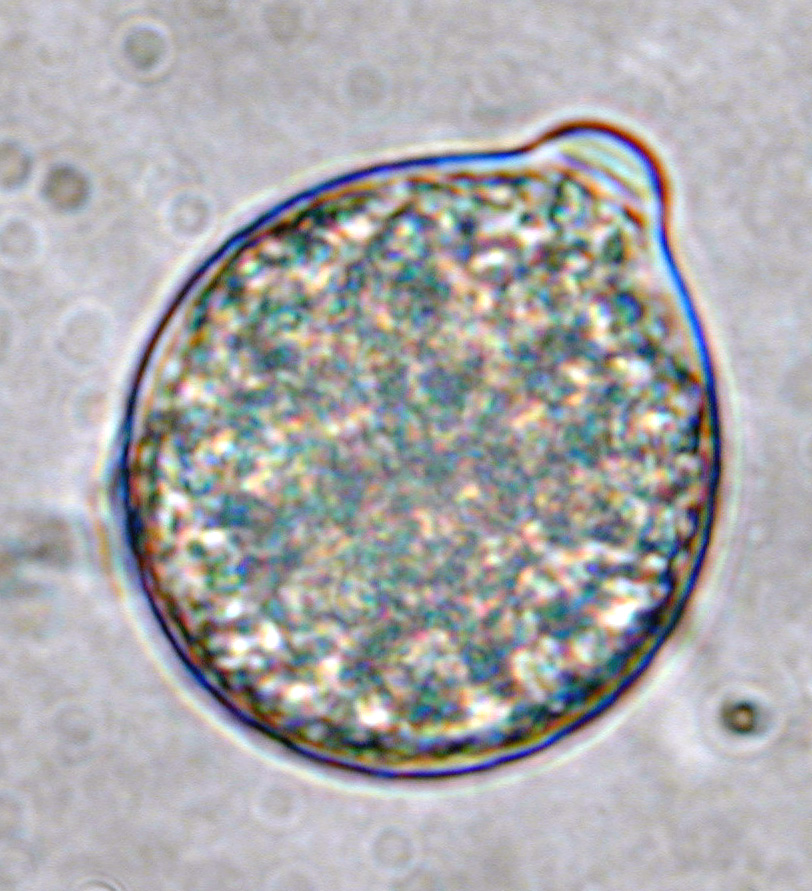 Phytophthora bishii (CPHST BL 6) asexual phase of the ex-type:nbsp;semipapillate persistent sporangium; photonbsp;by G. Abad, USDA-APHIS-PPQ.
Phytophthora bishii (CPHST BL 6) asexual phase of the ex-type: semipapillate persistent sporangium; photo by G. Abad, USDA-APHIS-PPQ.
 Phytophthora bishii nbsp;(CPHST BL 6) asexual phase of the ex-type: bipapillate persistent sporangium; photonbsp;by G. Abad, USDA-APHIS-PPQ.
Phytophthora bishii (CPHST BL 6) asexual phase of the ex-type: bipapillate persistent sporangium; photo by G. Abad, USDA-APHIS-PPQ.
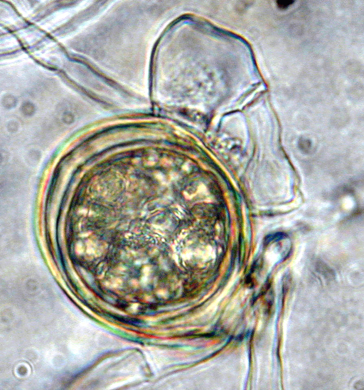 Phytophthora bishii nbsp;(CPHST BL 6) sexual phase of the ex-type: aplerotic oospore withnbsp;paragynousnbsp;antheridium;nbsp;photonbsp;by G. Abad, USDA-APHIS-PPQ.
Phytophthora bishii (CPHST BL 6) sexual phase of the ex-type: aplerotic oospore with paragynous antheridium; photo by G. Abad, USDA-APHIS-PPQ.
