
Gallery
Image Type
Morphological Structure
Clades
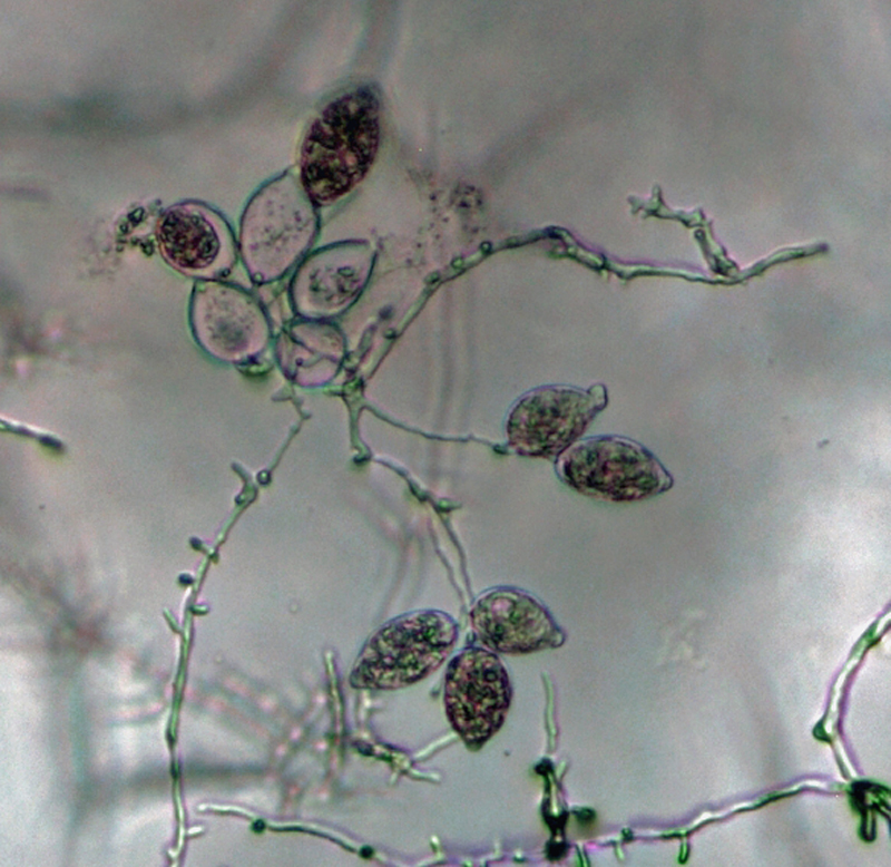 Phytophthora kernoviae nbsp;(CPHST BL 91 ex-type) asexual phase: sporangia produced in simple sympodial sporangiophores;nbsp;photonbsp;by Gloria Abad, USDA-APHIS-PPQ.
Phytophthora kernoviae (CPHST BL 91 ex-type) asexual phase: sporangia produced in simple sympodial sporangiophores; photo by Gloria Abad, USDA-APHIS-PPQ.
 Phytophthora kernoviae nbsp;(CPHST BL 91 ex-type) asexual phase:nbsp;sporangia produced in simple sympodial sporangiophores;nbsp;photonbsp;by Gloria Abad, USDA-APHIS-PPQ.
Phytophthora kernoviae (CPHST BL 91 ex-type) asexual phase: sporangia produced in simple sympodial sporangiophores; photo by Gloria Abad, USDA-APHIS-PPQ.
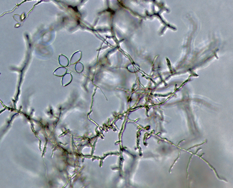
 Phytophthora kernoviae (selected specimen P10681) asexual phase: sporangia produced in close simple sympodial sporangiophores; photonbsp;by Gloria Abad, USDA-APHIS-PPQ.
Phytophthora kernoviae (selected specimen P10681) asexual phase: sporangia produced in close simple sympodial sporangiophores; photo by Gloria Abad, USDA-APHIS-PPQ.
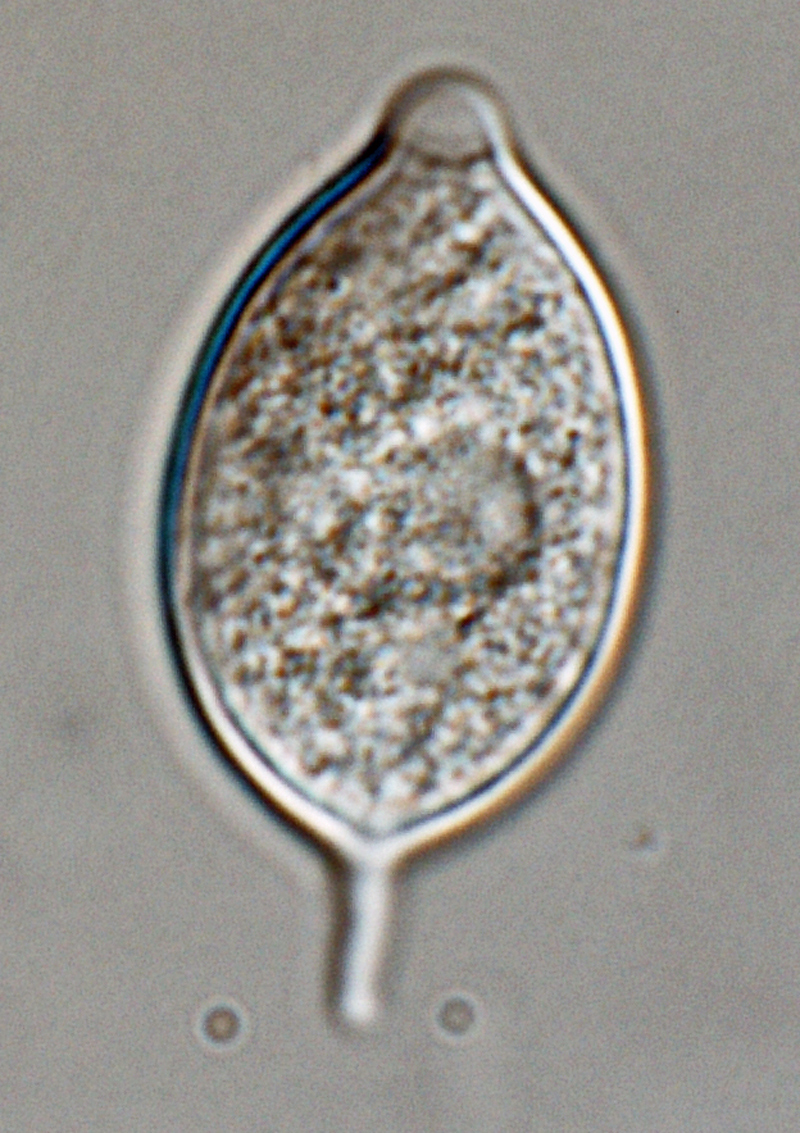
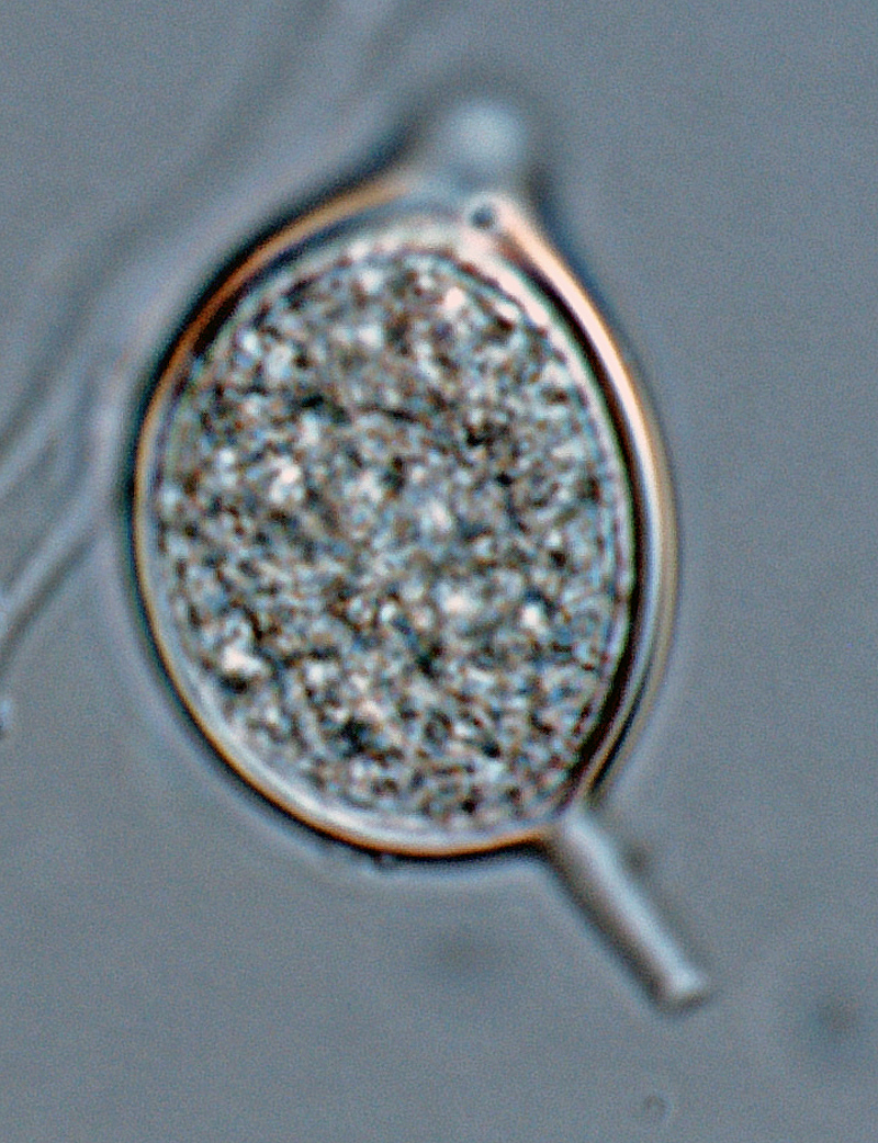
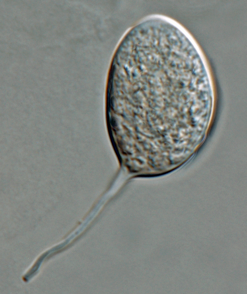
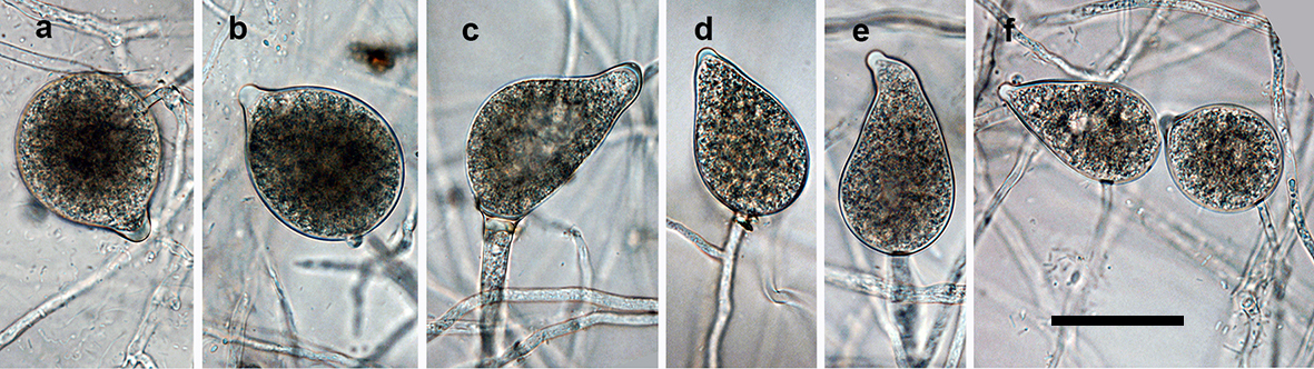
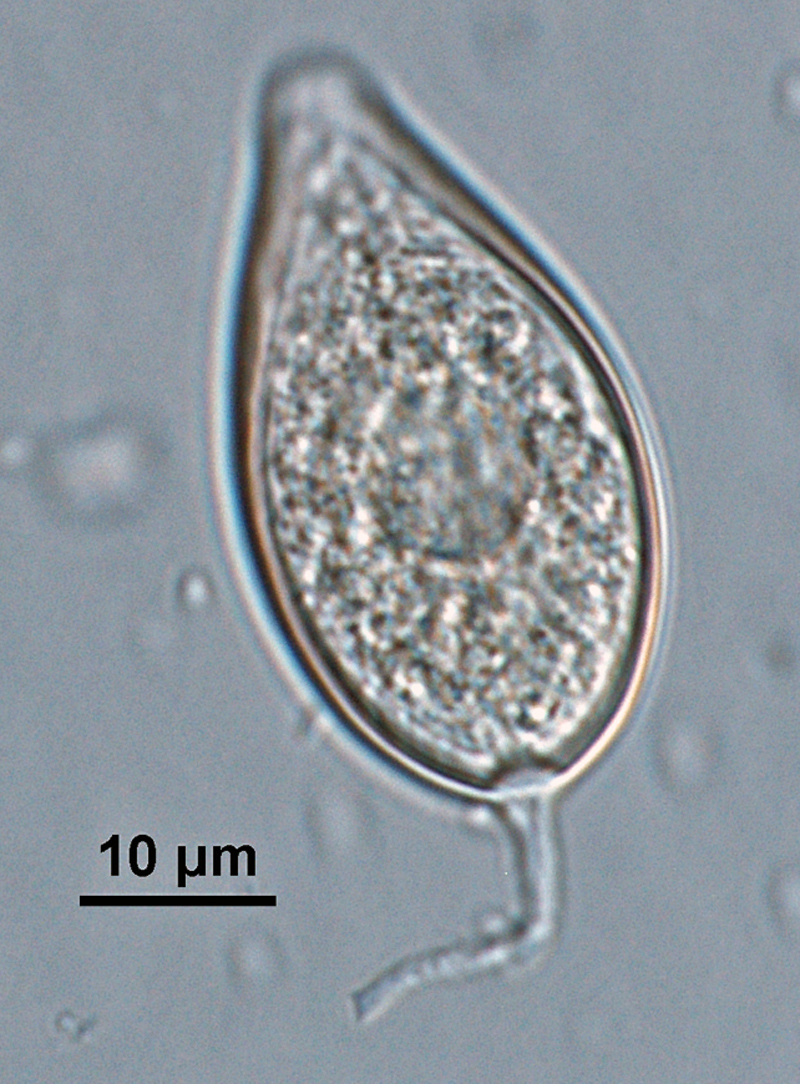 Phytophthora kernoviae (selected specimen P10681) asexual phase: papillatenbsp;ellipsoid sporangiumnbsp;with tapered topnbsp;and medium-length pedicel, formed on V8 agar flooded with soil extract; photo by Gloria Abad, USDA-APHIS-PPQ.
Phytophthora kernoviae (selected specimen P10681) asexual phase: papillate ellipsoid sporangium with tapered top and medium-length pedicel, formed on V8 agar flooded with soil extract; photo by Gloria Abad, USDA-APHIS-PPQ.
 Phytophthora kernoviae nbsp;(selected specimen P10681) asexual phase: zoospores in sporangium; photonbsp;by Gloria Abad, USDA-APHIS-PPQ.
Phytophthora kernoviae (selected specimen P10681) asexual phase: zoospores in sporangium; photo by Gloria Abad, USDA-APHIS-PPQ.
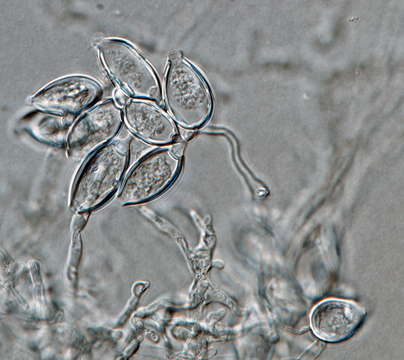
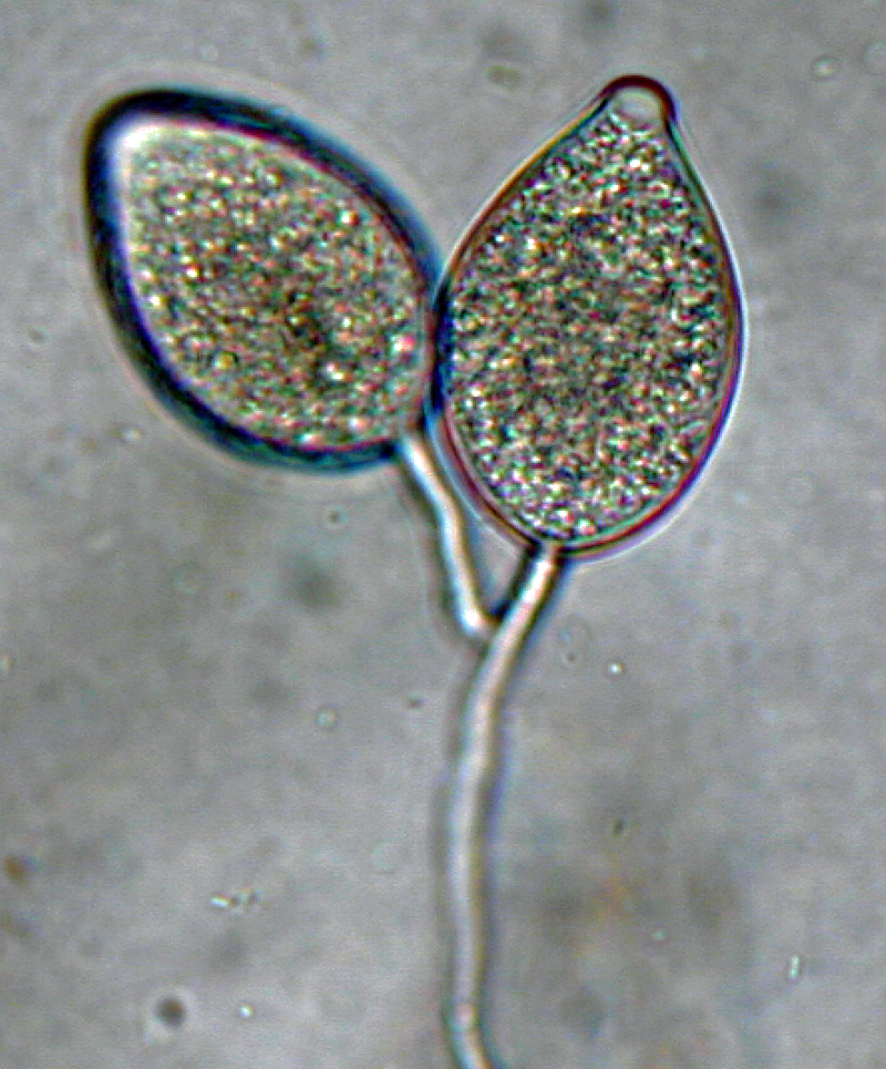
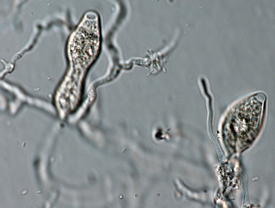 Phytophthora kernoviae (selected specimen P10681) asexual phase: papillate sporangianbsp;produced in unbranched sporangiophores; photo by Gloria Abad, USDA-APHIS-PPQ.
Phytophthora kernoviae (selected specimen P10681) asexual phase: papillate sporangia produced in unbranched sporangiophores; photo by Gloria Abad, USDA-APHIS-PPQ.
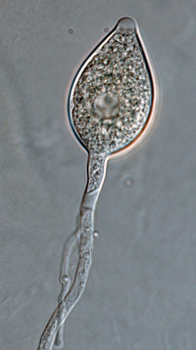
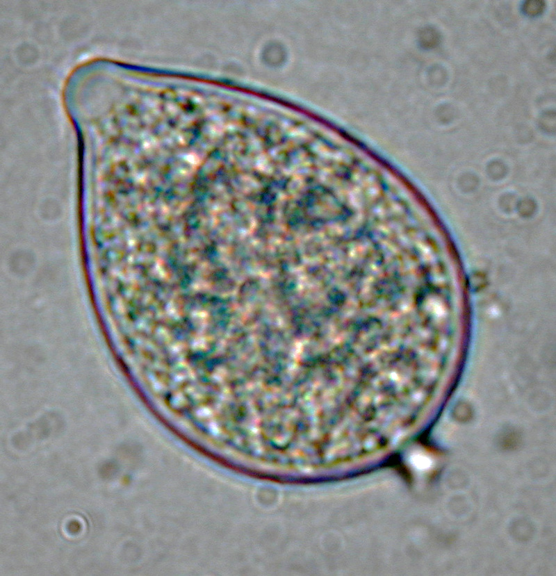
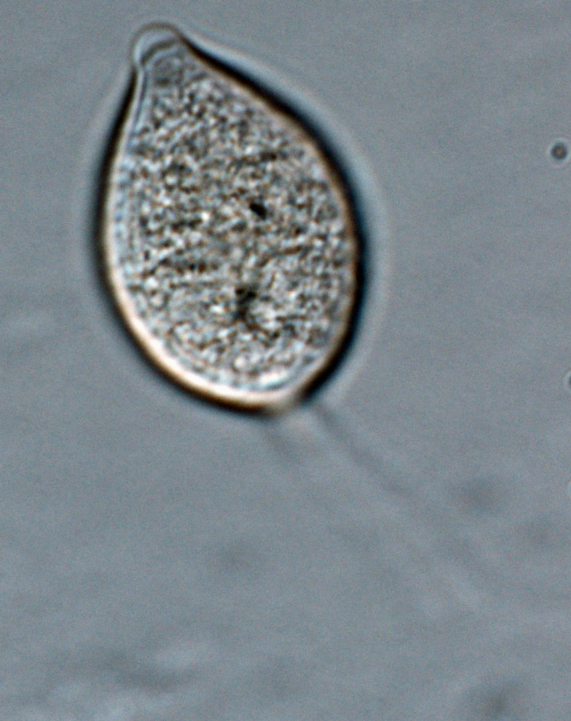
 Phytophthora kernoviae nbsp;(selected specimen P10681) asexual phase: papillatenbsp;ovoid sporangiumnbsp;with medium-length pedicel, formed on V8 agar flooded with soil extract; photo by Gloria Abad, USDA-APHIS-PPQ.
Phytophthora kernoviae (selected specimen P10681) asexual phase: papillate ovoid sporangium with medium-length pedicel, formed on V8 agar flooded with soil extract; photo by Gloria Abad, USDA-APHIS-PPQ.
 Phytophthora kernoviae (selected specimen P10681) asexual phase: sporangium borne in simple sympodial sporangiophore; photo by Gloria Abad, USDA-APHIS-PPQ.
Phytophthora kernoviae (selected specimen P10681) asexual phase: sporangium borne in simple sympodial sporangiophore; photo by Gloria Abad, USDA-APHIS-PPQ.
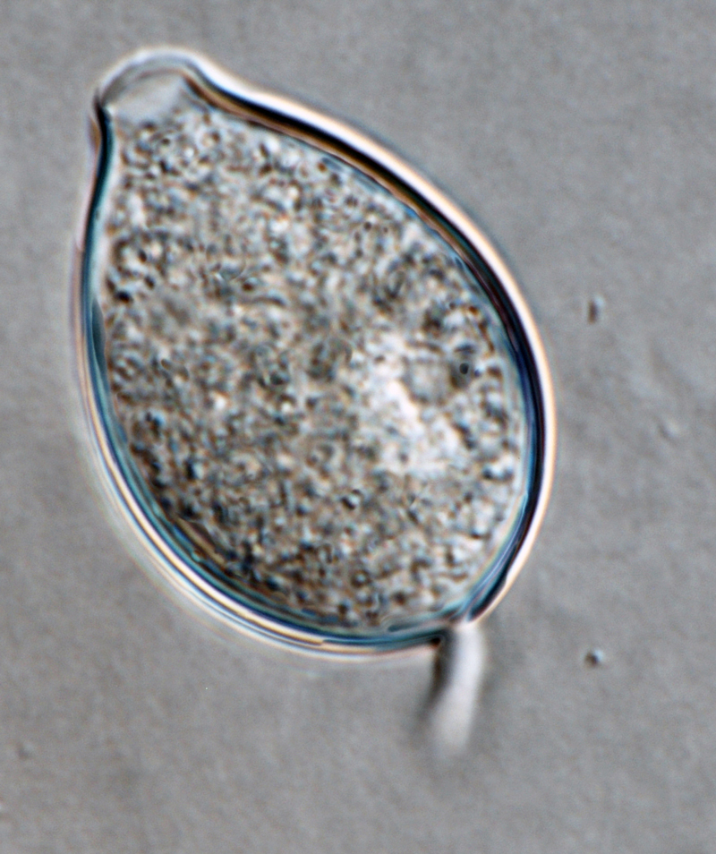
 Phytophthora kernoviae nbsp;(selected specimen P10681) asexual phase: papillatenbsp;irregular shaped sporangiumnbsp;with medium-length pedicel, formed on V8 agar flooded with soil extract; photo by Gloria Abad, USDA-APHIS-PPQ.
Phytophthora kernoviae (selected specimen P10681) asexual phase: papillate irregular shaped sporangium with medium-length pedicel, formed on V8 agar flooded with soil extract; photo by Gloria Abad, USDA-APHIS-PPQ.
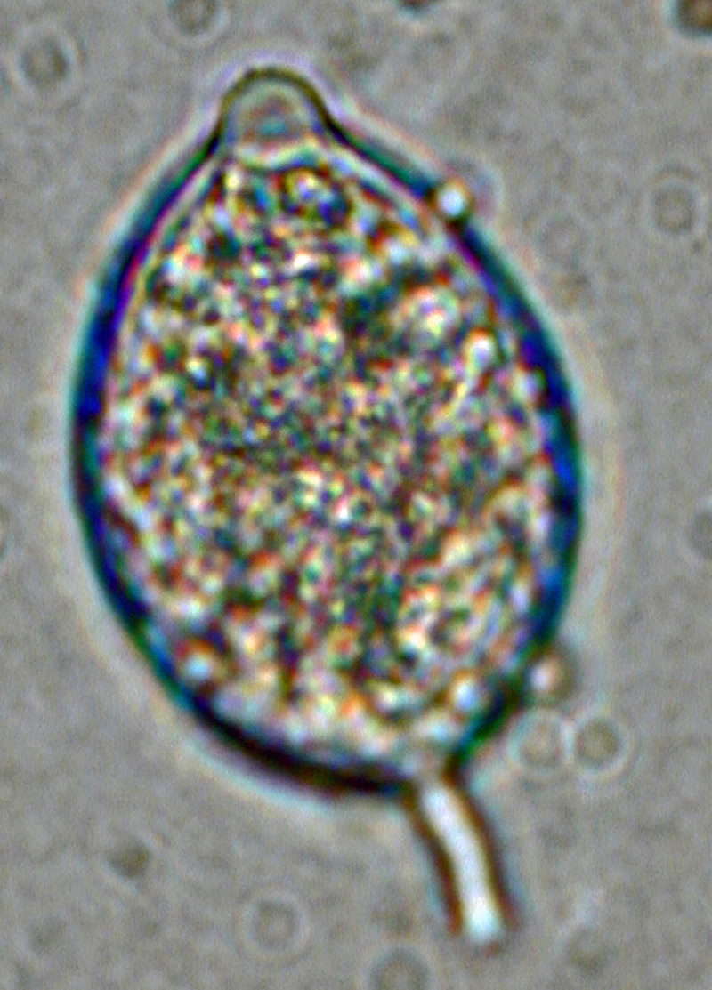
 Phytophthora kernoviae (selected specimen P10956) asexual phase: papillatenbsp;sporangium with short pedicel; photo by Gloria Abad, USDA-APHIS-PPQ.
Phytophthora kernoviae (selected specimen P10956) asexual phase: papillate sporangium with short pedicel; photo by Gloria Abad, USDA-APHIS-PPQ.
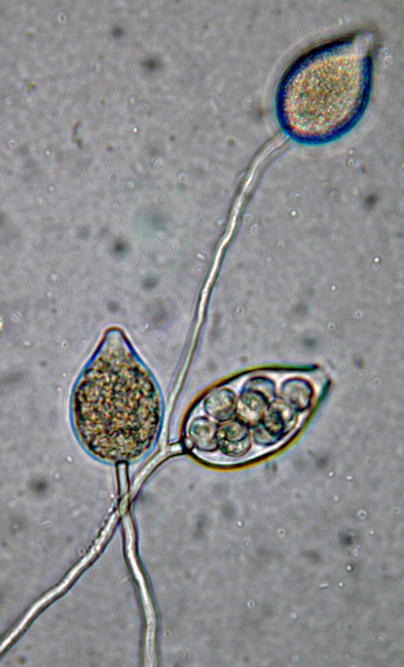
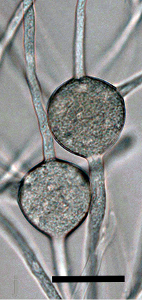
 Phytophthora kernoviae nbsp;(selected specimen P10956) asexual phase: papillatenbsp;sporangia borne in simple sympodial sporangiophore; photo by Gloria Abad, USDA-APHIS-PPQ.
Phytophthora kernoviae (selected specimen P10956) asexual phase: papillate sporangia borne in simple sympodial sporangiophore; photo by Gloria Abad, USDA-APHIS-PPQ.
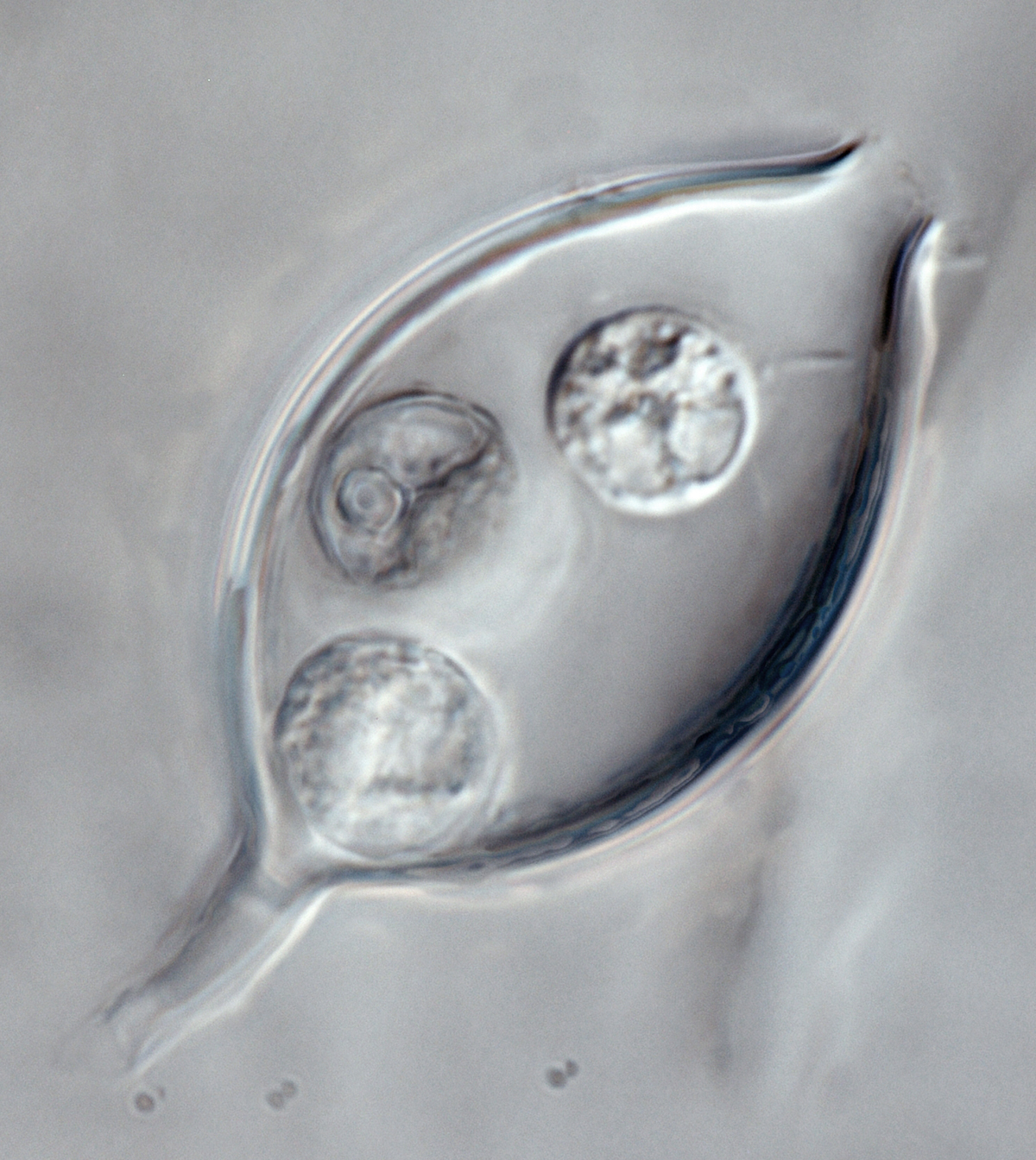
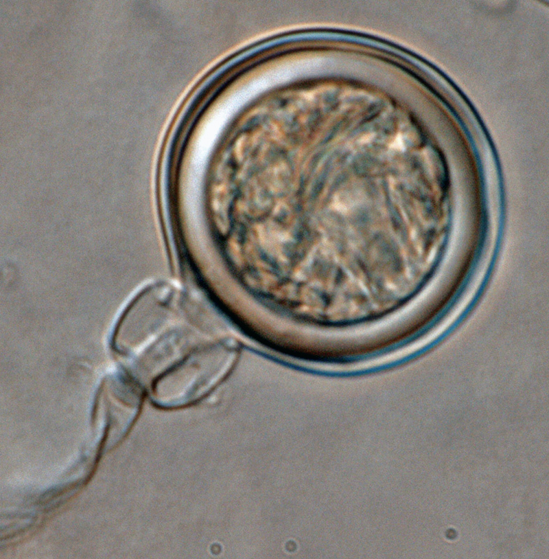
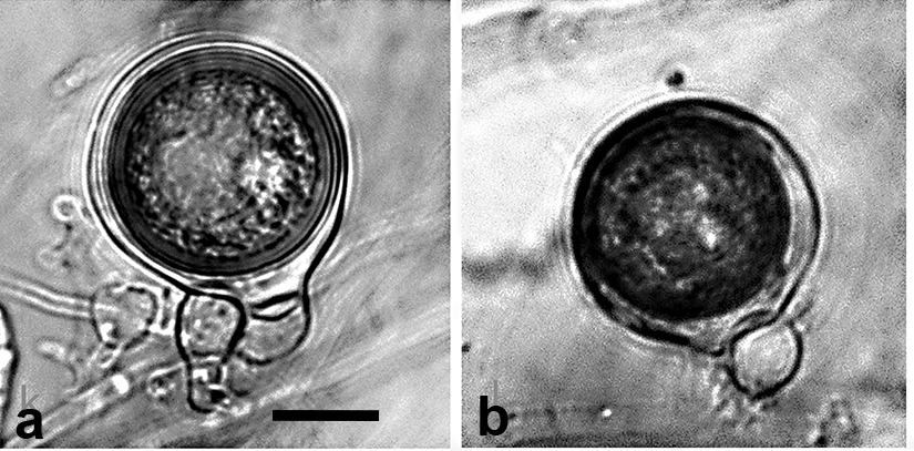
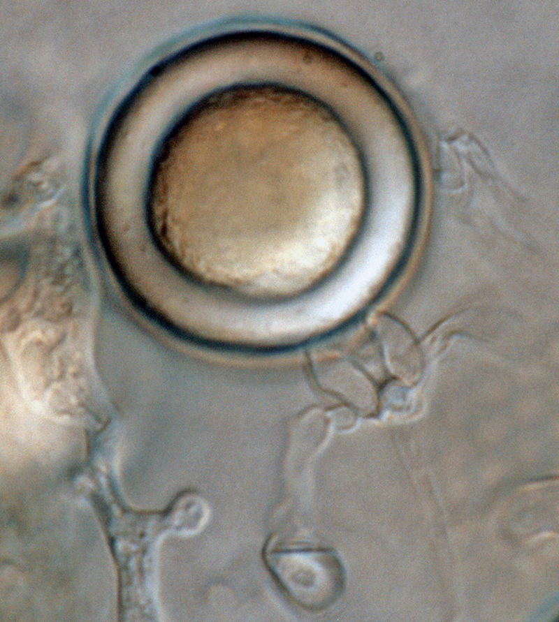 Phytophthora kernoviae (selected specimen P10956)nbsp;sexual phase: medium thick-walled plerotic oospore smooth-walled oogonium and amphigynous antheridium; photo by Gloria Abad, USDA-APHIS-PPQ.
Phytophthora kernoviae (selected specimen P10956) sexual phase: medium thick-walled plerotic oospore smooth-walled oogonium and amphigynous antheridium; photo by Gloria Abad, USDA-APHIS-PPQ.
 Phytophthora kernoviae (selected specimen P10956)nbsp;sexual phase:nbsp;smooth-walled oogonium with amphigynous antheridium; photo by Gloria Abad, USDA-APHIS-PPQ.
Phytophthora kernoviae (selected specimen P10956) sexual phase: smooth-walled oogonium with amphigynous antheridium; photo by Gloria Abad, USDA-APHIS-PPQ.
 Phytophthora kernoviae (selected specimen P10956) asexual phase: papillate sporangium with short pedicel; photo by Gloria Abad, USDA-APHIS-PPQ.
Phytophthora kernoviae (selected specimen P10956) asexual phase: papillate sporangium with short pedicel; photo by Gloria Abad, USDA-APHIS-PPQ.
 Phytophthora kernoviae nbsp;(selected specimen P10956) asexual phase: papillatenbsp;sporangia borne in simple sympodial sporangiophore; photo by Gloria Abad, USDA-APHIS-PPQ.
Phytophthora kernoviae (selected specimen P10956) asexual phase: papillate sporangia borne in simple sympodial sporangiophore; photo by Gloria Abad, USDA-APHIS-PPQ.
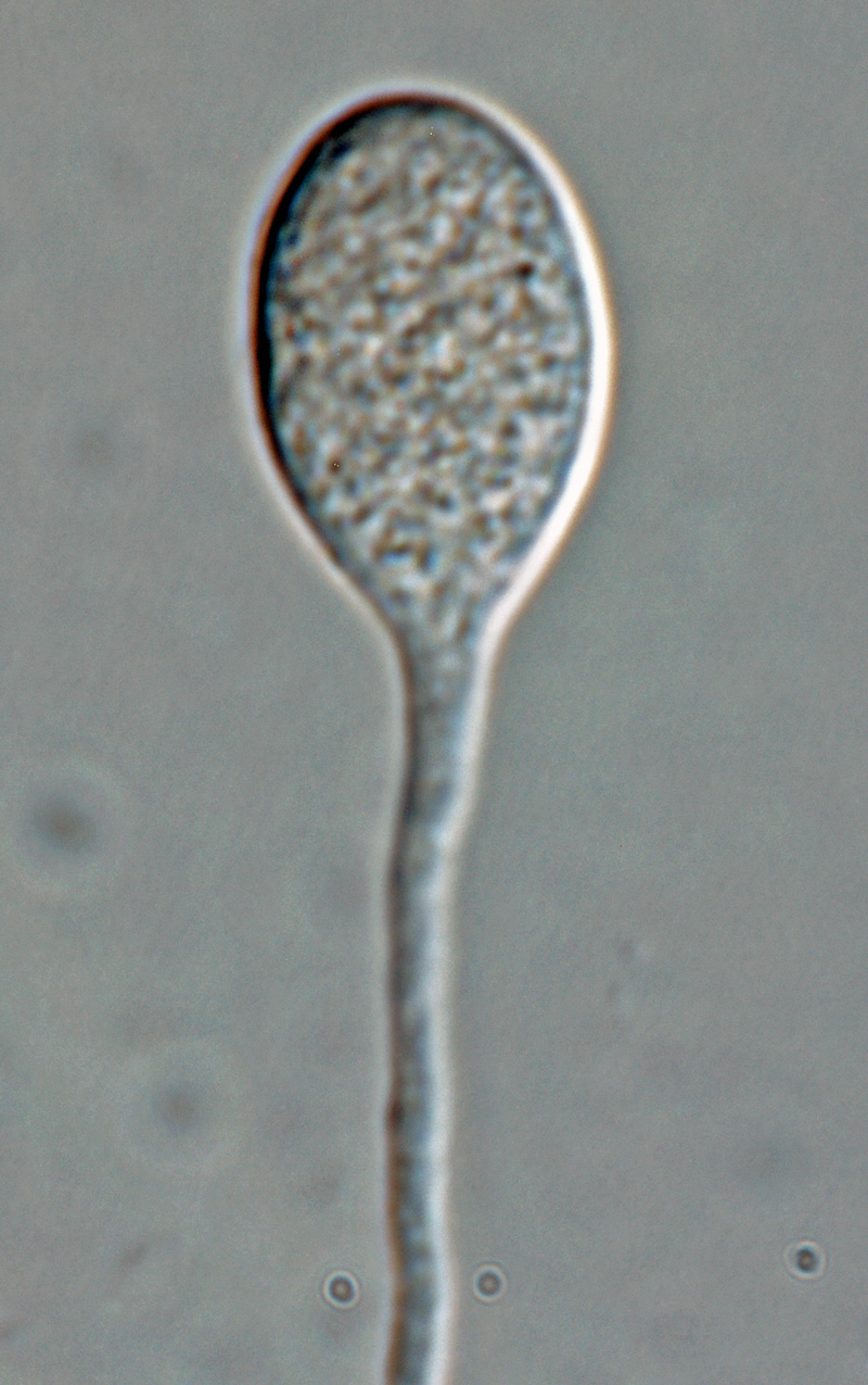 Phytophthora kernoviae nbsp;(selected specimen P10957) asexual phase: sporangiumnbsp;produced in unbranched sporangiophore; photonbsp;by Gloria Abad, USDA-APHIS-PPQ.
Phytophthora kernoviae (selected specimen P10957) asexual phase: sporangium produced in unbranched sporangiophore; photo by Gloria Abad, USDA-APHIS-PPQ.
 Phytophthora kernoviae nbsp;(selected specimen P10957) asexual phase:nbsp;sporangiumnbsp;with long pedicelnbsp;formed on V8 agar flooded with soil extract; photonbsp;by Gloria Abad, USDA-APHIS-PPQ.
Phytophthora kernoviae (selected specimen P10957) asexual phase: sporangium with long pedicel formed on V8 agar flooded with soil extract; photo by Gloria Abad, USDA-APHIS-PPQ.
.JPG) Phytophthora spp. in subclade 6a: portion of the seven-loci ML phylogeny featuring the type cultures of 212 described species (by T. Bourret). Notice the position of P. kwongonina Ex-type CBS 143060 = Samp;T BL187 . Gloria Abad, USDA Samp;T.
Phytophthora spp. in subclade 6a: portion of the seven-loci ML phylogeny featuring the type cultures of 212 described species (by T. Bourret). Notice the position of P. kwongonina Ex-type CBS 143060 = Samp;T BL187 . Gloria Abad, USDA Samp;T.
Phytophthora spp. in subclade 6a: portion of the seven-loci ML phylogeny featuring the type cultures of 212 described species (by T. Bourret). Notice the position of P. kwongonina Ex-type CBS 143060 = S&T BL187. Gloria Abad, USDA S&T.
.JPG) Phytophthora spp. in subclade 6a: Morphological Tabular key (PDF) and Tabular key legends (PDF) in IDphy2 KEY SECTION. Notice the data of P. kwongonina Ex-type CBS 143060 = Samp;T BL187 . Gloria Abad, USDA Samp;T.
Phytophthora spp. in subclade 6a: Morphological Tabular key (PDF) and Tabular key legends (PDF) in IDphy2 KEY SECTION. Notice the data of P. kwongonina Ex-type CBS 143060 = Samp;T BL187 . Gloria Abad, USDA Samp;T.
Phytophthora spp. in subclade 6a: Morphological Tabular key (PDF) and Tabular key legends (PDF) in IDphy2 KEY SECTION. Notice the data of P. kwongonina Ex-type CBS 143060 = S&T BL187. Gloria Abad, USDA S&T.
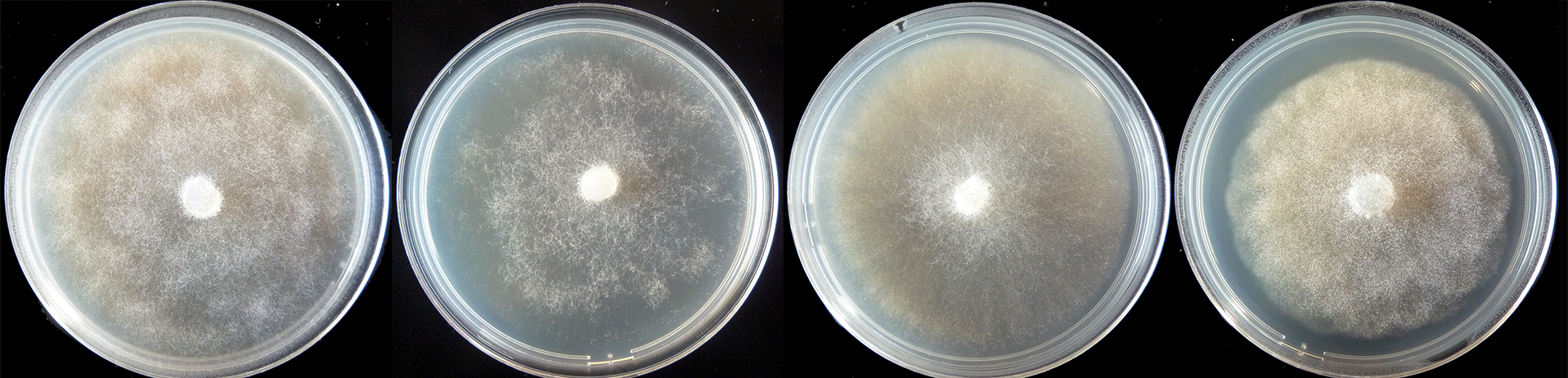 colony morphology after 5 d growth at 20ordm;C on carrot agar, V8 agar, malt extract agar, and potato-dextrose agar (from left to right)
colony morphology after 5 d growth at 20ºC on carrot agar, V8 agar, malt extract agar, and potato-dextrose agar (from left to right)
 persistent, nonpapillate, predominantly ovoid to elongated ovoid sporangia with nested and extended internal proliferation; scale bar = 25micro;m
persistent, nonpapillate, predominantly ovoid to elongated ovoid sporangia with nested and extended internal proliferation; scale bar = 25µm
 oogonia with wavy walls containing aplerotic oospores, with large ooplasts and thick walls which were pale on maturity; antheridia exclusively paragynous generally situated adjacent to the oogonial stalk; scale bar = 25micro;m
oogonia with wavy walls containing aplerotic oospores, with large ooplasts and thick walls which were pale on maturity; antheridia exclusively paragynous generally situated adjacent to the oogonial stalk; scale bar = 25µm
 spherical hyphal swellings with radiating hyphae which appear like small chlamydospores except that the wall doesnrsquo;t form; scale bar = 25micro;m
spherical hyphal swellings with radiating hyphae which appear like small chlamydospores except that the wall doesn’t form; scale bar = 25µm
.JPG) Phytophthora spp. in subclade 8b: portion of the seven-loci ML phylogeny featuring the type cultures of 212 described species (by T. Bourret). Notice the position of P. lactucae Ex-type BPIC 1985 = Samp;T BL 113 . Gloria Abad, USDA Samp;T.
Phytophthora spp. in subclade 8b: portion of the seven-loci ML phylogeny featuring the type cultures of 212 described species (by T. Bourret). Notice the position of P. lactucae Ex-type BPIC 1985 = Samp;T BL 113 . Gloria Abad, USDA Samp;T.
Phytophthora spp. in subclade 8b: portion of the seven-loci ML phylogeny featuring the type cultures of 212 described species (by T. Bourret). Notice the position of P. lactucae Ex-type BPIC 1985 = S&T BL 113. Gloria Abad, USDA S&T.
.JPG) Phytophthora spp. in subclade 8b: Morphological Tabular key (PDF) and Tabular key legends (PDF) in IDphy2 KEY SECTION. Notice the data of P. lactucae Ex-type BPIC 1985 = Samp;T BL 113 . Gloria Abad, USDA Samp;T.
Phytophthora spp. in subclade 8b: Morphological Tabular key (PDF) and Tabular key legends (PDF) in IDphy2 KEY SECTION. Notice the data of P. lactucae Ex-type BPIC 1985 = Samp;T BL 113 . Gloria Abad, USDA Samp;T.
Phytophthora spp. in subclade 8b: Morphological Tabular key (PDF) and Tabular key legends (PDF) in IDphy2 KEY SECTION. Notice the data of P. lactucae Ex-type BPIC 1985 = S&T BL 113. Gloria Abad, USDA S&T.
.JPG) Phytophthora spp. in subclade 6d: portion of the seven-loci ML phylogeny featuring the type cultures of 212 described species (by T. Bourret). Notice the position of P. lacustris Ex-type CABI IMI389725 (PA) = Samp;T BL 69 . Gloria Abad, USDA Samp;T.
Phytophthora spp. in subclade 6d: portion of the seven-loci ML phylogeny featuring the type cultures of 212 described species (by T. Bourret). Notice the position of P. lacustris Ex-type CABI IMI389725 (PA) = Samp;T BL 69 . Gloria Abad, USDA Samp;T.
Phytophthora spp. in subclade 6d: portion of the seven-loci ML phylogeny featuring the type cultures of 212 described species (by T. Bourret). Notice the position of P. lacustris Ex-type CABI IMI389725 (PA) = S&T BL 69. Gloria Abad, USDA S&T.
.JPG) Phytophthora spp. in subclade 6d: Morphological Tabular key (PDF) and Tabular key legends (PDF) in IDphy2 KEY SECTION. Notice the data of P. lacustris Ex-type CABI IMI389725 (PA) = Samp;T BL 69 . Gloria Abad, USDA Samp;T.
Phytophthora spp. in subclade 6d: Morphological Tabular key (PDF) and Tabular key legends (PDF) in IDphy2 KEY SECTION. Notice the data of P. lacustris Ex-type CABI IMI389725 (PA) = Samp;T BL 69 . Gloria Abad, USDA Samp;T.
Phytophthora spp. in subclade 6d: Morphological Tabular key (PDF) and Tabular key legends (PDF) in IDphy2 KEY SECTION. Notice the data of P. lacustris Ex-type CABI IMI389725 (PA) = S&T BL 69. Gloria Abad, USDA S&T.
 Phytophthora lacustris in riparian ecosystems in Italy, morphology, and position in partial phylogenetic tree; slide provided by Santa Olga Cacciola, University of Catania, Italy from presentation on ldquo; Phytophthora diversity in cultivated and natural environments in Europerdquo; at ICPP 2018 - 6th International Oomycetes Workshop, Boston, USA
Phytophthora lacustris in riparian ecosystems in Italy, morphology, and position in partial phylogenetic tree; slide provided by Santa Olga Cacciola, University of Catania, Italy from presentation on “Phytophthora diversity in cultivated and natural environments in Europe” at ICPP 2018 - 6th International Oomycetes Workshop, Boston, USA
.JPG) Phytophthora spp. in subclade 8c: portion of the seven-loci ML phylogeny featuring the type cultures of 212 described species (by T. Bourret). Notice the position of P. lateralis Ex-type CBS 168.42 = Samp;T BL 42 . Gloria Abad, USDA Samp;T.
Phytophthora spp. in subclade 8c: portion of the seven-loci ML phylogeny featuring the type cultures of 212 described species (by T. Bourret). Notice the position of P. lateralis Ex-type CBS 168.42 = Samp;T BL 42 . Gloria Abad, USDA Samp;T.
Phytophthora spp. in subclade 8c: portion of the seven-loci ML phylogeny featuring the type cultures of 212 described species (by T. Bourret). Notice the position of P. lateralis Ex-type CBS 168.42 = S&T BL 42. Gloria Abad, USDA S&T.
.JPG) Phytophthora spp. in subclade 8c: Morphological Tabular key (PDF) and Tabular key legends (PDF) in IDphy2 KEY SECTION. Notice the data of P. lateralis Ex-type CBS 168.42 = Samp;T BL 42 . Gloria Abad, USDA Samp;T.
Phytophthora spp. in subclade 8c: Morphological Tabular key (PDF) and Tabular key legends (PDF) in IDphy2 KEY SECTION. Notice the data of P. lateralis Ex-type CBS 168.42 = Samp;T BL 42 . Gloria Abad, USDA Samp;T.
Phytophthora spp. in subclade 8c: Morphological Tabular key (PDF) and Tabular key legends (PDF) in IDphy2 KEY SECTION. Notice the data of P. lateralis Ex-type CBS 168.42 = S&T BL 42. Gloria Abad, USDA S&T.
