IPM Images ITP Node
imageID
PPQ IDaids
Search IDaids
IDtools
LepIntercept
About
How to Use LepIntercept
Materials and methods
Authors and acknowledgments
Copyright, citation, and disclaimers
References
Larvae
Morphohology
Preserving and studying larvae
Microscope techniques
Examining larvae
Fact sheets
Keys
Tips for using the keys
Interactive key
Dichotomous keys
Gallery
Glossary
choose a page
Home
About
--How to Use LepIntercept
--Materials and methods
--Authors and acknowledgments
--Copyright, citation, and disclaimers
--References
Larvae
--Morphohology
--Preserving and studying larvae
--Microscope techniques
--Examining larvae
Fact sheets
Keys
--Tips for using the keys
--Interactive key
--Dichotomous keys
Gallery
Glossary
Your session is about to expire. You will be logged out in two minutes if no action is taken.
seconds until your session expires.
If you wish to keep working, click continue.
Continue
Your session HAS EXPIRED
To continue editing this content you will need to click the link from FSM again.
Close
Gallery
body part
abdomen
anal region
body entire
comb
crochets
head
hypopharyngeal complex
mandible
pinaculum
proleg
setae
shield
spinules
spiracle
thorax
view
dorsal
lateral
per page:
page:
20
40
60
80
1
2
3
4
5
6
7
8
9
10
11
12
13
14
15
16
17
18
of 18
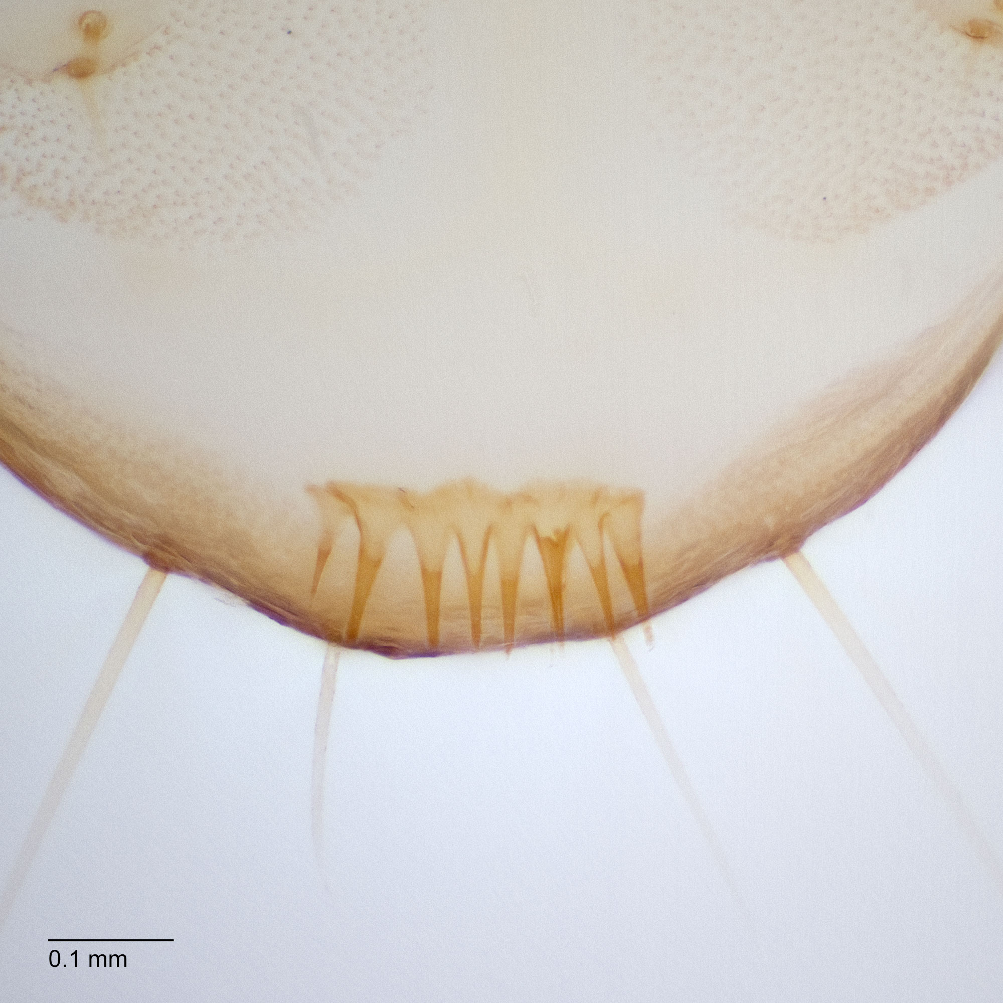
Fig. 4: Anal comb
Thaumatotibia leucotreta
Fig. 4: Anal comb
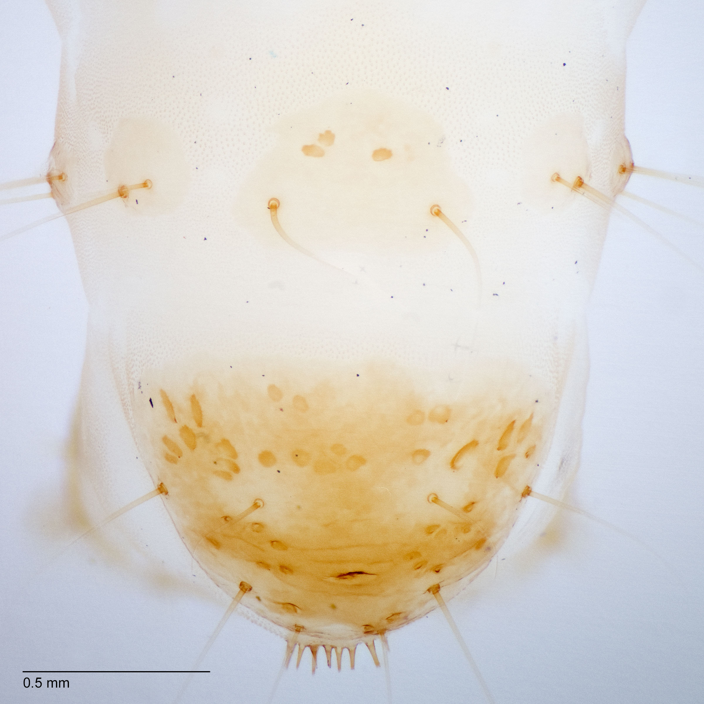
Fig. 5: A9, anal shield
Thaumatotibia leucotreta
Fig. 5: A9, anal shield
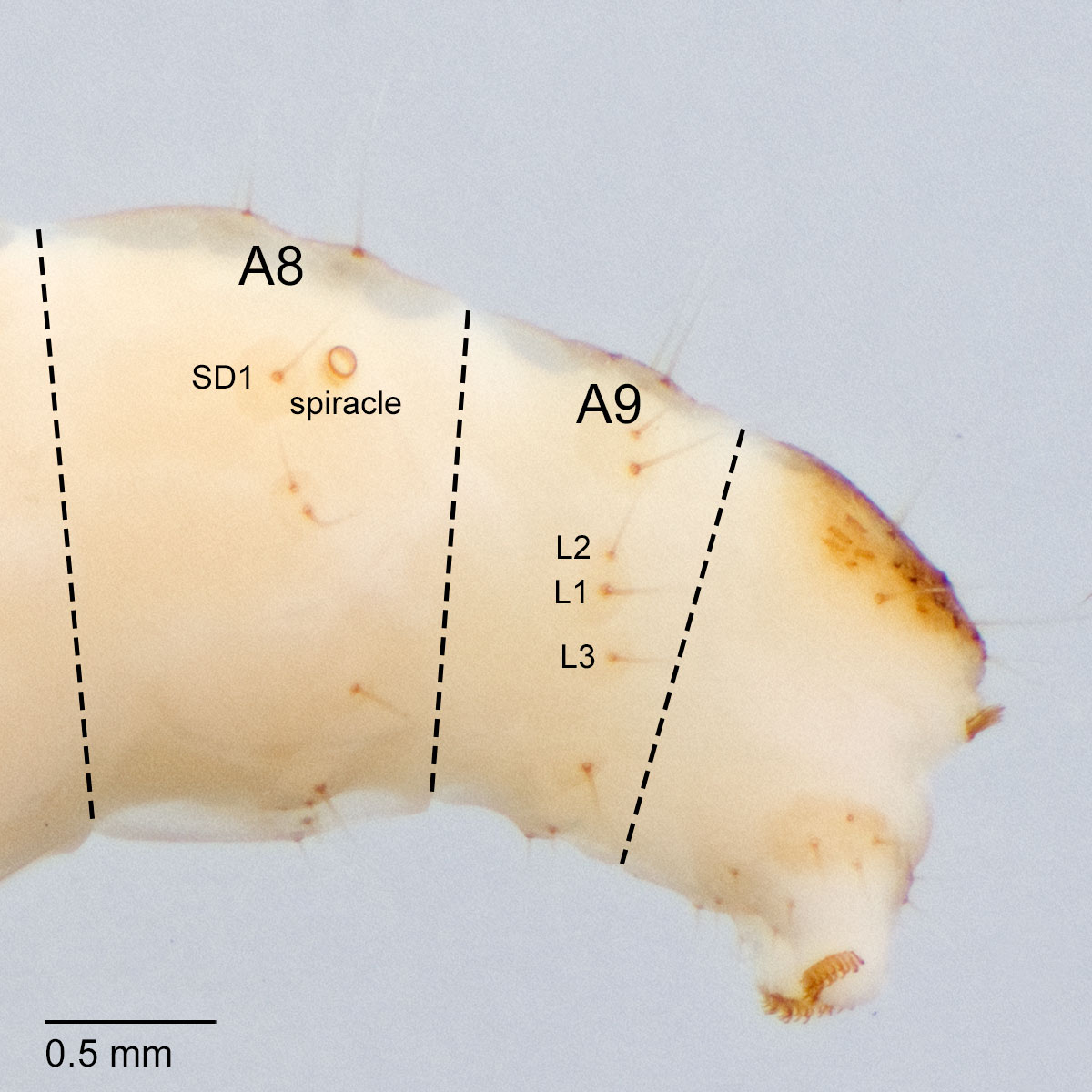
Fig. 6: A8 spiracle
Thaumatotibia leucotreta
Fig. 6: A8 spiracle
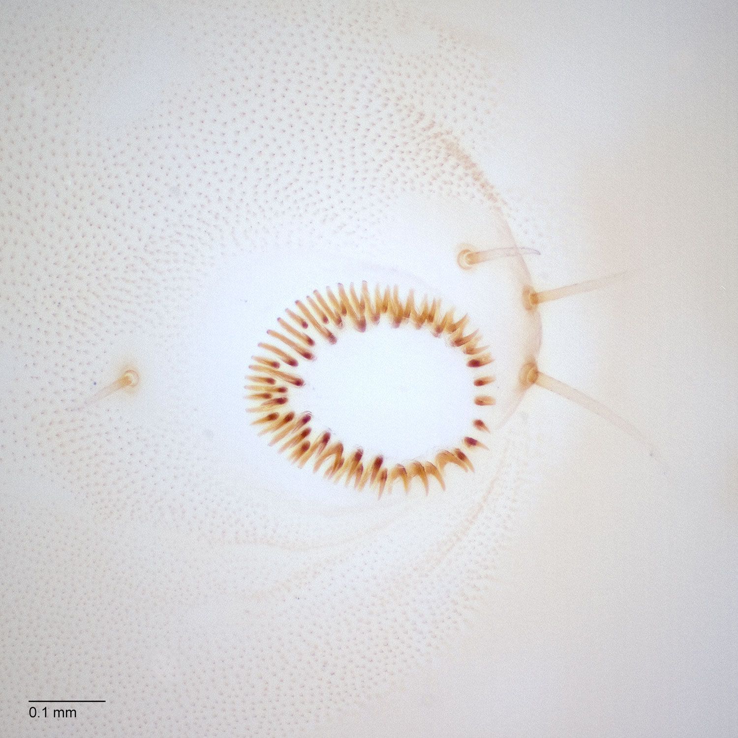
Fig. 7: Crochets
Thaumatotibia leucotreta
Fig. 7: Crochets
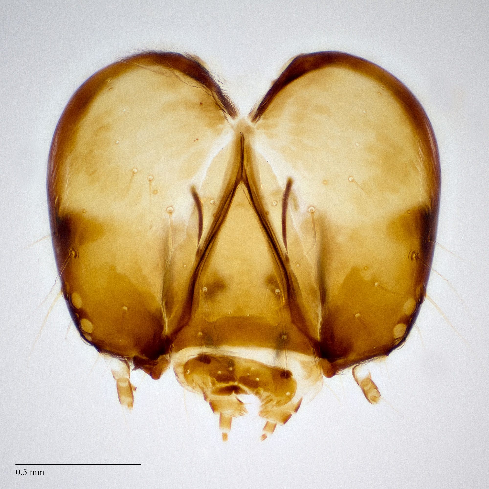
Fig. 8: Head
Thaumatotibia leucotreta
Fig. 8: Head
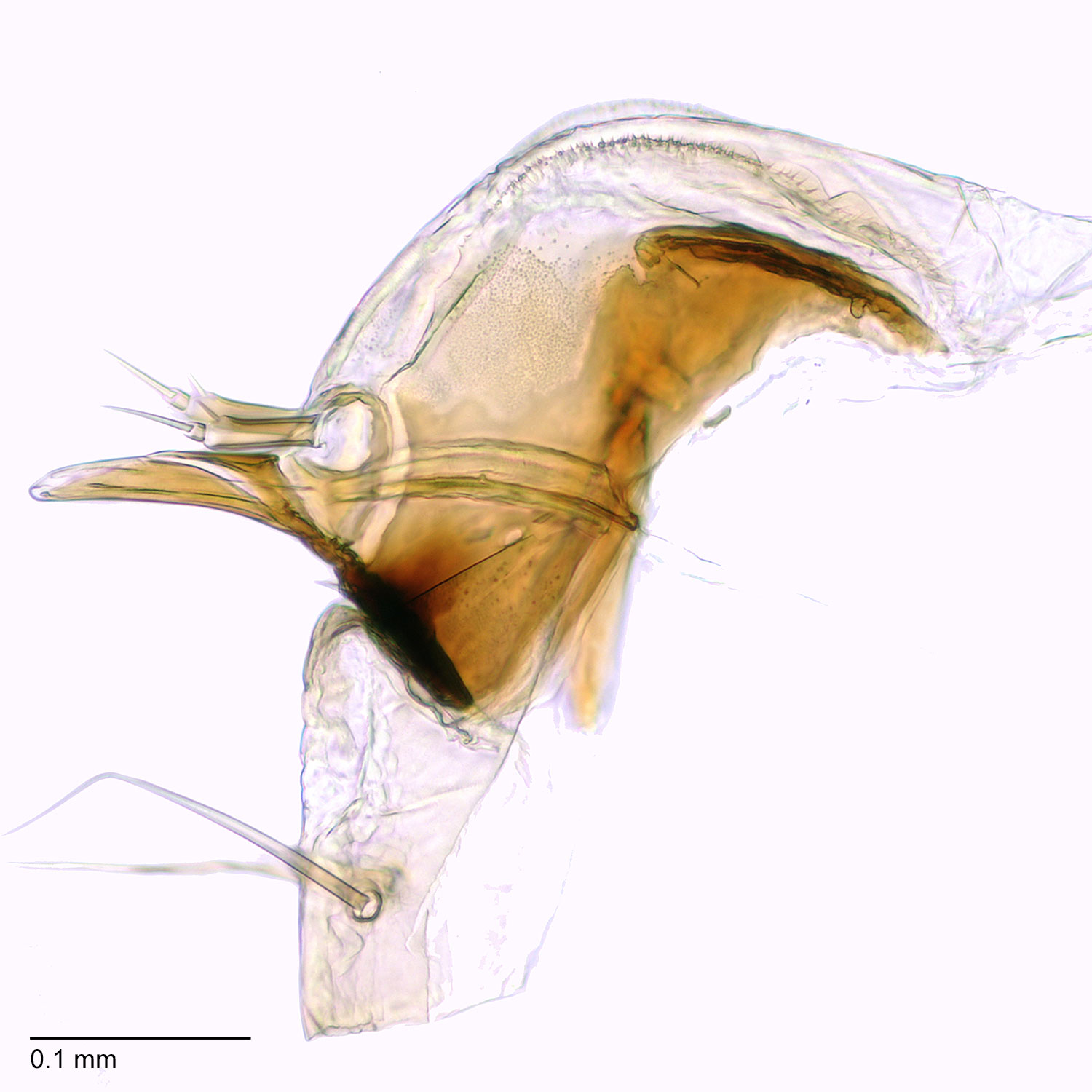
Fig. 9: Hypo. complex
Thaumatotibia leucotreta
Fig. 9: Hypo. complex
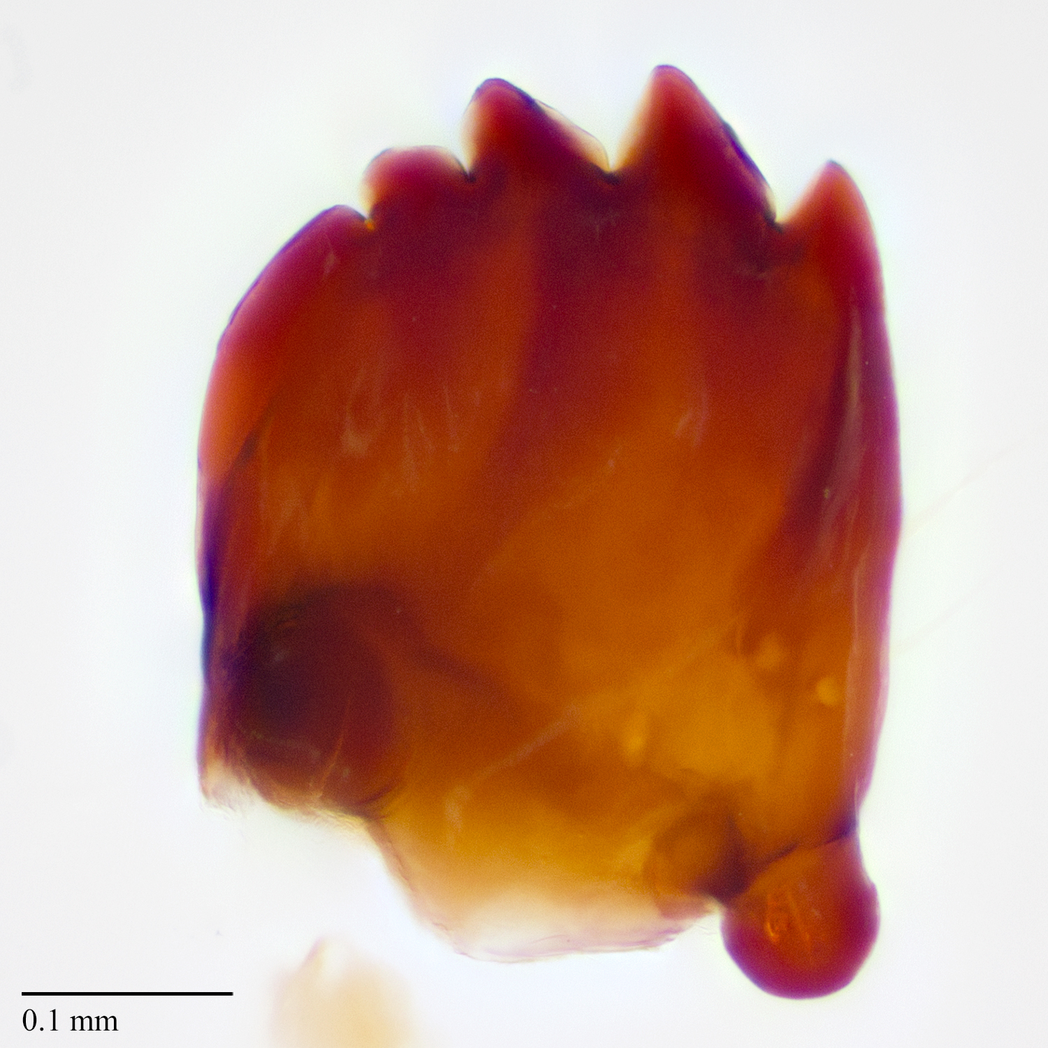
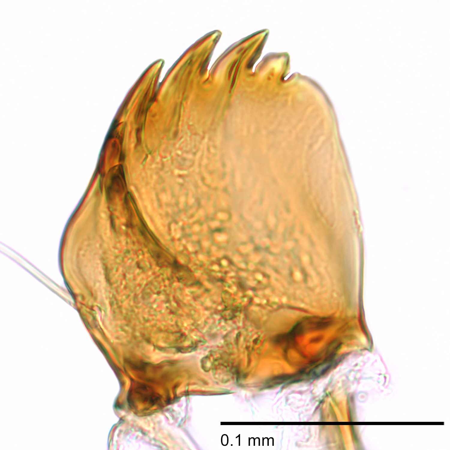
Fig. 10: Mandible
Thaumatotibia leucotreta
Fig. 10: Mandible
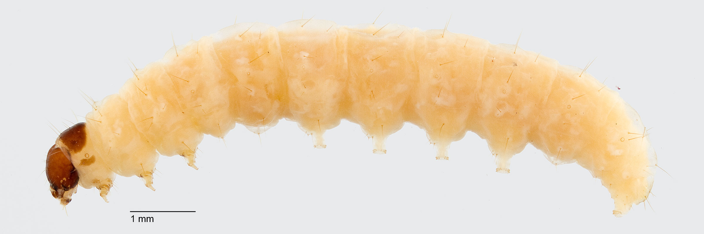
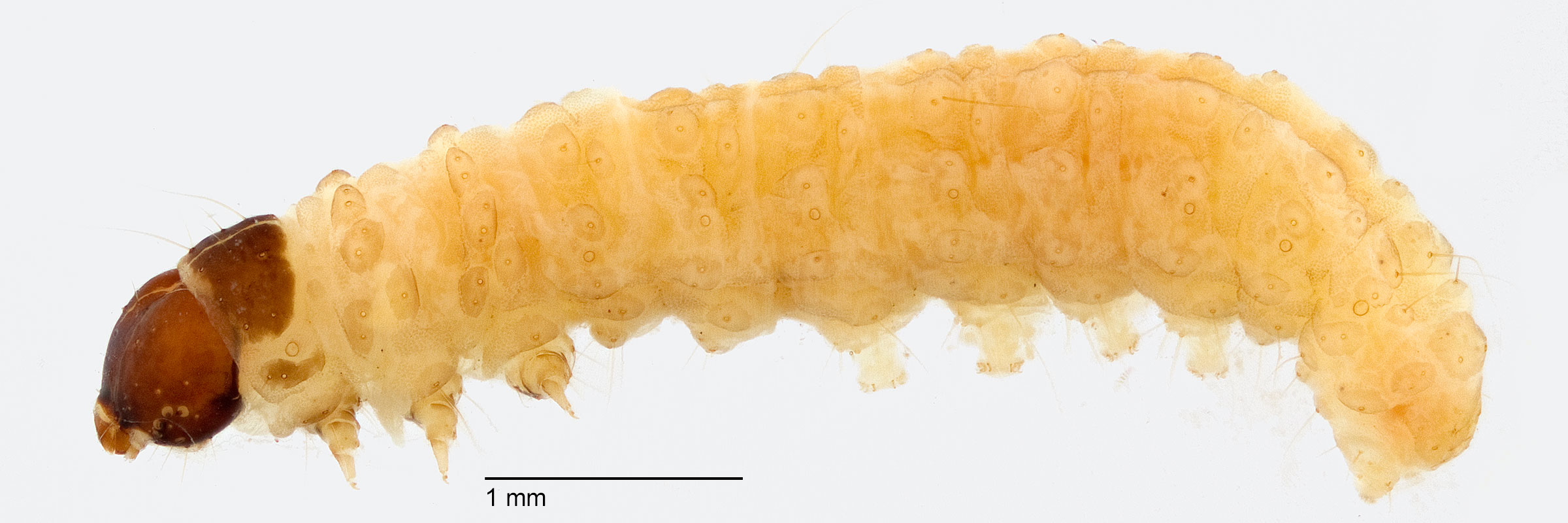
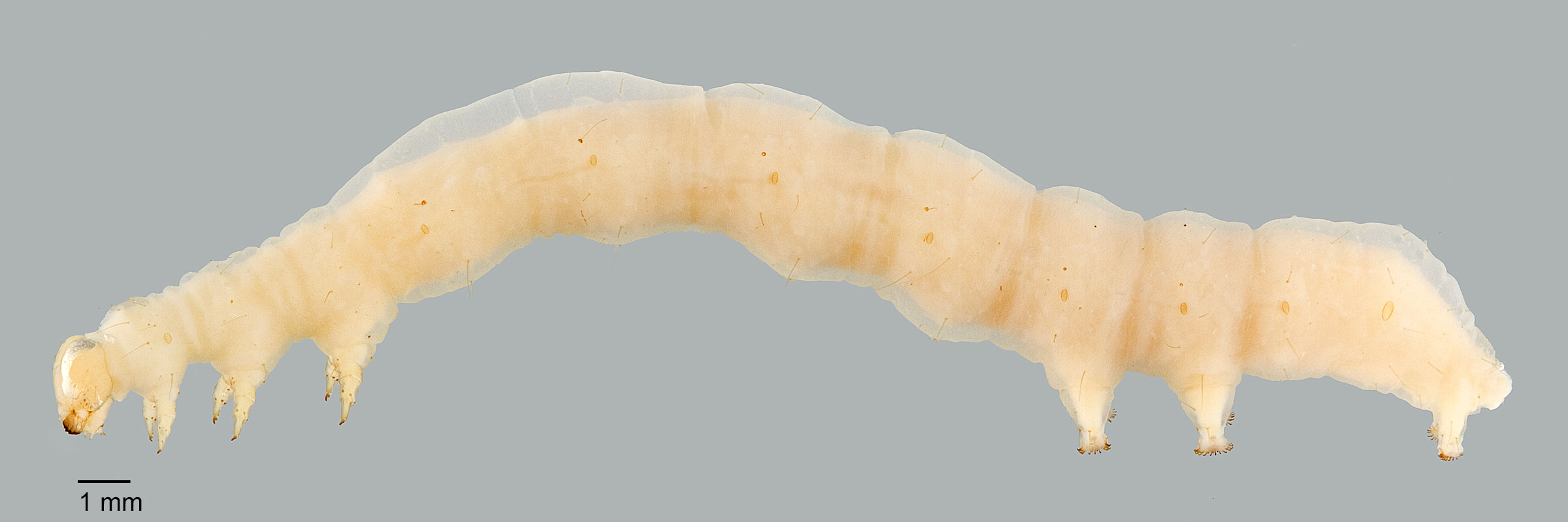
Fig. 1: Late instar, lateral view (India)
Trichophysetis duplifascialis
Fig. 1: Late instar, lateral view (India)
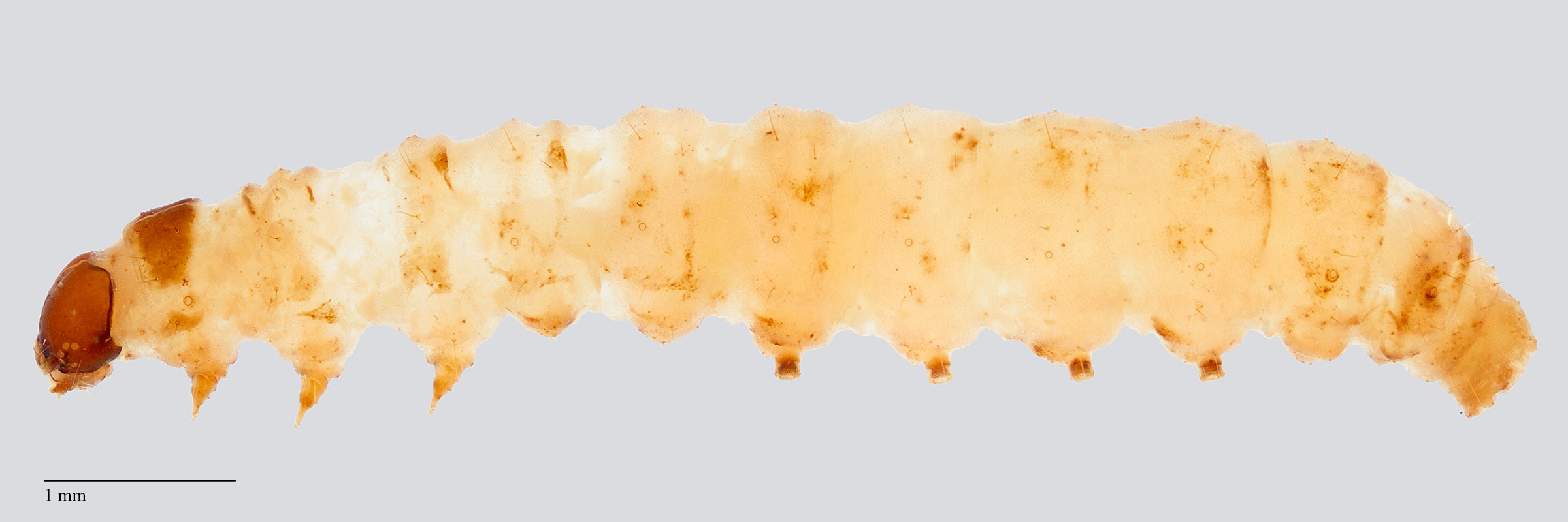
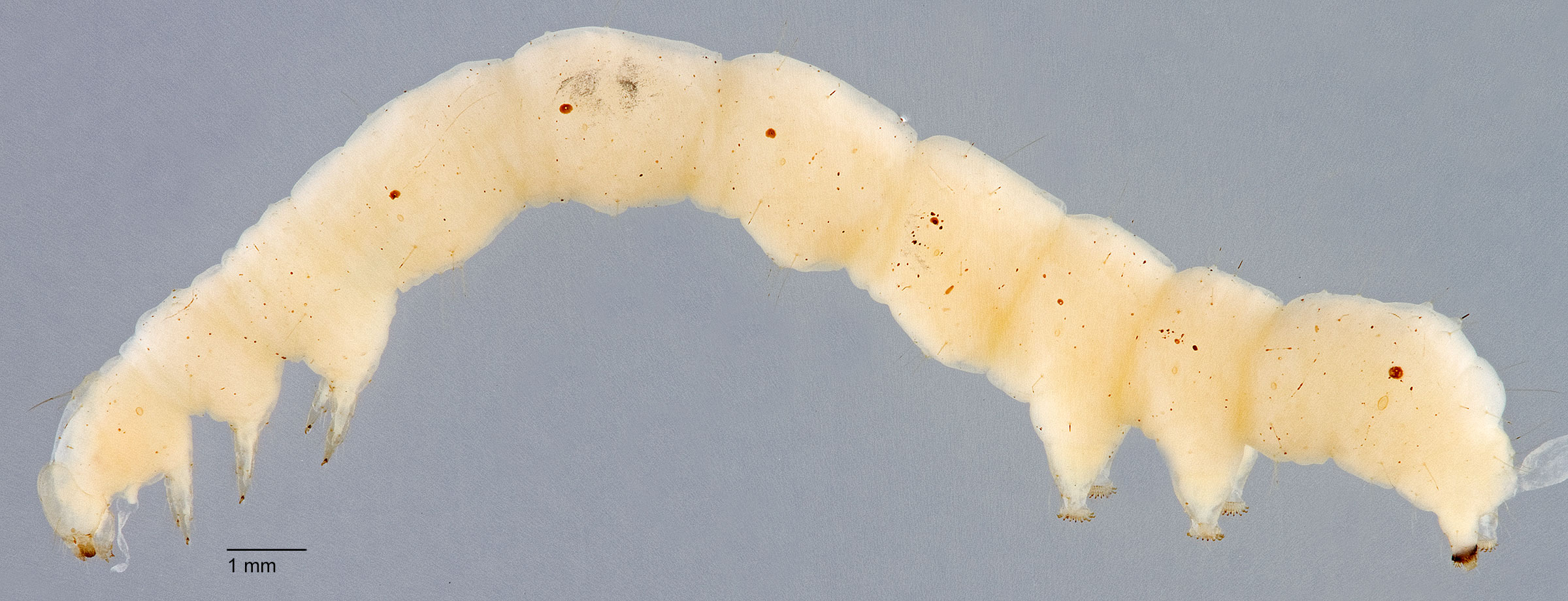
Fig. 2: Mid-instar, lateral view (Thailand)
Trichophysetis duplifascialis
Fig. 2: Mid-instar, lateral view (Thailand)
Fig. 3: Late instar, lateral view (India)
Trichophysetis duplifascialis
Fig. 3: Late instar, lateral view (India)
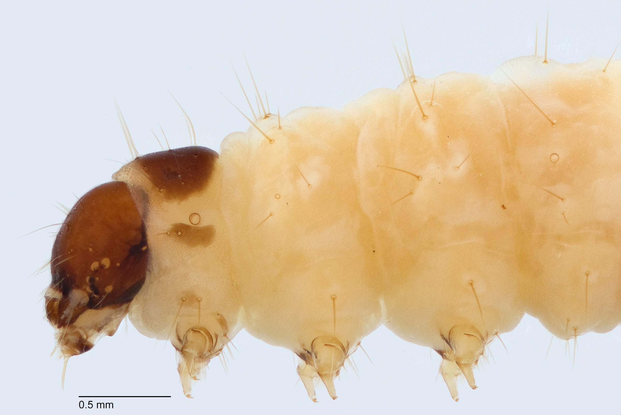
Fig. 4: Head and thorax, lateral view (India)
Trichophysetis duplifascialis
Fig. 4: Head and thorax, lateral view (India)
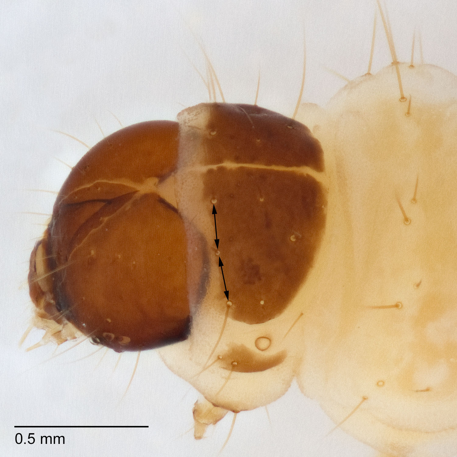
Fig. 5: P-t shield
Trichophysetis duplifascialis
Fig. 5: P-t shield
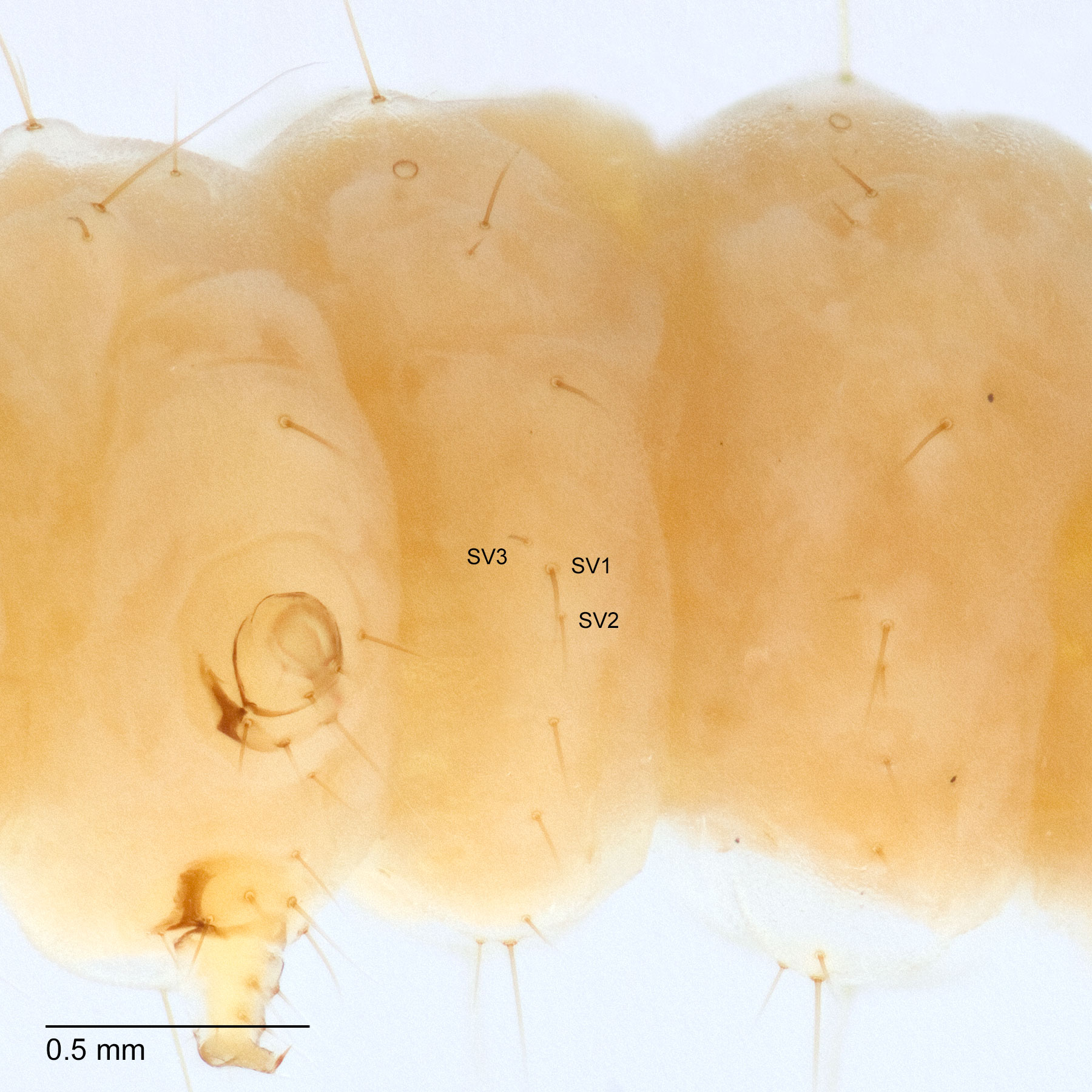
Fig. 6: SV group on A1
Trichophysetis duplifascialis
Fig. 6: SV group on A1
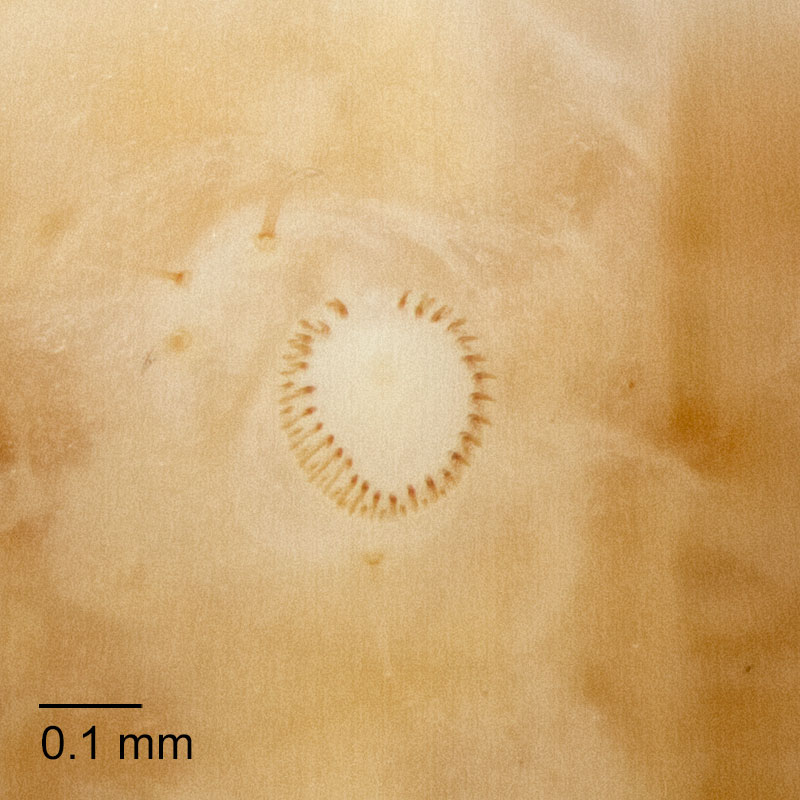
Fig. 7: Crochets
Trichophysetis duplifascialis
Fig. 7: Crochets
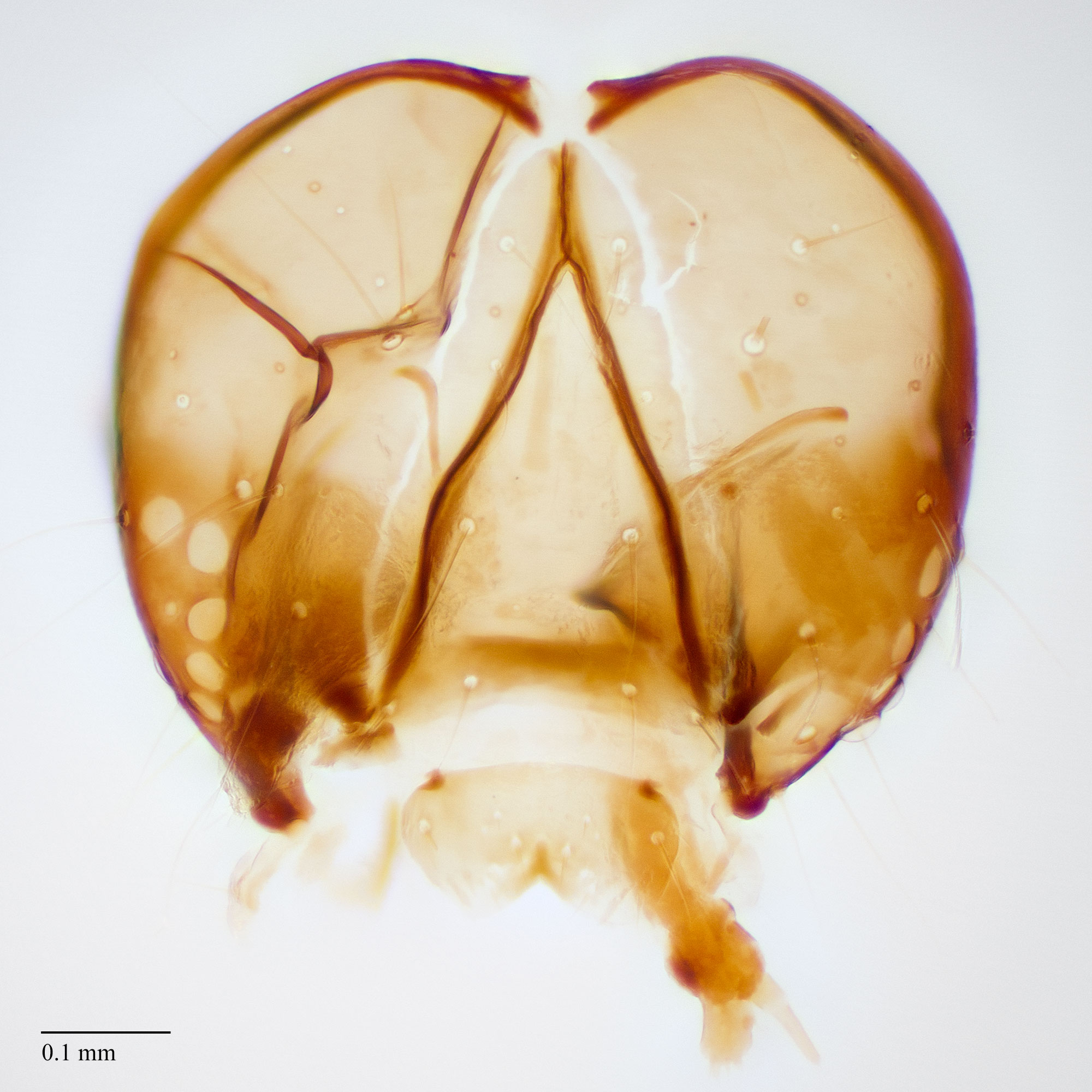
Fig. 8: Head
Trichophysetis duplifascialis
Fig. 8: Head
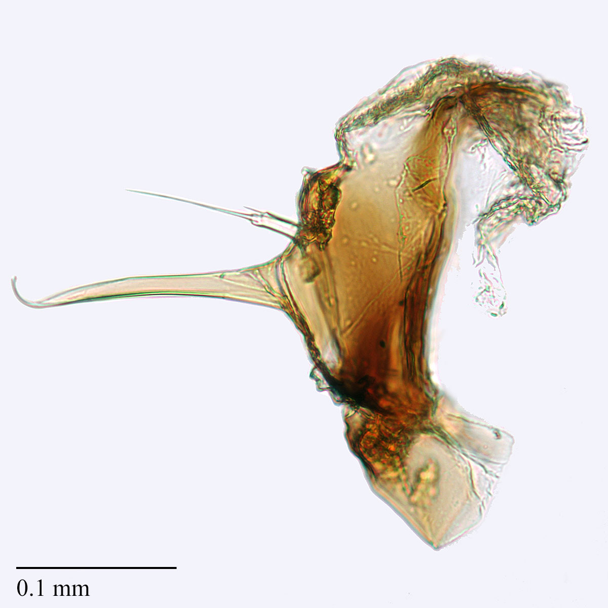
Fig. 9: Hypo. complex
Trichophysetis duplifascialis
Fig. 9: Hypo. complex
Fig. 10: Mandible
Trichophysetis duplifascialis
Fig. 10: Mandible
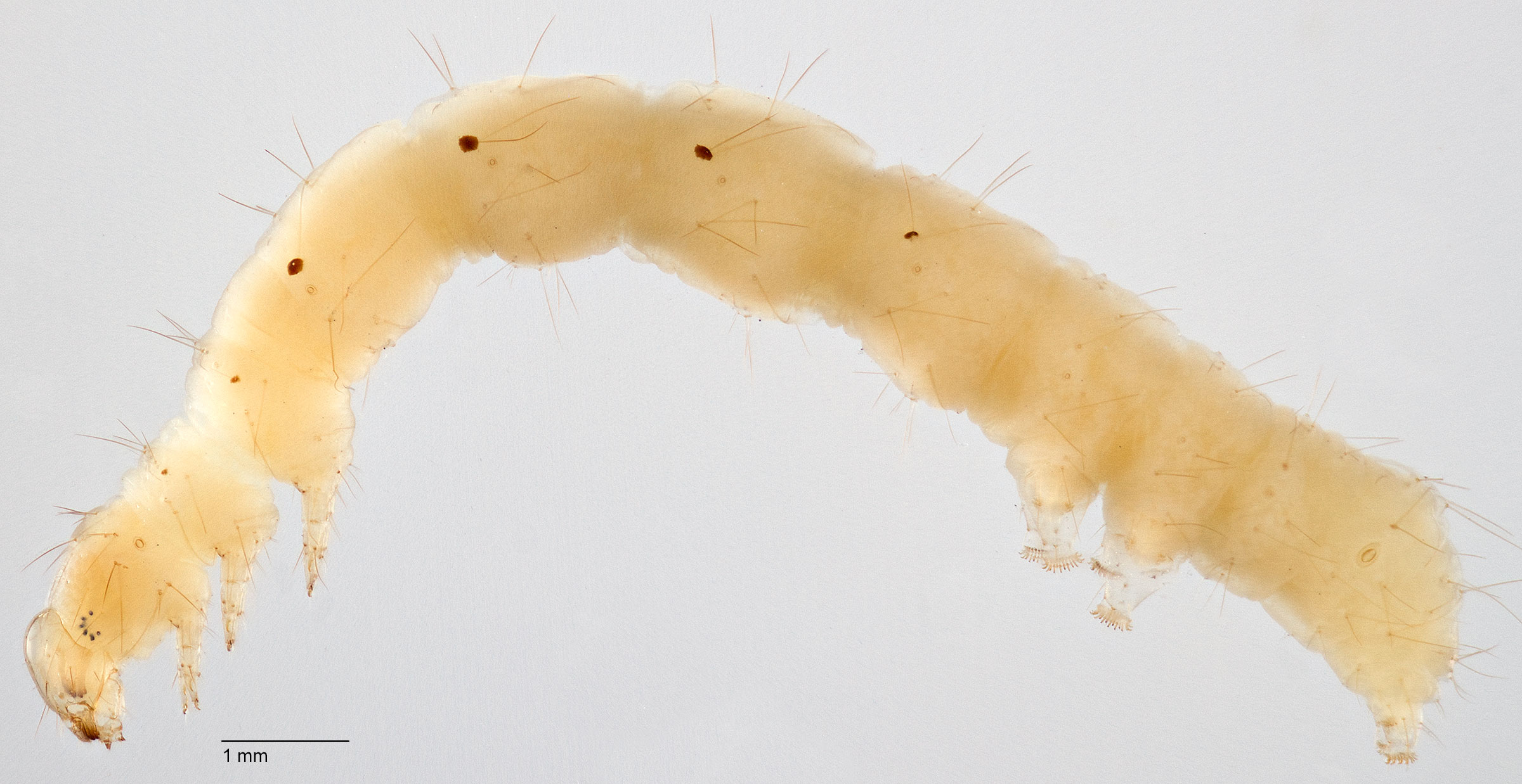
Fig. 1: Late instar, lateral view
Trichoplusia ni
Fig. 1: Late instar, lateral view
Fig. 2: Mid-instar, lateral view
Trichoplusia ni
Fig. 2: Mid-instar, lateral view
Fig. 3: Early instar, lateral view
Trichoplusia ni
Fig. 3: Early instar, lateral view