Anastrepha and
Toxotrypana:
descriptions, illustrations, and interactive keys

Anastrepha and
Toxotrypana:
|
|
Body. Setae red brown, or dark red brown.
Head. Frons with paired elongate brown mark along eye margin, not connected to mark on ocellar tubercle (usually), or with brown band or mark including ocellar tubercle and extending to eye margin (lateral mark rarely very narrowly connected posteriorly to mark on ocellar tubercle). Anterior or single orbital seta anterior or mesal to brown band, or at margin of brown band. Occiput without brown marks. Frontal setae 3–6 (usually 3–5). Orbital setae 1. Ocellar seta weak, small or absent. Gena with brown spot below eye. Facial carina in profile concave or flat on dorsal 2/3. Face with ventral part gradually tapered laterally; without brown markings. Antenna not extended to ventral margin of face.
Thorax. Mesonotum length 2.3–2.8 mm (2.37–2.66, n=6). Scutum nonmicrotrichose (except postsutural lateral margin, lateral to supra-alar seta). Scutellum disc mostly or entirely without microtrichia. Postpronotal, presutural supra-alar, dorsocentral, intra-alar and scutellar setae well developed, subequal to or longer than scutellum length; postpronotal seta on posterior half of postpronotal lobe. Acrostichal seta well developed. Basal scutellar seta strong, longer than scutellum. Katepisternal seta weak, no larger than postocellar seta, or absent. Mesonotum orange, or dark orange; with presutural lateral pale vitta on lateral margin of scutum and posterior part of notopleuron (including posterior corner). Scutum presutural dorsocentral pale vitta present and connected anteriorly with pale area of postpronotal lobe; with 5 (medial, dorsocentral, and sublateral) pale postsutural vittae; pale medial vitta narrow posteriorly, not expanded, or with posterior end bilobed (and connected to dorsocentral vitta); pale sublateral postsutural vitta extended posteriorly to intra-alar seta. Scutum posteriorly with pair of brown spots or markings. Scutum posteriorly with brown spot or vitta between postalar and intra-alar lines (often also with medial brown spot between acrostichal setae, sometimes extended to posterior margin); without brown vittae. Scutellum with at least basal third of sides and disk brown or orange, distinctly darker than apex (orange basally, with pair of dark brown spots laterally at midlength, sometimes narrowly connected, apex white). Mesopleuron mostly yellow to orange, with dark brown spots or bands on at least anepisternum, katepisternum and anepimeron. Subscutellum entirely yellow to orange, or yellow to orange, with medial black spot. Mediotergite entirely yellow to orange. Femora entirely yellow to orange. Fore femur with posterodorsal and ventral rows of well developed setae.
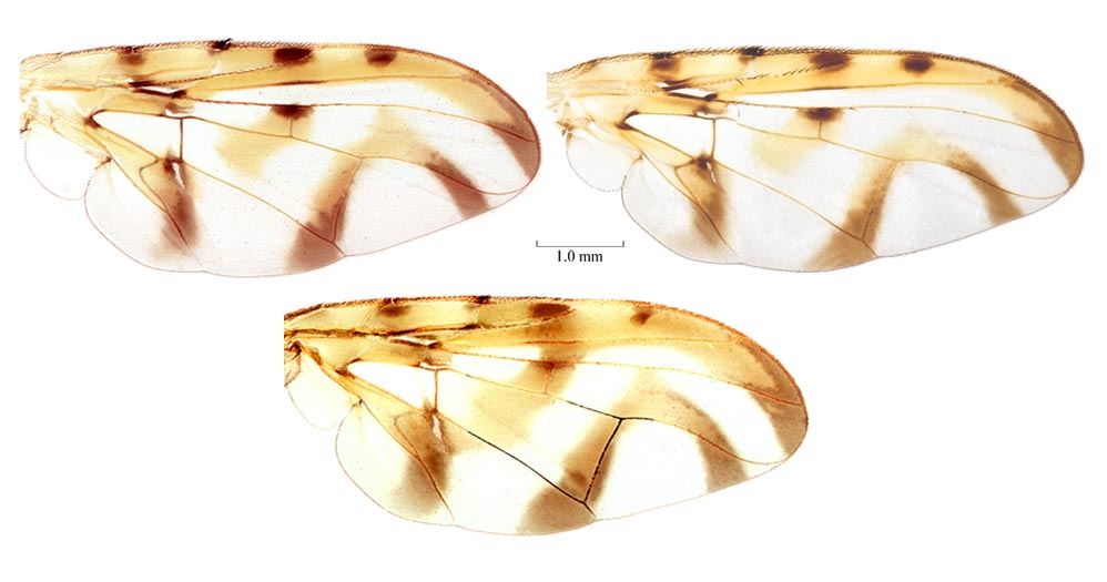
Wings. Wing pattern typical Anastrepha pattern (S-band complete or at most interrupted at crossvein r-m, C-band and at least proximal arm of V-band present), or with basal half of S-band divided into 3 parts by interruptions along veins R4+5 and Cu1 (i.e., a spot or partial band present in basal half of cell dm), C-band and at least proximal arm of V-band present. Cell c mostly or entirely infuscated to subhyaline, or paler posteriorly, without distinct subapical hyaline area. C-band broadly extending to vein M in cell br along cell bm; not covering base of cell r2+3. C-band and S-band connected along costal margin (connection extending at least to vein R2+3). S-band extended anteriorly to vein R4+5 and covering all of crossvein r-m. Cell bm entirely hyaline or infuscated only along subapical fold. S-band base without extension in middle of cell cu1 to posterior wing margin; without extension in cell a1 to or almost to posterior margin, or with extension in cell a1 to or almost to posterior margin. Cubital streak (isolated base of S-band) entirely covering cell bcu. Cubital streak (isolated base of S-band) bordering base of vein Cu1 but not extending to dm-cu, or extending at most slightly beyond bm-cu. Subapical hyaline area in radial cells distal to r-m extending anteriorly to vein R2+3, or extending into cell r2+3 but not reaching vein R2+3. S-band distal section without marginal hyaline band or spots in cell r2+3 or near apices of R2+3 or R4+5. S-band distally extended to apex of vein M, or not extended to apex of vein M. V-band proximal arm as dark as apical half of S-band; not connected anteriorly to S-band. V-band distal arm complete; connected to proximal arm of V band. Pterostigma ratio 3.6–5. Ratio of costa length between apices of Sc and R1/length between apices of R1 and R2+3 0.55–0.65. Vein R2+3 not sinuous; without accessory vein. Vein R4+5 distal to crossvein r-m more or less evenly curved or not strongly bowed medially. Vein M ratio (distance from bm-cu to r-m/distance from bm-cu to dm-cu) 0.55–0.6 (0.56–0.58). Cell bcu posteroapical lobe shorter than vein A1+Cu2. Costa in male with setulae on anterior margin between crossvein h and apex of vein R1 similar to other setulae. Crossvein dm-cu orientation with anterior end more distal than posterior end.
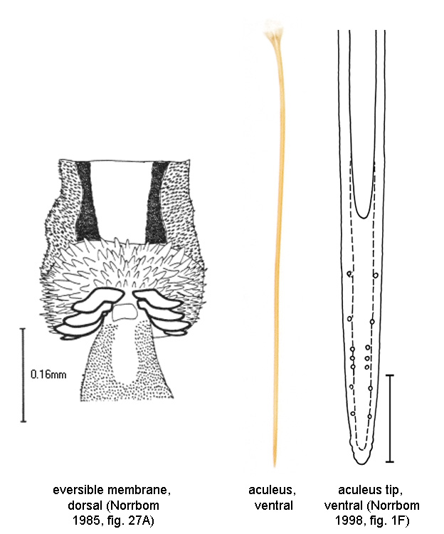
Abdomen. Abdomen ovate or parallel-sided, syntergite 1+2 gradually broadening or parallel-sided. Abdominal tergite with brown markings. Abdominal tergites 3–5 with sublateral or lateral dark brown spots or short bands, or at least with syntergite 1+2 with dark brown band. Epandrium posterodorsal margin evenly rounded. Lateral surstylus in posterior view very short, barely extended beyond prensisetae, rounded apically. Lateral surstylus in posterior view not boot-shaped. Lateral surstylus in lateral view short, apex blunt. Phallus length 0.1–0.3 mm; ratio (phallus length/mesonotum length) 0.02–0.15. Glans absent. Proctiger lateral and ventral sclerotized areas connected, lateral areas separate dorsally. Oviscape straight; length 2.75–3.3 mm (2.79–3.24, n=6); length ratio (oviscape length/mesonotum length) 1.1–1.35 (1.16–1.31, n=6). Eversible membrane with dorsobasal denticles mostly small and weak, apical row very large, stout, strongly sclerotized, and divided medially. Aculeus length 2.4–2.9 mm (2.5–2.8). Aculeus in ventral view more or less parallel-sided except extreme base. Aculeus tip length 0.11–0.15 mm; width 0.02–0.035 mm; lateral margins not curved dorsally; slender, needle-like, with circular cross-section; not flared outward at or proximal to base; without ridges or lobes; with minute serration, visible only with compound microscope, or with fine serrations; serrated part 0.1–0.2 times length of tip. Aculeus tip with serrations separated by less than width of serration. Spermathecae membranous; ovoid.
Sex of recorded specimens: male and female. Species group: daciformis group.
This species is not considered economically important. Its host plants are unknown, although it probably attacks fruits of species of Sapotaceae like other species of the daciformis species group. Refer to the Fruit Fly Databases for host plant information.
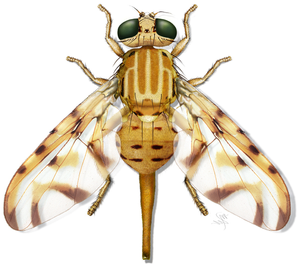
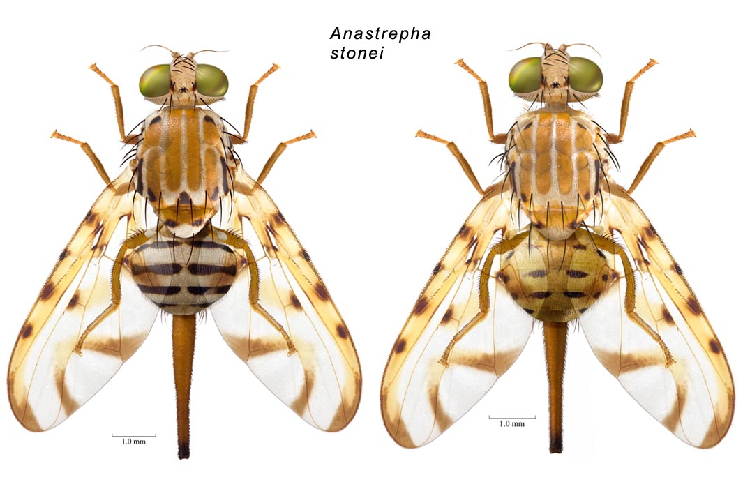
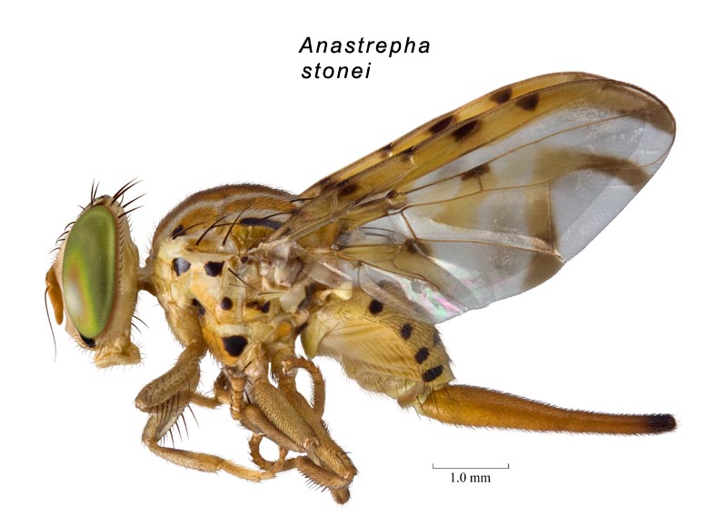
• Habitus, female (dorsal, painting). • Habitus, female (dorsal, photos). • Habitus, female (lateral). • Wings. • Terminalia, female.
Fruit Fly Databases for host plant, distribution, and nomenclatural information. Google search.