Anastrepha and
Toxotrypana:
descriptions, illustrations, and interactive keys

Anastrepha and
Toxotrypana:
|
|
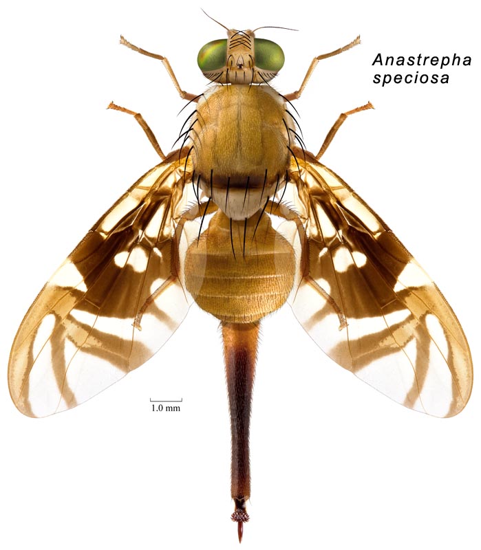
Body. Setae dark brown to black.
Head. Frons without brown markings except ocellar tubercle, or with brown band or mark including ocellar tubercle and extending to eye margin (mark U-shaped, narrow, connected only to posterior side of mark on ocellar tubercle). Occiput without brown marks. Frontal setae 2–5 (usually 3–4). Orbital setae 2. Ocellar seta weak, small or absent. Gena without brown spot. Facial carina in profile concave or flat on dorsal 2/3. Face with ventral part gradually tapered laterally; without brown markings.
Thorax. Mesonotum length 2.6–3.65 mm (2.63–3.61, n=10). Postpronotal lobe and notopleuron entirely microtrichose. Scutum mostly or entirely microtrichose. Scutellum disc entirely microtrichose. Postpronotal, presutural supra-alar, dorsocentral, intra-alar and scutellar setae well developed, subequal to or longer than scutellum length; postpronotal seta on posterior half of postpronotal lobe. Acrostichal seta well developed. Basal scutellar seta strong, longer than scutellum. Katepisternal seta well developed, at least half as large as anepimeral seta. Mesonotum yellow, or orange. Scutum presutural dorsocentral pale vitta absent; with 3 (both medial and sublateral) pale postsutural vittae; pale medial vitta with posterior end broadly quadrate; pale sublateral postsutural vitta not extended posteriorly to intra-alar seta. Scutum posteriorly with brown or orange brown band or other transverse marking or larger posteromedial mark. Scutum posteriorly with dark band or broad marking on posterior margin; scutal posterior brown band extended laterally to include intra-alar seta; without brown vittae. Scutellum entirely yellow or with dark markings only on extreme base of disk. Mesopleuron mostly yellow to orange, without brown markings. Subscutellum entirely yellow to orange. Mediotergite entirely yellow to orange.
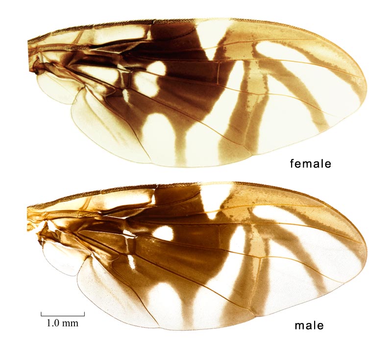
Wings. Wing length 6.2–8.2 mm (6.22–8.15, n=9). Wing pattern typical Anastrepha pattern (S-band complete or at most interrupted at crossvein r-m, C-band and at least proximal arm of V-band present). C-band broadly extending to vein M in cell br along cell bm; covering base of cell r2+3; yellow or orange area posterior to pterostigma small or absent, not extending beyond cell r1 nor distally beyond level of basal third of pterostigma. C-band and S-band connected (broadly along vein R4+5, cell r1 with basomarginal hyaline spot). Basal hyaline area between C-band and S-band extended to vein R4+5 (usually), or extended into cell br but not reaching vein R4+5 (small, at most 0.60 times as long as distal brown area of cell). Cell r1 basomarginal hyaline spot triangular to quadrate. Cell r1 basomarginal hyaline spot apex aligned proximal to crossvein r-m. S-band extended anteriorly to vein R4+5 and covering all of crossvein r-m. Cell bm entirely hyaline or infuscated only along subapical fold, or infuscated distally, or entirely infuscated. S-band posterior margin with distinct incision in cell cu1. S-band base with extension in middle of cell cu1 to or almost to posterior wing margin; without extension in cell a1 to or almost to posterior margin. Subapical hyaline area in radial cells distal to r-m extending anteriorly to vein R2+3. S-band distal section without marginal hyaline band or spots in cell r2+3 or near apices of R2+3 or R4+5. S-band distally not extended to apex of vein M. V-band proximal arm as dark as apical half of S-band; extending more than 1/3 distance from apex of vein Cu1 to apex of vein A1+Cu2, or extending less than 1/3 distance from apex of vein Cu1 to apex of vein A1+Cu2 ([confirm]); connected anteriorly to S-band along vein R4+5 or in cell r2+3 (connected broadly); not connected to S-band in cell dm. V-band distal arm complete; connected to proximal arm of V band. Apex of V-band not extended from vein R4+5 to vein M, hyaline area present between band and vein M. S-band distal section width ratio (width of S-band/width of cell r2+3, both measured perpendicular to costal margin at apex of vein R2+3) 0.8–1 (0.85–1.00, n=7). Area surrounding apex of lobe of cell bcu with microtrichia similar in density to area anterdistal to it along vein Cu1. Cell c: pterostigma ratio (cell c length/pterostigma length) 1.26–1.50, n=9. Vein R1 ratio (distance from wing base to apex of R1/wing length) 0.51–0.55, n=7. Vein R2+3 not sinuous; without accessory vein. Vein R4+5 distal to crossvein r-m more or less evenly curved or not strongly bowed medially. Vein M ratio (distance from bm-cu to r-m/distance from bm-cu to dm-cu) 0.67–0.71, n=9. Vein M curvature ratio (width of cell r4+5 at apex/width at level of dm-cu) 1.15–1.45 (1.21–1.41, n=9). Cell bcu ratio (length/anterior margin length along vein Cu) 1.32–1.64, n=8. Cell bcu posteroapical lobe shorter than vein A1+Cu2. Crossvein dm-cu orientation with anterior end more distal than posterior end.
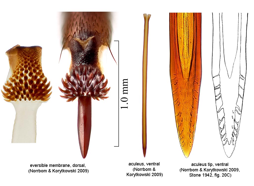
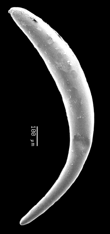
Abdomen. Abdomen ovate or parallel-sided, syntergite 1+2 gradually broadening or parallel-sided. Abdominal tergite without brown markings. Abdominal tergites evenly microtrichose. Epandrium posterodorsal margin evenly rounded. Phallus length 5.50–6.00, n=3; ratio (phallus length/mesonotum length) 1.65–2.00, n=3. Glans present; without spinules. Proctiger lateral and ventral sclerotized areas connected, lateral areas separate dorsally. Oviscape at least apex brown; straight; length 3.6–4.75 mm (3.62–4.70, n=7); length ratio (oviscape length/mesonotum length) 1.15–1.45 (1.23–1.38, n=7); spiracle ratio (distance from base to spiracle/oviscape length) 0.24–0.35, n=7. Eversible membrane with dorsobasal denticles all sclerotized, in continuous triangular to semicircular or suboval pattern. Eversible membrane with 40–60 denticles (large, hooklike dorsobasal denticles in triangular to semicircular pattern). Aculeus length 3.35–4.75 mm (3.40–4.70, n=7). Aculeus length/oviscape length 0.94--1.02, n=7. Aculeus in ventral view more or less parallel-sided except extreme base. Aculeus in lateral view straight or ventrally curved. Aculeus tip length 0.25–0.32 mm (0.26–0.31, n=7); width 0.1–0.14 mm (0.11–0.135, n=6); length/width in ventral view 2.10–2.50, n=6; depth (width in lateral view)/width (in ventral view) 0.50–0.62, n=4; lateral margins not curved dorsally; not flared outward at or proximal to base; without ridges or lobes; without elongate dorsolateral depressions apically; with minute serration, visible only with compound microscope, or with fine serrations; serrated part 0.27–0.39, n=6. Aculeus tip serrations not extending onto dorsal side basally. Aculeus tip with serrations separated by less than width of serration. Egg without lobe.
Data source: Stone 1942, Norrbom & Korytkowski 2008. Sex of recorded specimens: male and female. Species group: robusta group, speciosa clade.
This species is not considered economically important. Refer to the Fruit Fly Databases for host plant information.
• Habitus, female (dorsal). • Wings. • Terminalia, female. • Egg (SEM).
Fruit Fly Databases for host plant, distribution, and nomenclatural information. Google search.