Anastrepha and
Toxotrypana:
descriptions, illustrations, and interactive keys

Anastrepha and
Toxotrypana:
|
|
Body. Setae orange brown, or red brown.
Head. Frons without brown markings except ocellar tubercle. Occiput without brown marks. Frontal setae 3–4. Orbital setae 2, or 1 (rarely). Ocellar seta weak, small or absent. Gena without brown spot. Facial carina in profile concave or flat on dorsal 2/3. Face with ventral part gradually tapered laterally; without brown markings. Antenna not extended to ventral margin of face. Arista of male without preapical expansion. Palpus in lateral view evenly setulose.
Thorax. Mesonotum length 2.1–3 mm (2.16–2.95, n=13). Postpronotal lobe and notopleuron entirely microtrichose. Scutum mostly or entirely microtrichose. Scutellum disc entirely microtrichose. Postpronotal, presutural supra-alar, dorsocentral, intra-alar and scutellar setae well developed, subequal to or longer than scutellum length; postpronotal seta on posterior half of postpronotal lobe. Acrostichal seta well developed. Basal scutellar seta strong, longer than scutellum. Katepisternal seta weak, no larger than postocellar seta, or absent. Mesonotum yellow, or orange. Scutum with 3 (both medial and sublateral) pale postsutural vittae; pale medial vitta narrow posteriorly, not expanded, or with posterior end ovoid (usually), or with posterior end broadly quadrate; pale sublateral postsutural vitta extended posteriorly to intra-alar seta. Scutum posteriorly without brown or orange brown markings. Scutum without brown vittae. Scutellum entirely yellow or with dark markings only on extreme base of disk. Mesopleuron mostly yellow to orange, without brown markings. Subscutellum entirely yellow to orange. Mediotergite entirely yellow to orange. Femora entirely yellow to orange. Fore femur with posterodorsal and ventral rows of well developed setae.
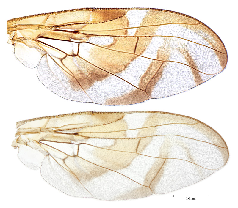
Wings. Wing pattern typical Anastrepha pattern (S-band complete or at most interrupted at crossvein r-m, C-band and at least proximal arm of V-band present). Cell c mostly or entirely infuscated to subhyaline, or paler posteriorly, without distinct subapical hyaline area. C-band broadly extending to vein M in cell br along cell bm; covering base of cell r2+3; yellow or orange area posterior to pterostigma broad, extending distally into cells r1 and r2+3 at least to level of midlength of pterostigma. C-band and S-band connected (broadly along vein R4+5, cell r1 with basomarginal hyaline spot). Basal hyaline area between C-band and S-band extended to vein R4+5. Cell r1 basomarginal hyaline spot triangular to quadrate. Cell r1 basomarginal hyaline spot apex aligned proximal to crossvein r-m. S-band extended anteriorly to vein R4+5 and covering all of crossvein r-m. Cell bm entirely hyaline or infuscated only along subapical fold. S-band posterior margin without incision in cell cu1. S-band base without extension in middle of cell cu1 to posterior wing margin; without extension in cell a1 to or almost to posterior margin. S-band middle section predominantly or entirely orange, often with brown margins. Subapical hyaline area in radial cells distal to r-m extending anteriorly to vein R2+3. S-band distal section without marginal hyaline band or spots in cell r2+3 or near apices of R2+3 or R4+5. S-band distally extended to apex of vein M, or not extended to apex of vein M. V-band proximal arm as dark as apical half of S-band; extending more than 1/3 distance from apex of vein Cu1 to apex of vein A1+Cu2; not connected anteriorly to S-band (usually), or connected anteriorly to S-band along vein R4+5 or in cell r2+3; not connected to S-band in cell dm. V-band distal arm complete; connected to proximal arm of V band. Apex of V-band not extended from vein R4+5 to vein M, hyaline area present between band and vein M. Area surrounding apex of lobe of cell bcu with microtrichia similar in density to area anterdistal to it along vein Cu1. Area between S-band and V-band entirely microtrichose in cells dm and cu1. Pterostigma ratio 3–3.7. Ratio of costa length between apices of Sc and R1/length between apices of R1 and R2+3 0.37–0.5. Vein R2+3 not sinuous; without accessory vein. Vein R4+5 distal to crossvein r-m more or less evenly curved or not strongly bowed medially. Cell bcu posteroapical lobe shorter than vein A1+Cu2. Costa in male with setulae on anterior margin between crossvein h and apex of vein R1 similar to other setulae. Crossvein dm-cu orientation with anterior end more distal than posterior end.
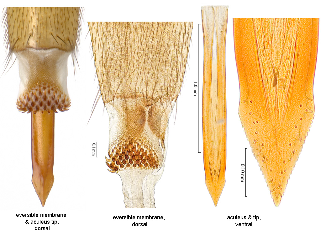
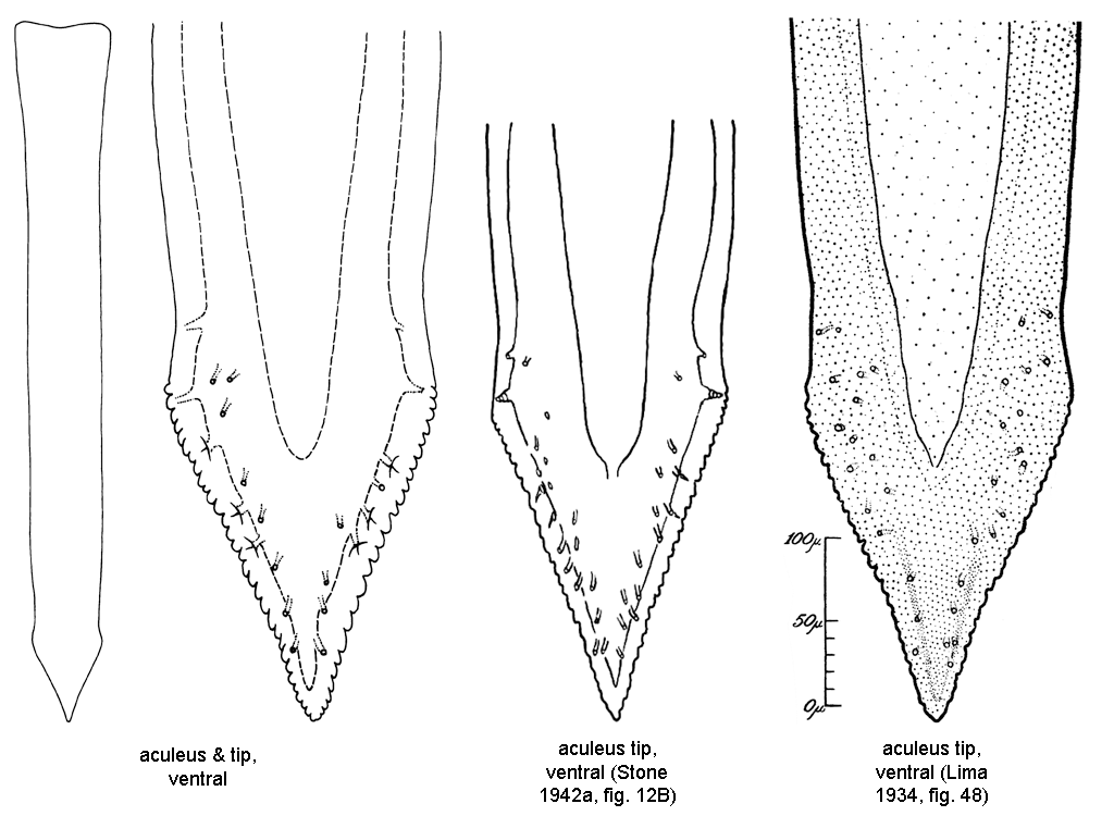
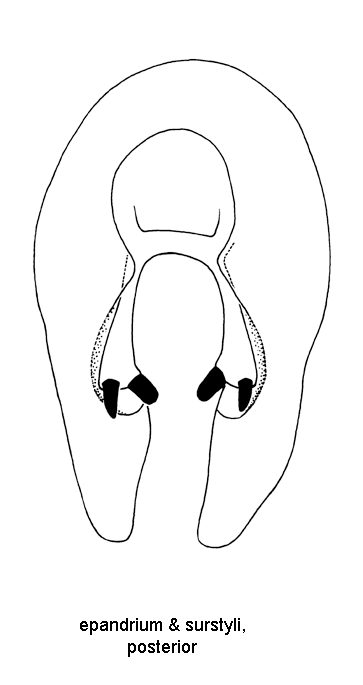
Abdomen. Abdomen ovate or parallel-sided, syntergite 1+2 gradually broadening or parallel-sided. Abdominal tergite without brown markings. Lateral surstylus in posterior view long, slightly tapered, somewhat truncate apically, or long, rounded apically. Lateral surstylus in posterior view not boot-shaped. Phallus length 2.3–2.9 mm (2.37–2.83, n=5); ratio (phallus length/mesonotum length) 0.85–1.1 (0.9–1.06, n=5). Glans present; without spinules. Proctiger lateral and ventral sclerotized areas separate, lateral areas separate dorsally. Oviscape straight; length 1.6–1.95 mm (1.66–1.89, n=8); length ratio (oviscape length/mesonotum length) 0.6–0.8 (0.63–0.77, n=8). Eversible membrane with dorsobasal denticles all sclerotized, in continuous triangular to semicircular or suboval pattern. Eversible membrane with relatively small. Aculeus length 1.45–1.8 mm (1.5–1.75, n=8). Aculeus length/oviscape length 0.82–0.96 (0.84–0.94, n=8). Aculeus in ventral view more or less parallel-sided except extreme base. Aculeus tip length/aculeus length 0.08–0.11 (0.09–0.10, n=8). Aculeus tip length 0.13–0.18 mm (0.14–0.17, n=8); width 0.13–0.18 mm (0.14–0.17, n=8); length/width in ventral view 0.85–1.1 (0.88–1.07, n=8); lateral margins not curved dorsally; triangular, or spatulate; without ridges or lobes; without elongate dorsolateral depressions apically; with medium sized serrations; serrated part 1.15–1.4 times length of tip (1.18–1.36, n=8). Aculeus tip serrations not extending onto dorsal side basally. Aculeus tip with serrations separated by less than width of serration. Spermathecae sclerotized; ovoid. Egg with long lobe on anterior end distal to micropyle (Dutra et al. 2011b).
Sex of recorded specimens: male and female. Species group: spatulata group.
This species is considered of some economical importance. It is unusual for Anastrepha species in breeding in the terminal buds and shoots (rather than fruit) of its host, yuca (Manihot esculenta Crantz). It does not damage the edible tubers of the plant, but reduces growth and increases bacterial stem rot. Host records from other plants are questionable. Refer to the Fruit Fly Databases for host plant information.
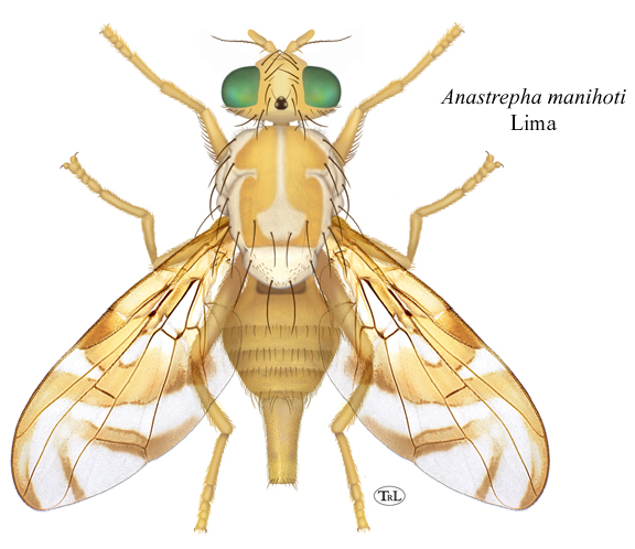
• Habitus, female (dorsal). • Wings. • Terminalia, female. • Terminalia, female (aculeus, line). • Terminalia, male.
Fruit Fly Databases for host plant, distribution, and nomenclatural information. Google search.