Anastrepha and
Toxotrypana:
descriptions, illustrations, and interactive keys

Anastrepha and
Toxotrypana:
|
|
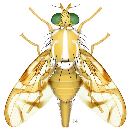
Head. Frons without brown markings except ocellar tubercle. Occiput without brown marks. Orbital setae 2. Ocellar seta weak, small or absent. Gena without brown spot. Facial carina in profile concave or flat on dorsal 2/3. Face with ventral part gradually tapered laterally; without brown markings. Antenna not extended to ventral margin of face.
Thorax. Mesonotum length 2.5–3.5 mm (2.66–3.33, n=18; 2.75–3.30, Stone 1942a). Postpronotal lobe and notopleuron entirely microtrichose. Scutum mostly or entirely microtrichose. Scutellum disc entirely microtrichose. Postpronotal, presutural supra-alar, dorsocentral, intra-alar and scutellar setae well developed, subequal to or longer than scutellum length; postpronotal seta on posterior half of postpronotal lobe. Acrostichal seta well developed. Basal scutellar seta strong, longer than scutellum. Mesonotum yellow, or orange. Scutum presutural dorsocentral pale vitta absent; with 3 (both medial and sublateral) pale postsutural vittae; pale medial vitta narrow posteriorly, not expanded, or with posterior end ovoid; pale sublateral postsutural vitta extended posteriorly to intra-alar seta. Scutum posteriorly without brown or orange brown markings, or with only single medial brown spot on scuto-scutellar suture (usually). Scutum without brown vittae. Scutellum entirely yellow or with dark markings only on extreme base of disk. Mesopleuron mostly yellow to orange, without brown markings. Subscutellum yellow to red brown medially, dark brown laterally. Mediotergite yellow to red brown medially, dark brown laterally. Femora entirely yellow to orange. Fore femur with posterodorsal and ventral rows of well developed setae.
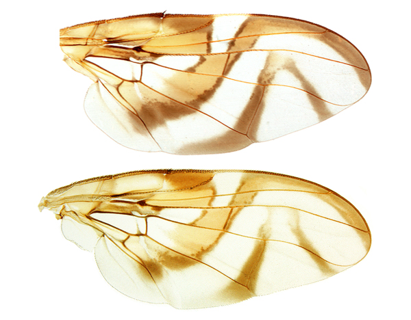
Wings. Wing pattern typical Anastrepha pattern (S-band complete or at most interrupted at crossvein r-m, C-band and at least proximal arm of V-band present). Cell c mostly or entirely infuscated to subhyaline, or paler posteriorly, without distinct subapical hyaline area. C-band broadly extending to vein M in cell br along cell bm; covering base of cell r2+3; yellow or orange area posterior to pterostigma broad, extending distally into cells r1 and r2+3 at least to level of midlength of pterostigma. C-band and S-band separated (by hyaline band from cell bm to costal margin in cell r1, narrowed along vein R4+5), or connected (along vein R4+5, cell r1 with basomarginal hyaline spot). Cell r1 basomarginal hyaline spot triangular to quadrate. Cell r1 basomarginal hyaline spot apex aligned proximal to crossvein r-m. S-band extended anteriorly to vein R4+5 and covering all of crossvein r-m. Cell bm entirely hyaline or infuscated only along subapical fold. S-band base without extension in middle of cell cu1 to posterior wing margin; without extension in cell a1 to or almost to posterior margin. S-band middle section predominantly or entirely orange, often with brown margins. Subapical hyaline area in radial cells distal to r-m extending anteriorly to vein R2+3. S-band distal section without marginal hyaline band or spots in cell r2+3 or near apices of R2+3 or R4+5. S-band distally not extended to apex of vein M. V-band proximal arm as dark as apical half of S-band; extending more than 1/3 distance from apex of vein Cu1 to apex of vein A1+Cu2; not connected anteriorly to S-band, or connected anteriorly to S-band along vein R4+5 or in cell r2+3; not connected to S-band in cell dm. V-band distal arm complete; isolated, not connected to proximal arm of V-band or to S-band, or connected to proximal arm of V band. Apex of V-band not extended from vein R4+5 to vein M, hyaline area present between band and vein M. S-band distal section width ratio (width of S-band/width of cell r2+3, both measured perpendicular to costal margin at apex of vein R2+3) 0.4–0.85 (0.45–0.66, n=5; 0.73–0.81 for Mesoamerican specimens, n=3). Area surrounding apex of lobe of cell bcu with microtrichia similar in density to area anterdistal to it along vein Cu1. Area between S-band and V-band entirely microtrichose in cells dm and cu1. Pterostigma ratio 3–3.7. Ratio of costa length between apices of Sc and R1/length between apices of R1 and R2+3 0.38–0.56. Vein R2+3 not sinuous; without accessory vein. Vein M curvature ratio (width of cell r4+5 at apex/width at level of dm-cu) 0.85–1 (0.90–0.97, n=8). Cell bcu posteroapical lobe shorter than vein A1+Cu2. Costa in male with setulae on anterior margin between crossvein h and apex of vein R1 similar to other setulae. Crossvein dm-cu orientation with anterior end more distal than posterior end.
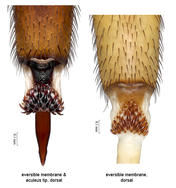
Abdomen. Abdomen ovate or parallel-sided, syntergite 1+2 gradually broadening or parallel-sided. Abdominal tergite without brown markings. Lateral surstylus in posterior view long, slightly tapered, somewhat truncate apically. Lateral surstylus in posterior view not boot-shaped. Phallus length 2.6–3.45 mm (2.75–3.33, n=6); ratio (phallus length/mesonotum length) 0.9–1.1 (0.93–1.06, n=6). Glans present; without spinules. Proctiger lateral and ventral sclerotized areas separate, lateral areas separate dorsally. Oviscape entirely yellow to orange brown; straight; length 1.65–2.15 mm (1.75–2.12, n=13; 1.65–2.10, Stone 1942a); length ratio (oviscape length/mesonotum length) 0.55–0.75 (0.59–0.70, n=12). Eversible membrane with dorsobasal denticles all sclerotized, in continuous triangular to semicircular or suboval pattern. Eversible membrane with 25–40 denticles (long hooklike dorsobasal denticles in 4–5 irregular rows in subtriangular pattern). Aculeus length 1.4–2.06 mm (1.60–2.06, n=26; 1.50–1.95, Stone 1942a; 1.4–1.9, Araujo & Zucchi 2006). Aculeus in ventral view more or less parallel-sided except extreme base. Aculeus tip length/aculeus length 0.11–0.17 (n=28). Aculeus tip length 0.2–0.3 mm (0.21–0.30, n=25; 0.20–0.30, Araujo & Zucchi 2006); width 0.12–0.15 mm (n=21); lateral margins not curved dorsally; gradually tapering, but with medial constriction; not flared outward at or proximal to base; without ridges or lobes; without elongate dorsolateral depressions apically; with medium sized serrations; serrated part 0.47–0.65 times length of tip (0.48–0.64, n=25). Aculeus tip serrations not extending onto dorsal side basally. Aculeus tip with serrations separated by less than width of serration. Spermathecae sclerotized; ovoid. Egg without lobe.
Other names for this species: Anastrepha braziliensis Greene, Anastrepha costarukmanii Capoor, Anastrepha fraterculus var. soluta Bezzi, Anastrepha lambayecae Korytkowski & Ojeda, Anastrepha peruviana Townsend, Anastrepha pseudofraterculus Capoor, Anastrepha scholae Capoor, Anthomyia frutalis Weyenbergh, Tephritis mellea Walker, Trypeta unicolor Loew. Data source: Stone 1942a. Data recording very inadequate. Sex of recorded specimens: male and female. Species group: fraterculus group.
This species, commonly known as the South American fruit fly, is a major pest of various cultivated fruits. It is considered a pest of quarantine significance by USDA-APHIS-PPQ and many other regulatory agencies. The main damage is caused by the larvae, which feed inside the fruit. Refer to the Fruit Fly Databases for host plant information.
• Habitus, female (dorsal). • Habitus, male (dorsal). • Wings. • Terminalia, female (eversible membrane). • Terminalia, female (aculeus). • Terminalia, male.
Fruit Fly Databases for host plant, distribution, and nomenclatural information. Google search.